HES1


Transcription factor HES1 (hairy and enhancer of split-1) is a protein that is encoded by the Hes1 gene, and is the mammalian homolog of the hairy gene in Drosophila.[5][6] HES1 is one of the seven members of the Hes gene family (HES1-7). Hes genes code nuclear proteins that suppress transcription.[7]
This protein belongs to the basic helix-loop-helix (bHLH) family of transcription factors. It is a transcriptional repressor of genes that require a bHLH protein for their transcription. The protein has a particular type of basic domain that contains a helix interrupting protein that binds to the N-box promoter region rather than the canonical enhancer box (E-box).[6] As a member of the bHLH family, it is a transcriptional repressor that influences cell proliferation and differentiation in embryogenesis.[7] HES1 regulates its own expression via a negative feedback loop, and oscillates with approximately 2-hour periodicity.[8]
Structure
There are three conserved domains in Hes genes that impart transcriptional functions: the bHLH domain, the Orange domain, and the WRPW motif. Hes genes differ from other bHLH factors in that they have a proline reside in the middle of the basic DNA binding region. This proline has been proposed to give Hes proteins unique DNA binding capacity. While most bHLH factors bind to the E-box consensus sequence (CANNTG) that is present in the promoter region of target genes, Hes factors bind more preferentially to the Class C site or N box (CACNAG).[7] The Orange domain serves to regulate the choice of bHLH heterodimer partners.[9] The C-terminal WRPW domain inhibits transcription.[10]
Interactions
Similarly to other HES proteins, Hes1 has been shown to interact with the co-repressors which Transducin-like E(spl) (TLE) genes and the Groucho-related gene (Grg), both homologs of the Drosophila groucho.[11] Because Groucho in Drosophila inhibits transcription by recruiting histone deacetylase, it is likely that a Hes-Groucho complex actively blocks transcription by disabling chromatin. Hes proteins also heterodimerize with bHLH repressors such as Hey1 and Hey2, a process which also blocks transcription. Hes factors also heterodimerize with bHLH activators such as E47, also known as Tcfe2a, and Mash1, also known as Ascl1, both of which are the mammalian homologs to proneural genes in Drosophila. The E47-Hes and Mash1-Hes heterodimer complexes cannot bind DNA, and therefore repress transcription.[7] Hes1 also interacts with TLE2[12] and Sirtuin 1.[13]
HES1 and stem cells
HES1 influences the maintenance of certain stem cells and progenitor cells. Specifically, HES1 influences the timing of differentiation by repressing bHLH activators, and determines binary cell fate. HES1 has been shown to play a large role in both the nervous, and digestive systems. HES1 has been shown to influence these two systems partially through the Notch signaling pathway.
Neural development
HES1 is expressed in both neuroepithelial cells and radial glial cells, both neural stem cells. Hes1 expression, along with that of Hes5, covers the majority of the developing embryo at embryonic day 10.5.[14] After this point, expression of Hes1 is limited to the subventricular zone. In HES1 knockout (KO) mice, Mash1 is compensatorily upregulated, and neurogenesis is accelerated. Indeed, if the expression of Hes1, Hes3, and Hes5 genes is inhibited, the expression of proneural genes increases, and while neurogenesis is accelerated, neural stem cells become prematurely depleted. Contrariwise, if these HES genes are overexpressed, neurogenesis is inhibited.[15] Thus HES1 genes are only involved in maintaining, not creating, neural stem cells.
Additionally, HES1 can guide neural stem cells down one of two paths of differentiation. HES1 can maintain neural stem cells expressing Pax6, but leads cells that are Pax6-negative to an astrocyte differentiation fate.[16] Epigenetic modifications such as DNA methylation also influence HES1's ability to direct differentiation. Demethylation of HES1 target sites in the promoter region of astrocyte-specific genes hastens astrocyte differentiation.[15] The oscillatory nature of Hes1 expression has a role in determining differentiation fate as well. HES1-high embryonic stem cells that received a differentiation signal often adopted a mesodermal fate, while HES1-low cells that received a differentiation signal differentiated into neuronal cells. These results were confirmed using quantitative PCR which showed that HES1-high cells showed high levels of Brachyury and Fgf5 expression (both of which are expressed highly in mesodermal cell types) with comparatively low levels genes expressed in neural cells such as Nestin. By contrast, HES1-low cells showed high levels of expression of genes involved in neural induction and low levels of expression of genes involved in mesodermal differentiation.[17] Cycling HES1 levels also contribute to the maintenance of neural progenitor cells by regulating Neurogenin2 (Ngn2) and Dll1 oscillations.[18] Hes1 levels fluctuate at different frequencies in different parts of the central nervous system: HES1 is continuously expressed at high levels in the boundaries, but vacillates in the compartments. This suggests that alternating HES1 levels may prompt differences in characteristics between anatomical elements of the central nervous system.[7]
Interactions with the Notch pathway
HES1 also plays an important role in the Notch signaling pathway.[19] In the absence of Notch signaling, RBPJ inhibits the expression of HES1. After Notch signals have been processed within the cell, however, the plasma membrane releases the intracellular domain of Notch, which moves to the nucleus where it associates with RBPJ. The binding causes a conformational change which leads co-repressors to disassociate and allows co-activators to bind. The new activating complex then prompts HES1 expression. Notch signaling activates HES1 expression. HES1 has been shown to target at least Notch ligands: Dll1, Jagged1 (Jag1), and Neurogenin-2.[15], [17] Dll1, as with other Notch ligands, has been shown to induce neural differentiation, and HES1 binding of Dll1 blocks neural differentiation and leads to the maintenance of the neural stem cells and neural progenitor cells.[20] Notch signaling also occurs in the intestinal crypt cells. Hyperactivated Notch causes a reduction in the number of secretory cell types (i.e. goblet cells, enteroendocrine cells, and Paneth cells). Deletion of the Notch pathway by removing the Notch expression controller, Rbpsuh, causes the production of nearly only goblet cells.[21]
Digestive system
HES1 has been shown to influence the differentiation decision of cells in the gastrointestinal tract. In pancreatic progenitor cells, HES1 expression inhibits the expression of Ptf1a, which controls exocrine cell differentiation, and Ngn3, which drives differentiation of endocrine cell types that will form the islets of Langerhans.[7] The absence of Hes1 in the developing intestine of mice promotes the increase of Math1 (a protein required for the production of intestinal secretory cell types), which leads to an increase of goblet, enteroendocrine, and Paneth cells. When Hes1 is deleted in mouse and zebrafish, surplus goblet cells and enteroendocrine cells are made while few enterocytes are made.[7], [21] Liver progenitor cells differentiate into two different cell types: hepatocytes and biliary epithelial cells. When Hes1 expression is low, hepatocytes form normally, but bile ducts are completely absent.[22] This phenotype resembles Alagille syndrome, a hallmark of which is mutations in Jagged1. Therefore, Hes-Notch interactions also play a role in digestive organ development.
References
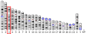
- 1 2 3 GRCh38: Ensembl release 89: ENSG00000114315 - Ensembl, May 2017
- 1 2 3 GRCm38: Ensembl release 89: ENSMUSG00000022528 - Ensembl, May 2017
- ↑ "Human PubMed Reference:".
- ↑ "Mouse PubMed Reference:".
- ↑ Feder JN, Li L, Jan LY, Jan YN (Jul 1994). "Genomic cloning and chromosomal localization of HRY, the human homolog to the Drosophila segmentation gene, hairy". Genomics. 20 (1): 56–61. doi:10.1006/geno.1994.1126. PMID 8020957.
- 1 2 "Entrez Gene: HES1 hairy and enhancer of split 1, (Drosophila)".
- 1 2 3 4 5 6 7 Kageyama R, Ohtsuka T, Kobayashi T (2007). "The Hes gene family: Repressors and oscillators that orchestrate embryogenesis". Development. 134 (7): 1243–1251. doi:10.1242/dev.000786. PMID 17329370.
- ↑ Hirata H, Yoshiura S, Ohtsuka T, Bessho Y, Harada T, Yoshikawa K, Kageyama R (October 2002). "Oscillatory Expression of the bHLH Factor Hes1 Regulated by a Negative Feedback Loop". Science. 298 (5594): 840–843. doi:10.1126/science.1074560. PMID 12399594.
- ↑ Taelman V, Van Wayenbergh R, Sölter M, Pichon B, Pieler T, Christophe D, Bellefroid EJ (2004). "Sequences downstream of the bHLH domain of the Xenopus hairy-related transcription factor-1 act as an extended dimerization domain that contributes to the selection of the partners". Developmental Biology. 276 (1): 47–63. doi:10.1016/j.ydbio.2004.08.019. PMID 15531363.
- ↑ Kang SA, Seol JH, Kim J (2005). "The conserved WRPW motif of Hes6 mediates proteasomal degradation". Biochemical and Biophysical Research Communications. 332 (1): 33–36. doi:10.1016/j.bbrc.2005.04.089. PMID 15896295.
- ↑ Paroush Z, Finley RL, Kidd T, Wainwright SM, Ingham PW, Brent R, Ish-Horowicz D (1994). "Groucho is required for Drosophila neurogenesis, segmentation, and sex determination and interacts directly with hairy-related bHLH proteins". Cell. 79 (5): 805–815. doi:10.1016/0092-8674(94)90070-1. PMID 8001118.
- ↑ Grbavec D, Lo R, Liu Y, Stifani S (December 1998). "Transducin-like Enhancer of split 2, a mammalian homologue of Drosophila Groucho, acts as a transcriptional repressor, interacts with Hairy/Enhancer of split proteins, and is expressed during neuronal development". Eur. J. Biochem. 258 (2): 339–49. doi:10.1046/j.1432-1327.1998.2580339.x. PMID 9874198.
- ↑ Takata T, Ishikawa F (January 2003). "Human Sir2-related protein SIRT1 associates with the bHLH repressors HES1 and HEY2 and is involved in HES1- and HEY2-mediated transcriptional repression". Biochem. Biophys. Res. Commun. 301 (1): 250–7. doi:10.1016/S0006-291X(02)03020-6. PMID 12535671.
- ↑ Hatakeyama J, Bessho Y, Katoh K, Ookawara S, Fujioka M, Guillemot F, Kageyama R (2004). "Hes genes regulate size, shape and histogenesis of the nervous system by control of the timing of neural stem cell differentiation". Development. 131 (22): 5539–5550. doi:10.1242/dev.01436. PMID 15496443.
- 1 2 3 Kageyama R, Ohtsuka T, Kobayashi T (2008). "Roles of Hes genes in neural development". Development, Growth & Differentiation. 50: S97–103. doi:10.1111/j.1440-169X.2008.00993.x. PMID 18430159.
- ↑ Sugimori M, Nagao M, Bertrand N, Parras CM, Guillemot F, Nakafuku M (2007). "Combinatorial actions of patterning and HLH transcription factors in the spatiotemporal control of neurogenesis and gliogenesis in the developing spinal cord". Development. 134 (8): 1617–1629. doi:10.1242/dev.001255. PMID 17344230.
- 1 2 Kobayashi T, Mizuno H, Imayoshi I, Furusawa C, Shirahige K, Kageyama R (2009). "The cyclic gene Hes1 contributes to diverse differentiation responses of embryonic stem cells". Genes & Development. 23 (16): 1870–1875. doi:10.1101/gad.1823109. PMC 2725939. PMID 19684110.
- ↑ Shimojo H, Ohtsuka T, Kageyama R (2008). "Oscillations in Notch Signaling Regulate Maintenance of Neural Progenitors". Neuron. 58 (1): 52–64. doi:10.1016/j.neuron.2008.02.014. PMID 18400163.
- ↑ Kageyama R, Ohtsuka T (1999). "The Notch-Hes pathway in mammalian neural development". Cell Research. 9 (3): 179–188. doi:10.1038/sj.cr.7290016. PMID 10520600.
- ↑ Lowell S, Benchoua A, Heavey B, Smith AG (2006). "Notch Promotes Neural Lineage Entry by Pluripotent Embryonic Stem Cells". PLoS Biology. 4 (5): e121. doi:10.1371/journal.pbio.0040121. PMC 1431581. PMID 16594731.
- 1 2 Crosnier C, Stamataki D, Lewis J (2006). "Organizing cell renewal in the intestine: Stem cells, signals and combinatorial control". Nature Reviews Genetics. 7 (5): 349–359. doi:10.1038/nrg1840. PMID 16619050.
- ↑ Kodama Y, Hijikata M, Kageyama R, Shimotohno K, Chiba T (2004). "The role of notch signaling in the development of intrahepatic bile ducts". Gastroenterology. 127 (6): 1775–1786. doi:10.1053/j.gastro.2004.09.004. PMID 15578515.
Further reading
- Takebayashi K, Sasai Y, Sakai Y, Watanabe T, Nakanishi S, Kageyama R (1994). "Structure, chromosomal locus, and promoter analysis of the gene encoding the mouse helix-loop-helix factor HES-1. Negative autoregulation through the multiple N box elements". J. Biol. Chem. 269 (7): 5150–6. PMID 7906273.
- Grbavec D, Stifani S (1996). "Molecular interaction between TLE1 and the carboxyl-terminal domain of HES-1 containing the WRPW motif". Biochem. Biophys. Res. Commun. 223 (3): 701–5. doi:10.1006/bbrc.1996.0959. PMID 8687460.
- Ström A, Castella P, Rockwood J, Wagner J, Caudy M (1998). "Mediation of NGF signaling by post-translational inhibition of HES-1, a basic helix-loop-helix repressor of neuronal differentiation". Genes Dev. 11 (23): 3168–81. doi:10.1101/gad.11.23.3168. PMC 316755. PMID 9389649.
- Votruba M, Payne A, Moore AT, Bhattacharya SS (1998). "Dominant optic atrophy: exclusion and fine genetic mapping of the candidate gene, HRY". Mamm. Genome. 9 (10): 784–7. doi:10.1007/s003359900867. PMID 9745030.
- Bae S, Bessho Y, Hojo M, Kageyama R (2000). "The bHLH gene Hes6, an inhibitor of Hes1, promotes neuronal differentiation". Development. 127 (13): 2933–43. PMID 10851137.
- Yao J, Lai E, Stifani S (2001). "The winged-helix protein brain factor 1 interacts with groucho and hes proteins to repress transcription". Mol. Cell. Biol. 21 (6): 1962–72. doi:10.1128/MCB.21.6.1962-1972.2001. PMC 86788. PMID 11238932.
- Iso T, Sartorelli V, Poizat C, Iezzi S, Wu HY, Chung G, Kedes L, Hamamori Y (2001). "HERP, a novel heterodimer partner of HES/E(spl) in Notch signaling". Mol. Cell. Biol. 21 (17): 6080–9. doi:10.1128/MCB.21.17.6080-6089.2001. PMC 87325. PMID 11486045.
- Dintilhac A, Bernués J (2002). "HMGB1 interacts with many apparently unrelated proteins by recognizing short amino acid sequences". J. Biol. Chem. 277 (9): 7021–8. doi:10.1074/jbc.M108417200. PMID 11748221.
- Jögi A, Persson P, Grynfeld A, Påhlman S, Axelson H (2002). "Modulation of basic helix-loop-helix transcription complex formation by Id proteins during neuronal differentiation". J. Biol. Chem. 277 (11): 9118–26. doi:10.1074/jbc.M107713200. PMID 11756408.
- Kunisato A, Chiba S, Nakagami-Yamaguchi E, Kumano K, Saito T, Masuda S, Yamaguchi T, Osawa M, Kageyama R, Nakauchi H, Nishikawa M, Hirai H (2003). "HES-1 preserves purified hematopoietic stem cells ex vivo and accumulates side population cells in vivo". Blood. 101 (5): 1777–83. doi:10.1182/blood-2002-07-2051. PMID 12406868.
- Strausberg RL, Feingold EA, Grouse LH, Derge JG, Klausner RD, Collins FS, Wagner L, Shenmen CM, Schuler GD, Altschul SF, Zeeberg B, Buetow KH, Schaefer CF, Bhat NK, Hopkins RF, Jordan H, Moore T, Max SI, Wang J, Hsieh F, Diatchenko L, Marusina K, Farmer AA, Rubin GM, Hong L, Stapleton M, Soares MB, Bonaldo MF, Casavant TL, Scheetz TE, Brownstein MJ, Usdin TB, Toshiyuki S, Carninci P, Prange C, Raha SS, Loquellano NA, Peters GJ, Abramson RD, Mullahy SJ, Bosak SA, McEwan PJ, McKernan KJ, Malek JA, Gunaratne PH, Richards S, Worley KC, Hale S, Garcia AM, Gay LJ, Hulyk SW, Villalon DK, Muzny DM, Sodergren EJ, Lu X, Gibbs RA, Fahey J, Helton E, Ketteman M, Madan A, Rodrigues S, Sanchez A, Whiting M, Madan A, Young AC, Shevchenko Y, Bouffard GG, Blakesley RW, Touchman JW, Green ED, Dickson MC, Rodriguez AC, Grimwood J, Schmutz J, Myers RM, Butterfield YS, Krzywinski MI, Skalska U, Smailus DE, Schnerch A, Schein JE, Jones SJ, Marra MA (2003). "Generation and initial analysis of more than 15,000 full-length human and mouse cDNA sequences". Proc. Natl. Acad. Sci. U.S.A. 99 (26): 16899–903. doi:10.1073/pnas.242603899. PMC 139241. PMID 12477932.
- Takata T, Ishikawa F (2003). "Human Sir2-related protein SIRT1 associates with the bHLH repressors HES1 and HEY2 and is involved in HES1- and HEY2-mediated transcriptional repression". Biochem. Biophys. Res. Commun. 301 (1): 250–7. doi:10.1016/S0006-291X(02)03020-6. PMID 12535671.
- Gratton MO, Torban E, Jasmin SB, Theriault FM, German MS, Stifani S (2003). "Hes6 promotes cortical neurogenesis and inhibits Hes1 transcription repression activity by multiple mechanisms". Mol. Cell. Biol. 23 (19): 6922–35. doi:10.1128/MCB.23.19.6922-6935.2003. PMC 193938. PMID 12972610.
- Thomsen JS, Kietz S, Ström A, Gustafsson JA (2004). "HES-1, a novel target gene for the aryl hydrocarbon receptor". Mol. Pharmacol. 65 (1): 165–71. doi:10.1124/mol.65.1.165. PMID 14722248.
- Kamakura S, Oishi K, Yoshimatsu T, Nakafuku M, Masuyama N, Gotoh Y (2004). "Hes binding to STAT3 mediates crosstalk between Notch and JAK-STAT signalling". Nat. Cell Biol. 6 (6): 547–54. doi:10.1038/ncb1138. PMID 15156153.
- Persson P, Stockhausen MT, Påhlman S, Axelson H (2005). "Ubiquilin-1 is a novel HASH-1-complexing protein that regulates levels of neuronal bHLH transcription factors in human neuroblastoma cells". Int. J. Oncol. 25 (5): 1213–21. doi:10.3892/ijo.25.5.1213. PMID 15492808.
- Aguilera C, Hoya-Arias R, Haegeman G, Espinosa L, Bigas A (2004). "Recruitment of IkappaBalpha to the hes1 promoter is associated with transcriptional repression". Proc. Natl. Acad. Sci. U.S.A. 101 (47): 16537–42. doi:10.1073/pnas.0404429101. PMC 534509. PMID 15536134.
- Fryer CJ, White JB, Jones KA (2005). "Mastermind recruits CycC:CDK8 to phosphorylate the Notch ICD and coordinate activation with turnover". Mol. Cell. 16 (4): 509–20. doi:10.1016/j.molcel.2004.10.014. PMID 15546612.
External links
- HES1+protein,+human at the US National Library of Medicine Medical Subject Headings (MeSH)
This article incorporates text from the United States National Library of Medicine, which is in the public domain.