RPS27A
40S ribosomal protein S27a is a protein that in humans is encoded by the RPS27A gene.[5][6]
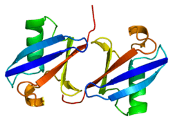
Ubiquitin, a highly conserved protein that has a major role in targeting cellular proteins for degradation by the 26S proteosome, is synthesized as a precursor protein consisting of either polyubiquitin chains or a single ubiquitin fused to an unrelated protein. This gene encodes a fusion protein consisting of ubiquitin at the N terminus and ribosomal protein S27a at the C terminus. When expressed in yeast, the protein is post-translationally processed, generating free ubiquitin monomer and ribosomal protein S27a. Ribosomal protein S27a is a component of the 40S subunit of the ribosome and belongs to the S27AE family of ribosomal proteins. It contains C4-type zinc finger domains and is located in the cytoplasm. Pseudogenes derived from this gene are present in the genome. As with ribosomal protein S27a, ribosomal protein L40 is also synthesized as a fusion protein with ubiquitin; similarly, ribosomal protein S30 is synthesized as a fusion protein with the ubiquitin-like protein fubi.[6]
References
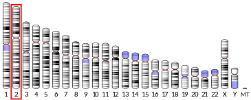
- 1 2 3 GRCh38: Ensembl release 89: ENSG00000143947 - Ensembl, May 2017
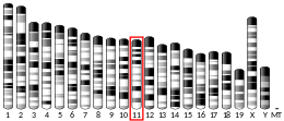
- 1 2 3 GRCm38: Ensembl release 89: ENSMUSG00000020460 - Ensembl, May 2017
- ↑ "Human PubMed Reference:".
- ↑ "Mouse PubMed Reference:".
- ↑ Kenmochi N, Kawaguchi T, Rozen S, Davis E, Goodman N, Hudson TJ, Tanaka T, Page DC (Aug 1998). "A map of 75 human ribosomal protein genes". Genome Res. 8 (5): 509–23. doi:10.1101/gr.8.5.509. PMID 9582194.
- 1 2 "Entrez Gene: RPS27A ribosomal protein S27a".
Further reading
- Wool IG, Chan YL, Glück A (1996). "Structure and evolution of mammalian ribosomal proteins". Biochem. Cell Biol. 73 (11–12): 933–47. doi:10.1139/o95-101. PMID 8722009.
- Adams SM, Sharp MG, Walker RA, et al. (1992). "Differential expression of translation-associated genes in benign and malignant human breast tumours". Br. J. Cancer. 65 (1): 65–71. doi:10.1038/bjc.1992.12. PMC 1977345. PMID 1370760.
- Pancré V, Pierce RJ, Fournier F, et al. (1991). "Effect of ubiquitin on platelet functions: possible identity with platelet activity suppressive lymphokine (PASL)". Eur. J. Immunol. 21 (11): 2735–41. doi:10.1002/eji.1830211113. PMID 1657614.
- Kanayama H, Tanaka K, Aki M, et al. (1992). "Changes in expressions of proteasome and ubiquitin genes in human renal cancer cells". Cancer Res. 51 (24): 6677–85. PMID 1660345.
- Monia BP, Ecker DJ, Jonnalagadda S, et al. (1989). "Gene synthesis, expression, and processing of human ubiquitin carboxyl extension proteins". J. Biol. Chem. 264 (7): 4093–103. PMID 2537304.
- Redman KL, Rechsteiner M (1989). "Identification of the long ubiquitin extension as ribosomal protein S27a". Nature. 338 (6214): 438–40. doi:10.1038/338438a0. PMID 2538756.
- Lund PK, Moats-Staats BM, Simmons JG, et al. (1985). "Nucleotide sequence analysis of a cDNA encoding human ubiquitin reveals that ubiquitin is synthesized as a precursor". J. Biol. Chem. 260 (12): 7609–13. PMID 2581967.
- Maruyama K, Sugano S (1994). "Oligo-capping: a simple method to replace the cap structure of eukaryotic mRNAs with oligoribonucleotides". Gene. 138 (1–2): 171–4. doi:10.1016/0378-1119(94)90802-8. PMID 8125298.
- Vladimirov SN, Ivanov AV, Karpova GG, et al. (1996). "Characterization of the human small-ribosomal-subunit proteins by N-terminal and internal sequencing, and mass spectrometry". Eur. J. Biochem. 239 (1): 144–9. doi:10.1111/j.1432-1033.1996.0144u.x. PMID 8706699.
- Suzuki Y, Yoshitomo-Nakagawa K, Maruyama K, et al. (1997). "Construction and characterization of a full length-enriched and a 5'-end-enriched cDNA library". Gene. 200 (1–2): 149–56. doi:10.1016/S0378-1119(97)00411-3. PMID 9373149.
- Kirschner LS, Stratakis CA (2000). "Structure of the human ubiquitin fusion gene Uba80 (RPS27a) and one of its pseudogenes". Biochem. Biophys. Res. Commun. 270 (3): 1106–10. doi:10.1006/bbrc.2000.2568. PMID 10772958.
- Petersen BO, Wagener C, Marinoni F, et al. (2000). "Cell cycle– and cell growth–regulated proteolysis of mammalian CDC6 is dependent on APC–CDH1". Genes Dev. 14 (18): 2330–43. doi:10.1101/gad.832500. PMC 316932. PMID 10995389.
- Bolton D, Evans PA, Stott K, Broadhurst RW (2002). "Structure and properties of a dimeric N-terminal fragment of human ubiquitin". J. Mol. Biol. 314 (4): 773–87. doi:10.1006/jmbi.2001.5181. PMID 11733996.
- Yoshihama M, Uechi T, Asakawa S, et al. (2002). "The Human Ribosomal Protein Genes: Sequencing and Comparative Analysis of 73 Genes". Genome Res. 12 (3): 379–90. doi:10.1101/gr.214202. PMC 155282. PMID 11875025.
- Strausberg RL, Feingold EA, Grouse LH, et al. (2003). "Generation and initial analysis of more than 15,000 full-length human and mouse cDNA sequences". Proc. Natl. Acad. Sci. U.S.A. 99 (26): 16899–903. doi:10.1073/pnas.242603899. PMC 139241. PMID 12477932.
- Cohen BD, Bariteau JT, Magenis LM, Dias JA (2003). "Regulation of follitropin receptor cell surface residency by the ubiquitin-proteasome pathway". Endocrinology. 144 (10): 4393–402. doi:10.1210/en.2002-0063. PMID 12960054.
- Ota T, Suzuki Y, Nishikawa T, et al. (2004). "Complete sequencing and characterization of 21,243 full-length human cDNAs". Nat. Genet. 36 (1): 40–5. doi:10.1038/ng1285. PMID 14702039.
- Li H, Seth A (2004). "An RNF11: Smurf2 complex mediates ubiquitination of the AMSH protein". Oncogene. 23 (10): 1801–8. doi:10.1038/sj.onc.1207319. PMID 14755250.
PDB gallery | |
|---|---|
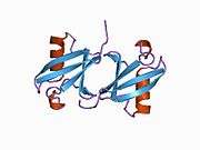 1aar: STRUCTURE OF A DIUBIQUITIN CONJUGATE AND A MODEL FOR INTERACTION WITH UBIQUITIN CONJUGATING ENZYME (E2)  1cmx: STRUCTURAL BASIS FOR THE SPECIFICITY OF UBIQUITIN C-TERMINAL HYDROLASES 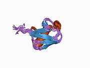 1d3z: UBIQUITIN NMR STRUCTURE  1f9j: STRUCTURE OF A NEW CRYSTAL FORM OF TETRAUBIQUITIN 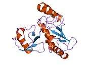 1fxt: STRUCTURE OF A CONJUGATING ENZYME-UBIQUITIN THIOLESTER COMPLEX  1g6j: STRUCTURE OF RECOMBINANT HUMAN UBIQUITIN IN AOT REVERSE MICELLES  1gjz: SOLUTION STRUCTURE OF A DIMERIC N-TERMINAL FRAGMENT OF HUMAN UBIQUITIN  1nbf: Crystal structure of a UBP-family deubiquitinating enzyme in isolation and in complex with ubiquitin aldehyde 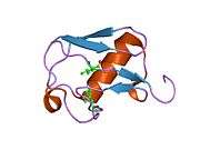 1ogw: SYNTHETIC UBIQUITIN WITH FLUORO-LEU AT 50 AND 67  1otr: Solution Structure of a CUE-Ubiquitin Complex  1p3q: Mechanism of Ubiquitin Recognition by the CUE Domain of VPS9 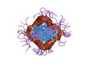 1q0w: Solution structure of Vps27 amino-terminal UIM-ubiquitin complex 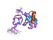 1q5w: Ubiquitin Recognition by Npl4 Zinc-Fingers  1s1q: TSG101(UEV) domain in complex with Ubiquitin  1sif: Crystal structure of a multiple hydrophobic core mutant of ubiquitin  1tbe: STRUCTURE OF TETRAUBIQUITIN SHOWS HOW MULTIUBIQUITIN CHAINS CAN BE FORMED 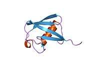 1ubi: SYNTHETIC STRUCTURAL AND BIOLOGICAL STUDIES OF THE UBIQUITIN SYSTEM. PART 1  1ubq: STRUCTURE OF UBIQUITIN REFINED AT 1.8 ANGSTROMS RESOLUTION  1ud7: SOLUTION STRUCTURE OF THE DESIGNED HYDROPHOBIC CORE MUTANT OF UBIQUITIN, 1D7  1uzx: A COMPLEX OF THE VPS23 UEV WITH UBIQUITIN  1v80: Solution structures of ubiquitin at 30 bar and 3 kbar  1v81: Solution structures of ubiquitin at 30 bar and 3 kbar  1wr1: The complex structure of Dsk2p UBA with ubiquitin  1wr6: Crystal structure of GGA3 GAT domain in complex with ubiquitin 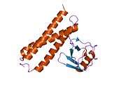 1wrd: Crystal structure of Tom1 GAT domain in complex with ubiquitin 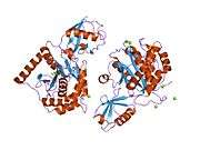 1xd3: Crystal structure of UCHL3-UbVME complex  1xqq: Simultaneous determination of protein structure and dynamics 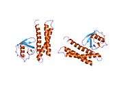 1yd8: COMPLEX OF HUMAN GGA3 GAT DOMAIN AND UBIQUITIN 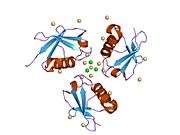 1yiw: X-ray Crystal Structure of a Chemically Synthesized Ubiquitin  1yj1: X-ray Crystal Structure of a Chemically Synthesized [D-Gln35]Ubiquitin  1yx5: Solution Structure of S5a UIM-1/Ubiquitin Complex 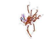 1yx6: Solution Structure of S5a UIM-2/Ubiquitin Complex 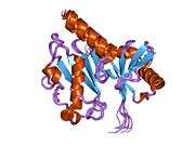 1zgu: Solution structure of the human Mms2-Ubiquitin complex  2ayo: Structure of USP14 bound to ubquitin aldehyde  2bgf: NMR STRUCTURE OF LYS48-LINKED DI-UBIQUITIN USING CHEMICAL SHIFT PERTURBATION DATA TOGETHER WITH RDCS AND 15N-RELAXATION DATA 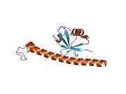 2c7m: HUMAN RABEX-5 RESIDUES 1-74 IN COMPLEX WITH UBIQUITIN  2c7n: HUMAN RABEX-5 RESIDUES 1-74 IN COMPLEX WITH UBIQUITIN 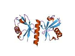 2d3g: Double sided ubiquitin binding of Hrs-UIM  2den: Solution Structure of the Ubiquitin-Associated Domain of Human BMSC-UbP and its Complex with Ubiquitin  2dx5: The complex structure between the mouse EAP45-GLUE domain and ubiquitin  2fcm: X-ray Crystal Structure of a Chemically Synthesized [D-Gln35]Ubiquitin with a Cubic Space Group  2fcn: X-ray Crystal Structure of a Chemically Synthesized [D-Val35]Ubiquitin with a Cubic Space Group 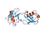 2fcq: X-ray Crystal Structure of a Chemically Synthesized Ubiquitin with a Cubic Space Group  2fcs: X-ray Crystal Structure of a Chemically Synthesized [L-Gln35]Ubiquitin with a Cubic Space Group  2fid: Crystal Structure of a Bovine Rabex-5 fragment complexed with ubiquitin  2fif: Crystal Structure of a Bovine Rabex-5 fragment complexed with ubiquitin  2fuh: Solution Structure of the UbcH5c/Ub Non-covalent Complex  2g3q: Solution Structure of Ede1 UBA-ubiquitin complex  2g45: Co-crystal structure of znf ubp domain from the deubiquitinating enzyme isopeptidase T (isot) in complex with ubiquitin  2gbj: Crystal Structure of the 9-10 8 Glycine Insertion Mutant of Ubiquitin. 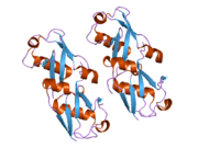 2gbk: Crystal Structure of the 9-10 MoaD Insertion Mutant of Ubiquitin 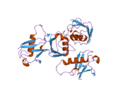 2gbm: Crystal Structure of the 35-36 8 Glycine Insertion Mutant of Ubiquitin 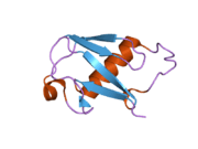 2gbn: Crystal Structure of the 35-36 8 Glycine Insertion Mutant of Ubiquitin 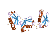 2gbr: Crystal Structure of the 35-36 MoaD Insertion Mutant of Ubiquitin  2gmi: Mms2/Ubc13~Ubiquitin 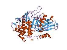 2hd5: USP2 in complex with ubiquitin 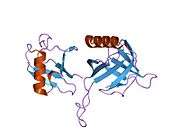 2hth: Structural basis for ubiquitin recognition by the human EAP45/ESCRT-II GLUE domain  2ibi: Covalent Ubiquitin-USP2 Complex  2j7q: CRYSTAL STRUCTURE OF THE UBIQUITIN-SPECIFIC PROTEASE ENCODED BY MURINE CYTOMEGALOVIRUS TEGUMENT PROTEIN M48 IN COMPLEX WITH A UBQUITIN-BASED SUICIDE SUBSTRATE  2nr2: The MUMO (minimal under-restraining minimal over-restraining) method for the determination of native states ensembles of proteins  2o6v: Crystal structure and solution NMR studies of Lys48-linked tetraubiquitin at neutral pH  2oob: crystal structure of the UBA domain from Cbl-b ubiquitin ligase in complex with ubiquitin |