Eukaryotic large ribosomal subunit (60S)
Ribosomal particles are denoted according to their sedimentation coefficients in Svedberg units. The 60S subunit is the large subunit of eukaryotic 80S ribosomes. It is structurally and functionally related to the 50S subunit of 70S prokaryotic ribosomes.[1][2][3][4][5][6] However, the 60S subunit is much larger than the prokaryotic 50S subunit and contains many additional protein segments, as well as ribosomal RNA expansion segments.
Overall structure
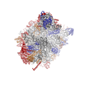
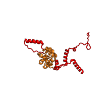
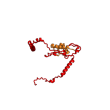
Characteristic features of the large subunit, shown below in the "Crown View", include the central protuberance (CP) and the two stalks, which are named according to their bacterial protein components (L1 stalk on the left as seen from the subunit interface and L7/L12 on the right). There are three binding sites for tRNA, the A-site, P-site and E-site (see article on protein translation for details). The core of the 60S subunit is formed by the 28S ribosomal RNA (abbreviated 28S rRNA), which is homologous to the prokaryotic 23S rRNA, which also contributes the active site (peptidyl transferase center, PTC) of the ribosome.[2][4] The rRNA core is decorated with dozens of proteins. In the figure "Crystal Structure of the Eukaryotic 60S Ribosomal Subunit from T. thermophila", the ribosomal RNA core is represented as a grey tube and expansion segments are shown in red. Proteins which have homologs in eukaryotes, archaea and bacteria are shown as blue ribbons. Proteins shared only between eukaryotes and archaea are shown as orange ribbons and proteins specific to eukaryotes are shown as red ribbons.

60S ribosomal proteins
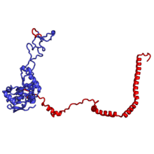
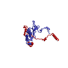
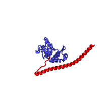
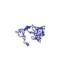
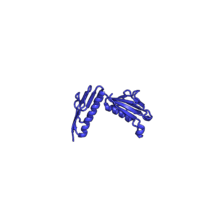
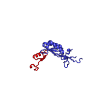
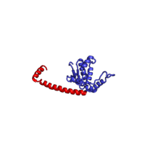
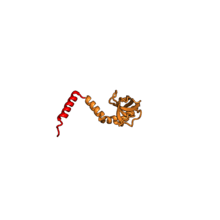
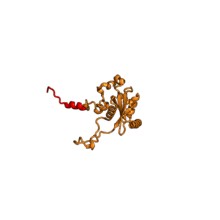
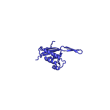
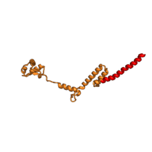
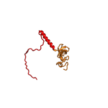
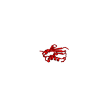
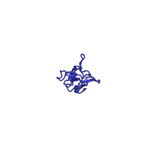
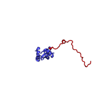
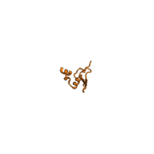
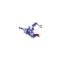
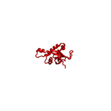
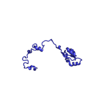
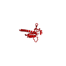
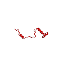
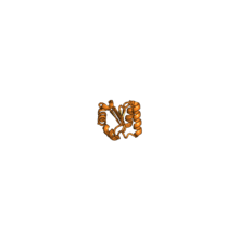
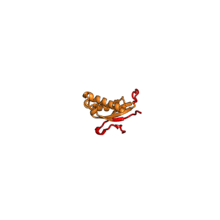
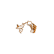
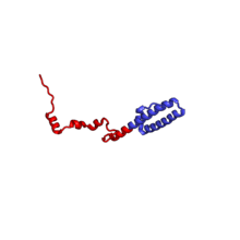
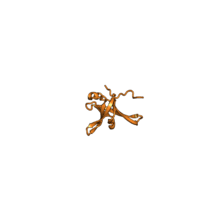
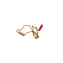
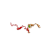
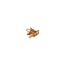
The table "60S ribosomal proteins" shows the individual protein folds of the 60S subunit colored by conservation as above. The eukaryote-specific extensions, ranging from a few residues or loops to very long alpha helices and additional domains, are highlighted in red.[2]
Historically, different nomenclatures have been used for ribosomal proteins. For instance, proteins have been numbered according to their migration properties in gel electrophoresis experiments. Therefore, different names may refer to homologous proteins from different organisms, while identical names do not necessarily denote homologous proteins. The table "60S ribosomal proteins" cross-references the human ribosomal protein names with yeast, bacterial, and archaeal homologs.[7] Further information can be found in the ribosomal protein gene database (RPG).[7]
| Structure (Eukaryotic)[8] | H. sapiens[7][9] | Amino acids[10] | Conservation[11] | S. cerevisiae[12] | Bacterial homolog (E. coli) | Archaeal homolog |
|---|---|---|---|---|---|---|
 | RPLP0 | 318 | EAB | P0 | L10 | L10 |
 | RPL3 | 404 | EAB | L3 | L3 | L3 |
 | RPL4 | 428 | EAB | L4 | L4 | L4 |
 | RPL5 | 298 | EAB | L5 | L18 | L18p |
 | RPL6 | 289 | E | L6 | n/a | n/a |
 | RPL7 | 254 | EAB | L7 | L30 | L30 |
 | RPL7A | 267 | EA | L8 | n/a | L7Ae |
 | RPL8 | 258 | EAB | L2 | L2 | L2 |
 | RPL9 | 193 | EAB | L9 | L6 | L6 |
 | RPL10 | 215 | EAB | L10 | L16 | L10e |
 | RPL11 | EAB | L11 | L5 | L5 | |
 | RPL13 | EA | L13 | n/a | L13e | |
 | RPL13A | 204 | EAB | L16 | L13 | L13 |
 | RPL14 | 221 | EA | L14 | n/a | L14e |
 | RPL15 | 205 | EA | L15 | n/a | L15e |
 | RPL17 | 185 | EAB | L17 | L22 | L22 |
 | RPL18 | 189 | EA | L18 | n/a | L18e |
 | RPL18A | 177 | EA | L20 | n/a | Lx |
 | RPL19 | 197 | EA | L19 | n/a | L19 |
 | RPL21 | 161 | EA | L21 | n/a | L21e |
 | RPL22 | 129 | E | L22 | n/a | n/a |
 | RPL23 | 141 | EAB | L23 | L14 | L14p |
 | RPL23A | 157 | EAB | L25 | L23 | L23 |
 | RPL24 | 158 | EA | L24 | n/a | L24e |
 | RPL26 | 146 | EAB | L26 | L24 | L24 |
 | RPL27 | 137 | E | L27 | n/a | n/a |
 | RPL27A | 149 | EAB | L28 | L15 | L15 |
 | RPL28 | E | n/a[2][3][13] | n/a | n/a | |
 | RPL29 | E | L29 | n/a | n/a | |
 | RPL30 | 116 | EA | L30 | n/a | L30e |
 | RPL31 | 126 | EA | L31 | n/a | L31e |
 | RPL32 | 136 | EA | L32 | n/a | L32e |
 | RPL34 | 118 | EA | L34 | n/a | L34e |
 | RPL35 | 124 | EAB | L35 | L29 | L29 |
 | RPL35A | EA | L33 | n/a | L35Ae | |
 | RPL36 | 106 | E | L36 | n/a | n/a |
 | RPL36A | 107 | EA | L42 | n/a | L44e |
 | RPL37 | 98 | EA | L37 | n/a | L37e |
 | RPL37A | EA | L43 | n/a | L37Ae | |
 | RPL38 | EA | L38 | n/a | L38e | |
 | RPL39 | 52 | EA | L39 | n/a | L37Ae |
 | RPL40 | 129 | EA | L40 | n/a | L40e |
See also
References
- ↑ 60S+Ribosome+Subunits at the US National Library of Medicine Medical Subject Headings (MeSH)
- 1 2 3 4 Klinge, S; Voigts-Hoffmann, F; Leibundgut, M; Arpagaus, S; Ban, N (2011). "Crystal structure of the eukaryotic 60S ribosomal subunit in complex with initiation factor 6". Science. 334 (6058): 941–8. doi:10.1126/science.1211204. PMID 22052974.
- 1 2 Ben-Shem, A; Garreau; de Loubresse, N; Melnikov, S; Jenner, L; Yusupova, G; Yusupov, M (Dec 2011). "The structure of the eukaryotic ribosome at 3.0 Å resolution". Science. 334 (6062): 1524–9. doi:10.1126/science.1212642. PMID 22096102.
- 1 2 Ban, N; Nissen, P; Hansen, J; Moore, PB; Steitz, TA (Aug 2000). "The complete atomic structure of the large ribosomal subunit at 2.4 A resolution". Science. 289 (5481): 905–20. doi:10.1126/science.289.5481.905. PMID 10937989.
- ↑ Cate, JH; Yusupov, MM; Yusupova, GZ; Earnest, TN; Noller, HF (Sep 1999). "X-ray crystal structures of 70S ribosome functional complexes". Science. 285 (5436): 2095–104. doi:10.1126/science.285.5436.2095. PMID 10497122.
- ↑ Yusupov, MM; Yusupova, GZ; Baucom, A; Lieberman, K; Earnest, TN; Cate, JH; Noller, HF (May 2001). "Crystal structure of the ribosome at 5.5 A resolution". Science. 292 (5518): 883–96. doi:10.1126/science.1060089. PMID 11283358.
- 1 2 3 Nakao, A; Yoshihama, M; Kenmochi, N (2004). "RPG: the Ribosomal Protein Gene database". Nucleic Acids Res. 32: D168–70. doi:10.1093/nar/gkh004. PMC 308739. PMID 14681386.
- ↑ Structure of the T. thermophila,' proteins from the structures of the large subunit PDBS 417, 4A19
- ↑ Nomenclature according to the ribosomal protein gene database, applies to H. sapiens and T. thermophila
- ↑ Yoshihama, Maki; Uechi, Tamayo; Asakawa, Shuichi; Kawasaki, Kauhiko (2002). "The Human Ribosomal Protein Genes: Sequencing and Comparative Analysis of 73 Genes". Genome Research. 12: 379–390. doi:10.1101/gr.214202. PMC 155282. PMID 11875025.
- ↑ EAB means conserved in eukaryotes, archaea and bacteria, EA means conserved in eukaryotes and archaea and E means eukaryote-specific protein
- ↑ Traditionally, ribosomal proteins were named according to their apparent molecular weight in gel electrophoresis, leading to different names for homologous proteins from different organisms. The RPG offers a unified nomenclature for ribosomal protein genes based on homology.
- ↑ RPL28 has no detectable homolog in yeast