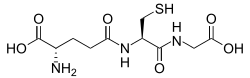
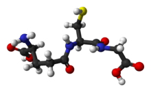
Glutathione
 | |
 | |
| Names | |
|---|---|
| IUPAC name
(2S)-2-Amino-4-{[(1R)-1-[(carboxymethyl)carbamoyl]-2-sulfanylethyl]carbamoyl}butanoic acid | |
| Other names
γ-L-Glutamyl-L-cysteinylglycine (2S)-2-Amino-5-[[(2R)-1-(carboxymethylamino)-1-oxo-3-sulfanylpropan-2-yl]amino]-5-oxopentanoic acid | |
| Identifiers | |
3D model (JSmol) |
|
| Abbreviations | GSH |
| ChEBI | |
| ChEMBL | |
| ChemSpider | |
| DrugBank | |
| ECHA InfoCard | 100.000.660 |
| KEGG | |
| MeSH | Glutathione |
PubChem CID |
|
| UNII | |
| |
| |
| Properties | |
| C10H17N3O6S | |
| Molar mass | 307.32 g·mol−1 |
| Melting point | 195 °C (383 °F; 468 K) |
| Freely soluble[1] | |
| Solubility in methanol, diethyl ether | Insoluble |
| Pharmacology | |
| V03AB32 (WHO) | |
Except where otherwise noted, data are given for materials in their standard state (at 25 °C [77 °F], 100 kPa). | |
| Infobox references | |
Glutathione (GSH) is an antioxidant in plants, animals, fungi, and some bacteria and archaea. Glutathione is capable of preventing damage to important cellular components caused by reactive oxygen species such as free radicals, peroxides, lipid peroxides, and heavy metals.[2] It is a tripeptide with a gamma peptide linkage between the carboxyl group of the glutamate side chain and the amine group of cysteine, and the carboxyl group of cysteine is attached by normal peptide linkage to a glycine.
Thiol groups are reducing agents, existing at a concentration around 5 mM in animal cells. Glutathione reduces disulfide bonds formed within cytoplasmic proteins to cysteines by serving as an electron donor. In the process, glutathione is converted to its oxidized form, glutathione disulfide (GSSG), also called L-(–)-glutathione.
Once oxidized, glutathione can be reduced back by glutathione reductase, using NADPH as an electron donor.[3] The ratio of reduced glutathione to oxidized glutathione within cells is often used as a measure of cellular oxidative stress.[4][5]
Biosynthesis
The biosynthesis pathway for glutathione is found in some bacteria, such as cyanobacteria and proteobacteria, but is missing in many other bacteria. Most eukaryotes, including humans, synthesize glutathione, but some do not, such as Leguminosae, Entamoeba, and Giardia. The only archaea that make glutathione are halobacteria.[6][7]
Glutathione is not an essential nutrient for humans, since it can be synthesized in the body from the amino acids L-cysteine, L-glutamic acid, and glycine; it does not have to be present as a supplement in the diet. The sulfhydryl group (SH) of cysteine serves as a proton donor and is responsible for its biological activity. Cysteine is the rate-limiting factor in cellular glutathione biosynthesis, since this amino acid is relatively rare in foods.
Cells make glutathione in two adenosine triphosphate-dependent steps:
- First, gamma-glutamylcysteine is synthesized from L-glutamate and cysteine via the enzyme gamma-glutamylcysteine synthetase (glutamate cysteine ligase, GCL). This reaction is the rate-limiting step in glutathione synthesis.[8]
- Second, glycine is added to the C-terminal of gamma-glutamylcysteine via the enzyme glutathione synthetase.
Animal glutamate cysteine ligase (GCL) is a heterodimeric enzyme composed of a catalytic and a modulatory subunit. The catalytic subunit is necessary and sufficient for all GCL enzymatic activity, whereas the modulatory subunit increases the catalytic efficiency of the enzyme. Mice lacking the catalytic subunit (i.e., lacking all de novo GSH synthesis) die before birth.[9] Mice lacking the modulatory subunit demonstrate no obvious phenotype, but exhibit marked decrease in GSH and increased sensitivity to toxic insults.[10][11][12]
While all animal cells are capable of synthesizing glutathione, glutathione synthesis in the liver has been shown to be essential. GCLC knockout mice die within a month of birth due to the absence of hepatic GSH synthesis.[13][14] Major transport into the blood stream is driven by an electrochemical gradient, specifically through the transport proteins RcGshT and RsGshT.[15] Similarly, glutathione S-conjugates, synthesized hepatically, feature preferential secretion into bile.[14][16]
The plant glutamate cysteine ligase (GCL) is a redox-sensitive homodimeric enzyme, conserved in the plant kingdom.[17] In an oxidizing environment, intermolecular disulfide bridges are formed and the enzyme switches to the dimeric active state. The midpoint potential of the critical cysteine pair is -318 mV. In addition to the redox-dependent control, the plant GCL enzyme is feedback inhibited by glutathione.[18] GCL is exclusively located in plastids, and glutathione synthetase (GS) is dual-targeted to plastids and cytosol, thus GSH and gamma-glutamylcysteine are exported from the plastids.[19] Both glutathione biosynthesis enzymes are essential in plants; knock-outs of GCL and GS are lethal to embryo and seedling.[20]
Function
Glutathione exists in both reduced (GSH) and oxidized (GSSG) states. In the reduced state, the thiol group of cysteine is able to donate a reducing equivalent (H++ e−) to other molecules, such as reactive oxygen species to neutralize them, or to protein cysteines to maintain their reduced forms. With donating an electron, glutathione itself becomes reactive and readily reacts with another reactive glutathione to form glutathione disulfide (GSSG). Such a reaction is probable due to the relatively high concentration of glutathione in cells (up to 7 mM in the liver).[21]
Generally, interactions between GSH and other molecules with higher relative electrophilicity deplete GSH levels within the cell. An exception to this case involves the sensitivity of GSH to the electrophilic compound's relative concentration. In high concentrations, the organic molecule diethyl maleate fully depleted GSH levels in cells. However, in low concentrations, a minor decrease in cellular GSH levels was followed by a two-fold increase.[22][23]
GSH can be regenerated from GSSG by the enzyme glutathione reductase (GSR):[3] NADPH reduces FAD present in GSR to produce a transient FADH-anion. This anion then quickly breaks a disulfide bond (Cys58 – Cys63) and leads to Cys63's nucleophilically attacking the nearest sulfide unit in the GSSG molecule (promoted by His467), which creates a mixed disulfide bond (GS-Cys58) and a GS-anion. His467 of GSR then protonates the GS-anion to form the first GSH. Next, Cys63 nucleophilically attacks the sulfide of Cys58, releasing a GS-anion, which, in turn, picks up a solvent proton and is released from the enzyme, thereby creating the second GSH. So, for every GSSG and NADPH, two reduced GSH molecules are gained, which can again act as antioxidants scavenging reactive oxygen species in the cell.
In healthy cells and tissue, more than 90% of the total glutathione pool is in the reduced form (GSH) and less than 10% exists in the disulfide form (GSSG). An increased GSSG-to-GSH ratio is considered indicative of oxidative stress.[24]
Glutathione participates in thiol protection and redox regulation of cellular thiol proteins under oxidative stress by protein S-glutathionylation, a redox-regulated post-translational thiol modification.
Glutathione has multiple functions:
- It maintains levels of reduced glutaredoxin and glutathione peroxidase.[25]
- It is one of the major endogenous antioxidants produced by the cells, participating directly in the neutralization of free radicals and reactive oxygen compounds, as well as maintaining exogenous antioxidants such as vitamins C and E in their reduced (active) forms.[26][27][28]
- Regulation of the nitric oxide cycle is critical for life, but can be problematic if unregulated.[29] Glutathione enhances the function of citrulline as part of the nitric oxide cycle.
- It is used in metabolic and biochemical reactions such as DNA synthesis and repair, protein synthesis, prostaglandin synthesis, amino acid transport, and enzyme activation. Thus, every system in the body can be affected by the state of the glutathione system, especially the immune system, the nervous system, the gastrointestinal system, and the lungs.
- It has a vital function in iron metabolism. Yeast cells depleted of GSH or containing toxic levels of GSH show an intense iron starvation-like response and impairment of the activity of extramitochondrial ISC enzymes thus inhibiting oxidative endoplasmic reticulum folding, followed by death.[30]
- It has roles in progression of the cell cycle, including cell death.[5] GSH levels regulate redox changes to nuclear proteins necessary for the initiation of cell differentiation. Differences in GSH levels also determine the expressed mode of cell death, being either apoptosis or cell necrosis. Manageably low levels result in the systematic breakage of the cell whereas excessively low levels result in rapid cell death.[31]
Function in animals
GSH is known as a substrate in conjugation reactions, which is catalyzed by glutathione S-transferase enzymes in cytosol, microsomes, and mitochondria. However, GSH is also capable of participating in nonenzymatic conjugation with some chemicals.
In the case of N-acetyl-p-benzoquinone imine (NAPQI), the reactive cytochrome P450-reactive metabolite formed by paracetamol (acetaminophen), which becomes toxic when GSH is depleted by an overdose of acetaminophen, glutathione is an essential antidote to overdose. Glutathione conjugates to NAPQI and helps to detoxify it. In this capacity, it protects cellular protein thiol groups, which would otherwise become covalently modified; when all GSH has been spent, NAPQI begins to react with the cellular proteins, killing the cells in the process. The preferred treatment for an overdose of this painkiller is the administration (usually in atomized form) of N-acetyl-L-cysteine (often as a preparation called Mucomyst[32]), which is processed by cells to L-cysteine and used in the de novo synthesis of GSH.
Glutathione (GSH) participates in leukotriene synthesis and is a cofactor for the enzyme glutathione peroxidase. It is also important as a hydrophilic molecule that is added to lipophilic toxins and waste in the liver during biotransformation before they can become part of the bile. Glutathione is also needed for the detoxification of methylglyoxal, a toxin produced as a byproduct of metabolism.
This detoxification reaction is carried out by the glyoxalase system. Glyoxalase I (EC 4.4.1.5) catalyzes the conversion of methylglyoxal and reduced glutathione to S-D-lactoyl-glutathione. Glyoxalase II (EC 3.1.2.6) catalyzes the hydrolysis of S-D-lactoyl-glutathione to glutathione and D-lactic acid.
Glutathione, along with oxidized glutathione (GSSG) and S-nitrosoglutathione (GSNO), have been found to bind to the glutamate recognition site of the NMDA and AMPA receptors (via their γ-glutamyl moieties), and may be endogenous neuromodulators.[33][34][35] At millimolar concentrations, they may also modulate the redox state of the NMDA receptor complex.[34] Glutathione has been found to bind to and activate ionotropic receptors that are different from any other excitatory amino acid receptor, and which may constitute glutathione receptors, potentially making it a neurotransmitter.[36] Glutathione is also able to activate the purinergic P2X7 receptor from Müller glia, inducing acute calcium transient signals and GABA release from both retinal neurons and glial cells.[37][38]
Function in plants
In plants, glutathione is crucial for biotic and abiotic stress management. It is a pivotal component of the glutathione-ascorbate cycle, a system that reduces poisonous hydrogen peroxide.[39] It is the precursor of phytochelatins, glutathione oligomers that chelate heavy metals such as cadmium.[40] Glutathione is required for efficient defence against plant pathogens such as Pseudomonas syringae and Phytophthora brassicae.[41] Adenylyl-sulfate reductase, an enzyme of the sulfur assimilation pathway, uses glutathione as an electron donor. Other enzymes using glutathione as a substrate are glutaredoxins. These small oxidoreductases are involved in flower development, salicylic acid, and plant defence signalling.[42] In a recent report it is shown that seeds of Cassia occidentalis plants which contains multiple anthraquinones are capable of forming conjugates with glutathione. It was also found that Rhein have the most cytotoxic response with maximum oxidization of glutathione followed by emodin and aloe-emodin.[43]
Bioavailability and supplementation
Systemic bioavailability of orally consumed glutathione is poor because the molecule, a tripeptide, is the substrate of proteases (peptidases) of the alimentary canal, and due to the absence of a specific carrier of glutathione at the level of cell membrane.[44][45]
Because direct supplementation of glutathione is not always successful, supply of the raw nutritional materials used to generate GSH, such as cysteine and glycine, may be more effective at increasing glutathione levels. Other antioxidants such as ascorbic acid (vitamin C) may also work synergistically with glutathione, preventing depletion of either. The glutathione-ascorbate cycle, which works to detoxify hydrogen peroxide (H2O2), is one very specific example of this phenomenon.
Additionally, compounds such as N-acetylcysteine[46] (NAC) and alpha lipoic acid[47] (ALA, not to be confused with the unrelated alpha-linolenic acid) are both capable of helping to regenerate glutathione levels. NAC in particular is commonly used to treat overdose of acetaminophen, a type of potentially fatal poisoning which is harmful in part due to severe depletion of glutathione levels.
Calcitriol (1,25-dihydroxyvitamin D3), the active metabolite of vitamin D3, after being synthesized from calcifediol in the kidney, increases glutathione levels in the brain and appears to be a catalyst for glutathione production.[48] About ten days are needed for the body to process vitamin D3 into calcitriol.[49]
S-adenosylmethionine (SAMe), a cosubstrate involved in methyl group transfer, has also been shown to increase cellular glutathione content in persons suffering from a disease-related glutathione deficiency.[50][51][52]
Low glutathione is commonly observed in wasting and negative nitrogen balance, as seen in cancer, HIV/AIDS, sepsis, trauma, burns, and athletic overtraining. Low levels are also observed in periods of starvation. These effects are hypothesized to be influenced by the higher glycolytic activity associated with cachexia, which result from reduced levels of oxidative phosphorylation.[53][54]
Methods to determine glutathione
Small molecule-based glutathione probes
Ellman's reagent and monobromobimane
Reduced glutathione may be visualized using Ellman's reagent or bimane derivatives such as monobromobimane. The monobromobimane method is more sensitive. In this procedure, cells are lysed and thiols extracted using a HCl buffer. The thiols are then reduced with dithiothreitol and labelled by monobromobimane. Monobromobimane becomes fluorescent after binding to GSH. The thiols are then separated by HPLC and the fluorescence quantified with a fluorescence detector.
Monochlorobimane
Monochlorobimane can be used to quantify glutathione in vivo. The quantification is done by confocal laser scanning microscopy after application of the dye to living cells.[55] This quantification process relies on measuring the rates of fluorescence changes and is limited to plant cells.
5-Chloromethylfluorescein diacetate (CMFDA)
CMFDA was initially used as a cell tracker. Unfortunately, it has also been mistakenly used as a glutathione probe. Unlike monochlorobimane, whose fluorescence increases upon reacting with glutathione, the fluorescence increase of CMFDA is due to the hydrolysis of the acetate groups inside cells. Although CMFDA may react with glutathione in cells, the fluorescence increase does not reflect the reaction. Therefore, studies using CMFDA as a glutathione probe should be revisited and reinterpreted.[56][57]
ThiolQuant Green
The major limitation of these bimane-based probes and many other reported probes is that these probes are based on irreversible chemical reactions with glutathione, which renders these probes incapable of monitoring the real-time glutathione dynamics. Recently, the first reversible reaction based fluorescent probe-ThiolQuant Green (TQG)-for glutathione was reported.[58] ThiolQuant Green can not only perform high resolution measurements of glutathione levels in single cells using a confocal microscope, but also be applied in flow cytometry to perform bulk measurements.
RealThiol
The RealThiol (RT) probe is the second-generation reversible reaction-based GSH probe developed by the Wang group. A few key features of RealThiol: 1) it has a much faster forward and backward reaction kinetics compared to ThiolQuant Green, which enables real-time monitoring of GSH dynamics in live cells; 2) only micromolar to sub-micromolar RealThiol is needed for staining in cell-based experiments, which induces minimal perturbation to GSH level in cells; 3) a high-quantum-yield coumarin fluorophore was implemented so that background noise can be minimized; and 4) equilibrium constant of the reaction between RealThiol and GSH has been fine-tuned to respond to physiologically relevant concentration of GSH.[59] RealThiol can be used to perform measurements of glutathione levels in single cells using a high-resolution confocal microscope, as well as be applied in flow cytometry to perform bulk measurements in high throughput manner.
Organelle-targeted RT probe has also been developed. A mitochondria targeted version, MitoRT, was reported and demonstrated in monitoring the dynamic of mitochondrial glutathione both on confocoal microscope and FACS based analysis.[60]
Protein-based glutathione probes
Another approach, which allows measurement of the glutathione redox potential at a high spatial and temporal resolution in living cells, is based on redox imaging using the redox-sensitive green fluorescent protein (roGFP)[61] or redox-sensitive yellow fluorescent protein (rxYFP)[62] GSSG because its very low physiological concentration is difficult to measure accurately unless the procedure is carefully executed and monitored and the occurrence of interfering compounds is properly addressed. GSSG concentration ranges from 10 to 50 μM in all solid tissues, and from 2 to 5 μM in blood (13–33 nmol per gram Hb). GSH-to-GSSG ratio ranges from 100 to 700.[63]
Other biological implications
Lead
The sulphur-rich aspect of glutathione results in it forming relatively strong complexes with lead(II).[64]
Cancer
Once a tumor has been established, elevated levels of glutathione may act to protect cancerous cells by conferring resistance to chemotherapeutic drugs.[65] The antineoplastic mustard drug canfosfamide was modelled on the structure of glutathione.
Cystic fibrosis
Several studies have been completed on the effectiveness of introducing inhaled glutathione to people with cystic fibrosis with mixed results.[66][67]
Alzheimer's disease
While extracellular amyloid beta (Aβ) plaques, neurofibrillary tangles (NFT), inflammation in the form of reactive astrocytes and microglia, and neuronal loss are all consistent pathological features of Alzheimer's disease (AD), a mechanistic link between these factors is yet to be clarified. Although the majority of past research has focused on fibrillar Aβ, soluble oligomeric Aβ species are now considered to be of major pathological importance in AD. Upregulation of GSH may be protective against the oxidative and neurotoxic effects of oligomeric Aβ.
Uses
Winemaking
The content of glutathione in must, the first raw form of wine, determines the browning, or caramelizing effect, during the production of white wine by trapping the caffeoyltartaric acid quinones generated by enzymic oxidation as grape reaction product.[68] Its concentration in wine can be determined by UPLC-MRM mass spectrometry.[69]
Cosmetics
Glutathione plays an important role in preventing oxidative damage to the skin.[70] In addition to its many recognized biological functions, glutathione has also been associated with skin lightening ability.[71] The role of glutathione as a skin whitener was discovered as a side effect of large doses of glutathione.[72] Glutathione utilizes different mechanisms to exert its action as a skin whitening agent at various levels of melanogenesis. It inhibits melanin synthesis by means of stopping the neurotransmitter precursor L-DOPA's ability to interact with tyrosinase in the process of melanin production.[73] Glutathione inhibits the actual production as well as agglutination of melanin by interrupting the function of L-DOPA. Another study found that glutathione inhibits melanin formation by direct inactivation of the enzyme tyrosinase by binding and chelating copper within the enzyme's active site.[74] Glutathione's antioxidant property allows it to inhibit melanin synthesis by quenching of free radicals and peroxides that contribute to tyrosinase activation and melanin formation.[75] Its antioxidant property also protects the skin from UV radiation and other environmental as well as internal stressors that generate free radicals that cause skin damage and hyperpigmentation.[76] In most mammals, melanin formation consists of eumelanin (brown-black pigment) and pheomelanin ( yellow-red pigment) as either mixtures or co-polymers.[77] Increase in glutathione level may induce the pigment cell to produce pheomelanin instead of eumelanin pigments.[78] A research by Te-Sheng Chang found lowest levels of reduced glutathione to be associated with eumelanin type pigmentation, whereas the highest ones were associated with the pheomelanin.[71] As a result, it is reasonable to assume that depletion of glutathione would result in eumelanin formation. Prota [79] observed that decreased glutathione concentration led to the conversion of L-Dopaquinone to Dopachrome, increasing the formation of brown-black pigment (eumelanin).
Importance of gamma-glutamylcysteine as a precursor for glutathione synthesis
Gamma-glutamylcysteine (GGC) is the immediate precursor to GSH. GGC supplementation would circumvent feedback inhibitory control of GCL by the end product GSH. Accordingly, a method of elevating GSH levels with the notable advantage of bypassing negative feedback inhibition has been described. Because of this, GGC has been the focus of therapeutic efforts since Puri and Meister 1983. The first documented use of GGC in brains appears to be Pileblad and Magnusson, 1992. Astroglia cells are capable of utilising GGC.[80] Direct delivery of the GSH precursor GGC to brain has been reported to effectively replenish levels of GSH in the brain.[81]
Most of the work done on GGC has been preclinical, based on in vivo animal models, or in vitro brain cultures. In order for the therapeutic value of GGC elevation against AD to be vindicated, two empirical hurdles have to be cleared. The first is to demonstrate that delivery of GGC into the brain can indeed increase GSH.[81] The second is to demonstrate that the increase in GGC can indeed reduce oxidative stress in the brain,[82] a condition frequently linked with cognitive decline.
See also
- Reductive stress
- Glutathione synthetase deficiency
- Ophthalmic acid
- roGFP, a tool to measure the cellular glutathione redox potential
- Glutathione-ascorbate cycle
- Bacterial glutathione transferase
- Thioredoxin, a cysteine-containing small proteins with very similar functions as reducing agents
- Glutaredoxin, an antioxidant protein that uses reduced glutathione as a cofactor and is reduced nonenzymatically by it
- Bacillithiol
- Mycothiol
- Gamma-L-Glutamyl-L-cysteine
References
- 1 2 Merck Index, 11th Edition, 4369
- ↑ Pompella A, Visvikis A, Paolicchi A, De Tata V, Casini AF (October 2003). "The changing faces of glutathione, a cellular protagonist". Biochemical Pharmacology. 66 (8): 1499–503. doi:10.1016/S0006-2952(03)00504-5. PMID 14555227.
- 1 2 Couto N, Malys N, Gaskell SJ, Barber J (June 2013). "Partition and turnover of glutathione reductase from Saccharomyces cerevisiae: a proteomic approach". Journal of Proteome Research. 12 (6): 2885–94. doi:10.1021/pr4001948. PMID 23631642.
- ↑ Pastore A, Piemonte F, Locatelli M, Lo Russo A, Gaeta LM, Tozzi G, Federici G (August 2001). "Determination of blood total, reduced, and oxidized glutathione in pediatric subjects". Clinical Chemistry. 47 (8): 1467–9. PMID 11468240.
- 1 2 Lu SC (May 2013). "Glutathione synthesis". Biochimica et Biophysica Acta. 1830 (5): 3143–53. doi:10.1016/j.bbagen.2012.09.008. PMC 3549305. PMID 22995213. (Subscription required (help)).
- ↑ Copley SD, Dhillon JK (29 April 2002). "Lateral gene transfer and parallel evolution in the history of glutathione biosynthesis genes". Genome Biology. 3 (5): research0025. doi:10.1186/gb-2002-3-5-research0025. PMC 115227. PMID 12049666.
- ↑ Wonisch W, Schaur RJ (2001). "Chapter 2: Chemistry of Glutathione". In Grill D, Tausz T, De Kok L. Significance of glutathione in plant adaptation to the environment. Springer. ISBN 1-4020-0178-9 – via Google Books.
- ↑ White CC, Viernes H, Krejsa CM, Botta D, Kavanagh TJ (July 2003). "Fluorescence-based microtiter plate assay for glutamate-cysteine ligase activity". Analytical Biochemistry. 318 (2): 175–80. doi:10.1016/S0003-2697(03)00143-X. PMID 12814619. (Subscription required (help)).
- ↑ Dalton TP, Dieter MZ, Yang Y, Shertzer HG, Nebert DW (December 2000). "Knockout of the mouse glutamate cysteine ligase catalytic subunit (Gclc) gene: embryonic lethal when homozygous, and proposed model for moderate glutathione deficiency when heterozygous". Biochemical and Biophysical Research Communications. 279 (2): 324–9. doi:10.1006/bbrc.2000.3930. PMID 11118286. (Subscription required (help)).
- ↑ Yang Y, Dieter MZ, Chen Y, Shertzer HG, Nebert DW, Dalton TP (December 2002). "Initial characterization of the glutamate-cysteine ligase modifier subunit Gclm(-/-) knockout mouse. Novel model system for a severely compromised oxidative stress response". The Journal of Biological Chemistry. 277 (51): 49446–52. doi:10.1074/jbc.M209372200. PMID 12384496.
- ↑ Giordano G, Afsharinejad Z, Guizzetti M, Vitalone A, Kavanagh TJ, Costa LG (March 2007). "Organophosphorus insecticides chlorpyrifos and diazinon and oxidative stress in neuronal cells in a genetic model of glutathione deficiency". Toxicology and Applied Pharmacology. 219 (2–3): 181–9. doi:10.1016/j.taap.2006.09.016. PMID 17084875.
- ↑ McConnachie LA, Mohar I, Hudson FN, Ware CB, Ladiges WC, Fernandez C, Chatterton-Kirchmeier S, White CC, Pierce RH, Kavanagh TJ (October 2007). "Glutamate cysteine ligase modifier subunit deficiency and gender as determinants of acetaminophen-induced hepatotoxicity in mice". Toxicological Sciences. 99 (2): 628–36. doi:10.1093/toxsci/kfm165. PMID 17584759.
- ↑ Chen Y, Yang Y, Miller ML, Shen D, Shertzer HG, Stringer KF, Wang B, Schneider SN, Nebert DW, Dalton TP (May 2007). "Hepatocyte-specific Gclc deletion leads to rapid onset of steatosis with mitochondrial injury and liver failure". Hepatology. 45 (5): 1118–28. doi:10.1002/hep.21635. PMID 17464988.
- 1 2 Sies H (1999). "Glutathione and its role in cellular functions". Free Radical Biology & Medicine. 27 (9–10): 916–21. doi:10.1016/S0891-5849(99)00177-X. PMID 10569624.
- ↑ Li L, Lee TK, Ballatori N (1997-08-01). "Functional re-evaluation of the putative glutathione transporters, RcGshT and RsGshT". The Yale Journal of Biology and Medicine. 70 (4): 301–10. PMC 2589333. PMID 9626750.
- ↑ Lee TK, Li L, Ballatori N (1997-08-01). "Hepatic glutathione and glutathione S-conjugate transport mechanisms". The Yale Journal of Biology and Medicine. 70 (4): 287–300. PMC 2589341. PMID 9626749.
- ↑ Hothorn M, Wachter A, Gromes R, Stuwe T, Rausch T, Scheffzek K (September 2006). "Structural basis for the redox control of plant glutamate cysteine ligase". The Journal of Biological Chemistry. 281 (37): 27557–65. doi:10.1074/jbc.M602770200. PMID 16766527.
- ↑ Hicks LM, Cahoon RE, Bonner ER, Rivard RS, Sheffield J, Jez JM (August 2007). "Thiol-based regulation of redox-active glutamate-cysteine ligase from Arabidopsis thaliana". The Plant Cell. 19 (8): 2653–61. doi:10.1105/tpc.107.052597. PMC 2002632. PMID 17766407.
- ↑ Wachter A, Wolf S, Steininger H, Bogs J, Rausch T (January 2005). "Differential targeting of GSH1 and GSH2 is achieved by multiple transcription initiation: implications for the compartmentation of glutathione biosynthesis in the Brassicaceae". The Plant Journal. 41 (1): 15–30. doi:10.1111/j.1365-313X.2004.02269.x. PMID 15610346.
- ↑ Pasternak M, Lim B, Wirtz M, Hell R, Cobbett CS, Meyer AJ (March 2008). "Restricting glutathione biosynthesis to the cytosol is sufficient for normal plant development". The Plant Journal. 53 (6): 999–1012. doi:10.1111/j.1365-313X.2007.03389.x. PMID 18088327.
- ↑ Kaplowitz N (1981-01-01). "The importance and regulation of hepatic glutathione". The Yale Journal of Biology and Medicine. 54 (6): 497–502. PMC 2596047. PMID 7342494.
- ↑ Bannai S, Tateishi N (1986). "Role of membrane transport in metabolism and function of glutathione in mammals". The Journal of Membrane Biology. 89 (1): 1–8. doi:10.1007/BF01870891. PMID 2870192.
- ↑ Bannai S (February 1984). "Induction of cystine and glutamate transport activity in human fibroblasts by diethyl maleate and other electrophilic agents". The Journal of Biological Chemistry. 259 (4): 2435–40. PMID 6142042.
- ↑ Halprin KM, Ohkawara A (1967). "The measurement of glutathione in human epidermis using glutathione reductase". The Journal of Investigative Dermatology. 48 (2): 149–52. doi:10.1038/jid.1967.24. PMID 6020678.
- ↑ Grant CM (2001). "Role of the glutathione/glutaredoxin and thioredoxin systems in yeast growth and response to stress conditions". Molecular Microbiology. 39 (3): 533–41. doi:10.1046/j.1365-2958.2001.02283.x. PMID 11169096.
- ↑ Dringen R (December 2000). "Metabolism and functions of glutathione in brain". Progress in Neurobiology. 62 (6): 649–71. doi:10.1016/s0301-0082(99)00060-x. PMID 10880854.
- ↑ Scholz, RW. Graham KS. Gumpricht E. Reddy CC. (1989). "Mechanism of interaction of vitamin E and glutathione in the protection against membrane lipid peroxidation". Ann NY Acad Sci. 570: 514–7. doi:10.1111/j.1749-6632.1989.tb14973.x.
- ↑ Hughes RE (1964). "Reduction of dehydroascorbic acid by animal tissues". Nature. 203 (4949): 1068–9. doi:10.1038/2031068a0.
- ↑ Ha SB, Smith AP, Howden R, Dietrich WM, Bugg S, O'Connell MJ, Goldsbrough PB, Cobbett CS (June 1999). "Phytochelatin synthase genes from Arabidopsis and the yeast Schizosaccharomyces pombe". The Plant Cell. 11 (6): 1153–64. doi:10.1105/tpc.11.6.1153. JSTOR 3870806. PMC 144235. PMID 10368185.
- ↑ Kumar C, Igbaria A, D'Autreaux B, Planson AG, Junot C, Godat E, Bachhawat AK, Delaunay-Moisan A, Toledano MB (May 2011). "Glutathione revisited: a vital function in iron metabolism and ancillary role in thiol-redox control". The EMBO Journal. 30 (10): 2044–56. doi:10.1038/emboj.2011.105. PMC 3098478. PMID 21478822.
- ↑ Hall AG (March 1999). "Review: The role of glutathione in the regulation of apoptosis". European Journal of Clinical Investigation. 29 (3): 238–45. doi:10.1046/j.1365-2362.1999.00447.x. PMID 10202381.
- ↑ "Pharmaceutical Information – MUCOMYST". RxMed. Retrieved 2014-02-13.
- ↑ Steullet P, Neijt HC, Cuénod M, Do KQ (February 2006). "Synaptic plasticity impairment and hypofunction of NMDA receptors induced by glutathione deficit: relevance to schizophrenia". Neuroscience. 137 (3): 807–19. doi:10.1016/j.neuroscience.2005.10.014. PMID 16330153.
- 1 2 Varga V, Jenei Z, Janáky R, Saransaari P, Oja SS (September 1997). "Glutathione is an endogenous ligand of rat brain N-methyl-D-aspartate (NMDA) and 2-amino-3-hydroxy-5-methyl-4-isoxazolepropionate (AMPA) receptors". Neurochemical Research. 22 (9): 1165–71. doi:10.1023/A:1027377605054. PMID 9251108.
- ↑ Janáky R, Ogita K, Pasqualotto BA, Bains JS, Oja SS, Yoneda Y, Shaw CA (September 1999). "Glutathione and signal transduction in the mammalian CNS". Journal of Neurochemistry. 73 (3): 889–902. doi:10.1046/j.1471-4159.1999.0730889.x. PMID 10461878.
- ↑ Oja SS, Janáky R, Varga V, Saransaari P (2000). "Modulation of glutamate receptor functions by glutathione". Neurochemistry International. 37 (2–3): 299–306. doi:10.1016/S0197-0186(00)00031-0. PMID 10812215.
- ↑ Freitas HR, Ferraz G, Ferreira GC, Ribeiro-Resende VT, Chiarini LB, do Nascimento JL, Matos Oliveira KR, Pereira Tde L, Ferreira LG, Kubrusly RC, Faria RX, Herculano AM, Reis RA (2016-04-14). "Glutathione-Induced Calcium Shifts in Chick Retinal Glial Cells". PLOS ONE. 11 (4): e0153677. doi:10.1371/journal.pone.0153677. PMC 4831842. PMID 27078878.
- ↑ Freitas HR, Reis RA (2017-01-01). "Glutathione induces GABA release through P2X7R activation on Müller glia". Neurogenesis. 4 (1): e1283188. doi:10.1080/23262133.2017.1283188. PMC 5305167. PMID 28229088.
- ↑ Noctor G, Foyer CH (June 1998). "ASCORBATE AND GLUTATHIONE: Keeping Active Oxygen Under Control". Annual Review of Plant Physiology and Plant Molecular Biology. 49 (1): 249–279. doi:10.1146/annurev.arplant.49.1.249. PMID 15012235.
- ↑ Ha SB, Smith AP, Howden R, Dietrich WM, Bugg S, O'Connell MJ, Goldsbrough PB, Cobbett CS (June 1999). "Phytochelatin synthase genes from Arabidopsis and the yeast Schizosaccharomyces pombe". The Plant Cell. 11 (6): 1153–64. doi:10.1105/tpc.11.6.1153. PMC 144235. PMID 10368185.
- ↑ Parisy V, Poinssot B, Owsianowski L, Buchala A, Glazebrook J, Mauch F (January 2007). "Identification of PAD2 as a gamma-glutamylcysteine synthetase highlights the importance of glutathione in disease resistance of Arabidopsis" (PDF). The Plant Journal. 49 (1): 159–72. doi:10.1111/j.1365-313X.2006.02938.x. PMID 17144898.
- ↑ Rouhier N, Lemaire SD, Jacquot JP (2008). "The role of glutathione in photosynthetic organisms: emerging functions for glutaredoxins and glutathionylation". Annual Review of Plant Biology. 59 (1): 143–66. doi:10.1146/annurev.arplant.59.032607.092811. PMID 18444899.
- ↑ Panigrahi GK, Verma N, Singh N, Asthana S, Gupta SK, Tripathi A, Das M (January 2018). "Interaction of anthraquinones of Cassia occidentalis seeds with DNA and Glutathione". Toxicology Reports. 5: 164–172. doi:10.1016/j.toxrep.2017.12.024. PMID 29326881.
- ↑ Allen J, Bradley RD (September 2011). "Effects of oral glutathione supplementation on systemic oxidative stress biomarkers in human volunteers". Journal of Alternative and Complementary Medicine. 17 (9): 827–33. doi:10.1089/acm.2010.0716. PMC 3162377. PMID 21875351.
- ↑ Witschi A, Reddy S, Stofer B, Lauterburg BH (1992). "The systemic availability of oral glutathione". European Journal of Clinical Pharmacology. 43 (6): 667–9. doi:10.1007/bf02284971. PMID 1362956.
- ↑ "Acetylcysteine Monograph for Professionals - Drugs.com".
- ↑ Zhang J, Zhou X, Wu W, Wang J, Xie H, Wu Z (2017). "Regeneration of glutathione by α-lipoic acid via Nrf2/ARE signaling pathway alleviates cadmium-induced HepG2 cell toxicity". Environ Toxicol Pharmacol. 51: 30–37. doi:10.1016/j.etap.2017.02.022. PMID 28262510.
- ↑ Garcion E, Wion-Barbot N, Montero-Menei CN, Berger F, Wion D (April 2002). "New clues about vitamin D functions in the nervous system". Trends in Endocrinology and Metabolism. 13 (3): 100–5. doi:10.1016/S1043-2760(01)00547-1. PMID 11893522.
- ↑ van Groningen L, Opdenoordt S, van Sorge A, Telting D, Giesen A, de Boer H (April 2010). "Cholecalciferol loading dose guideline for vitamin D-deficient adults". European Journal of Endocrinology. 162 (4): 805–11. doi:10.1530/EJE-09-0932. PMID 20139241.
- ↑ Lieber CS (November 2002). "S-adenosyl-L-methionine: its role in the treatment of liver disorders". The American Journal of Clinical Nutrition. 76 (5): 1183S–7S. PMID 12418503.
- ↑ Vendemiale G, Altomare E, Trizio T, Le Grazie C, Di Padova C, Salerno MT, Carrieri V, Albano O (May 1989). "Effects of oral S-adenosyl-L-methionine on hepatic glutathione in patients with liver disease". Scandinavian Journal of Gastroenterology. 24 (4): 407–15. doi:10.3109/00365528909093067. PMID 2781235.
- ↑ Loguercio C, Nardi G, Argenzio F, Aurilio C, Petrone E, Grella A, Del Vecchio Blanco C, Coltorti M (September 1994). "Effect of S-adenosyl-L-methionine administration on red blood cell cysteine and glutathione levels in alcoholic patients with and without liver disease". Alcohol and Alcoholism. 29 (5): 597–604. doi:10.1093/oxfordjournals.alcalc.a045589. PMID 7811344.
- ↑ Dröge W, Holm E (November 1997). "Role of cysteine and glutathione in HIV infection and other diseases associated with muscle wasting and immunological dysfunction". FASEB Journal. 11 (13): 1077–89. PMID 9367343.
- ↑ Tateishi N, Higashi T, Shinya S, Naruse A, Sakamoto Y (January 1974). "Studies on the regulation of glutathione level in rat liver". Journal of Biochemistry. 75 (1): 93–103. doi:10.1093/oxfordjournals.jbchem.a130387. PMID 4151174.
- ↑ Meyer AJ, May MJ, Fricker M (July 2001). "Quantitative in vivo measurement of glutathione in Arabidopsis cells". The Plant Journal. 27 (1): 67–78. doi:10.1046/j.1365-313x.2001.01071.x. PMID 11489184.
- ↑ Sebastià J, Cristòfol R, Martín M, Rodríguez-Farré E, Sanfeliu C (January 2003). "Evaluation of fluorescent dyes for measuring intracellular glutathione content in primary cultures of human neurons and neuroblastoma SH-SY5Y". Cytometry. Part A. 51 (1): 16–25. doi:10.1002/cyto.a.10003. PMID 12500301.
- ↑ Lantz RC, Lemus R, Lange RW, Karol MH (April 2001). "Rapid reduction of intracellular glutathione in human bronchial epithelial cells exposed to occupational levels of toluene diisocyanate". Toxicological Sciences. 60 (2): 348–55. doi:10.1093/toxsci/60.2.348. PMID 11248147.
- ↑ Jiang X, Yu Y, Chen J, Zhao M, Chen H, Song X, Matzuk AJ, Carroll SL, Tan X, Sizovs A, Cheng N, Wang MC, Wang J (March 2015). "Quantitative imaging of glutathione in live cells using a reversible reaction-based ratiometric fluorescent probe". ACS Chemical Biology. 10 (3): 864–74. doi:10.1021/cb500986w. PMC 4371605. PMID 25531746.
- ↑ Jiang X, Chen J, Bajić A, Zhang C, Song X, Carroll SL, Cai ZL, Tang M, Xue M, Cheng N, Schaaf CP, Li F, MacKenzie KR, Ferreon AC, Xia F, Wang MC, Maletić-Savatić M, Wang J (July 2017). "Quantitative imaging of glutathione". Nature Communications. 8: 16087. doi:10.1038/ncomms16087. PMC 5511354. PMID 28703127.
- ↑ Chen J, Jiang X, Zhang C, MacKenzie KR, Stossi F, Palzkill T, Wang MC, Wang J (2017). "Reversible Reaction-Based Fluorescent Probe for Real-Time Imaging of Glutathione Dynamics in Mitochondria". ACS Sensors. 2 (9): 1257–1261. doi:10.1021/acssensors.7b00425. PMC 5771714. PMID 28809477.
- ↑ Meyer AJ, Brach T, Marty L, Kreye S, Rouhier N, Jacquot JP, Hell R (December 2007). "Redox-sensitive GFP in Arabidopsis thaliana is a quantitative biosensor for the redox potential of the cellular glutathione redox buffer". The Plant Journal. 52 (5): 973–86. doi:10.1111/j.1365-313X.2007.03280.x. PMID 17892447.
- ↑ Maulucci G, Labate V, Mele M, Panieri E, Arcovito G, Galeotti T, Østergaard H, Winther JR, De Spirito M, Pani G (October 2008). "High-resolution imaging of redox signaling in live cells through an oxidation-sensitive yellow fluorescent protein". Science Signaling. 1 (43): pl3. doi:10.1126/scisignal.143pl3. PMID 18957692.
- ↑ Giustarini D, Dalle-Donne I, Milzani A, Fanti P, Rossi R (September 2013). "Analysis of GSH and GSSG after derivatization with N-ethylmaleimide". Nature Protocols. 8 (9): 1660–9. doi:10.1038/nprot.2013.095. PMID 23928499.
- ↑ Farkas E, Buglyó P (2017). "Chapter 8. Lead(II) Complexes of Amino Acids, Peptides, and Other Related Ligands of Biological Interest". In Astrid S, Helmut S, Sigel RK. Lead: Its Effects on Environment and Health. Metal Ions in Life Sciences. 17. de Gruyter. pp. 201–240. doi:10.1515/9783110434330-008.
- ↑ Balendiran GK, Dabur R, Fraser D (2004). "The role of glutathione in cancer". Cell Biochemistry and Function. 22 (6): 343–52. doi:10.1002/cbf.1149. PMID 15386533.
- ↑ Visca A, Bishop CT, Hilton SC, Hudson VM. "Improvement in clinical markers in CF patients using a reduced glutathione regimen: an uncontrolled, observational study. J Cyst Fibros 2008
- ↑ Bishop C, Hudson VM, Hilton SC, Wilde C (January 2005). "A pilot study of the effect of inhaled buffered reduced glutathione on the clinical status of patients with cystic fibrosis". Chest. 127 (1): 308–17. doi:10.1378/chest.127.1.308. PMID 15653998.
- ↑ Rigaud J, Cheynier V, Souquet J, Moutounet M (1991). "Influence of must composition on phenolic oxidation kinetics". Journal of the Science of Food and Agriculture. 57 (1): 55–63. doi:10.1002/jsfa.2740570107.
- ↑ Vallverdú-Queralt A, Verbaere A, Meudec E, Cheynier V, Sommerer N (January 2015). "Straightforward method to quantify GSH, GSSG, GRP, and hydroxycinnamic acids in wines by UPLC-MRM-MS". Journal of Agricultural and Food Chemistry. 63 (1): 142–9. doi:10.1021/jf504383g. PMID 25457918.
- ↑ Jansen AH, Russell BJ, Chernick V (October 1975). "Respiratory effects of H+ and dinitrophenol injections into the brain stem subarachnoid space of fetal lambs". Canadian Journal of Physiology and Pharmacology. 53 (5): 726–33. doi:10.1139/y75-101. PMID 134.
- 1 2 Libíková H, Pogády J, Wiedermann V, Breier S (November 1975). "Search for herpetic antibodies in the cerebrospinal fluid in senile dementia and mental retardation". Acta Virologica. 19 (6): 493–5. PMC 2443094. PMID 1996.
- ↑ Prasad S, Srivastava S, Singh M, Shukla Y (2009). "Clastogenic effects of glyphosate in bone marrow cells of swiss albino mice". Journal of Toxicology. 2009: 308985. doi:10.1155/2009/308985. PMC 2809416. PMID 20107585.
- ↑ Matsuki M, Watanabe T, Ogasawara A, Mikami T, Matsumoto T (August 2008). "[Inhibitory mechanism of melanin synthesis by glutathione]". Yakugaku Zasshi. 128 (8): 1203–7. doi:10.1248/yakushi.128.1203. PMID 18670186.
- ↑ Scott DM, Mazurkiewicz M, Leeman P (January 1976). "The long-term monitoring of ventilation rhythms of the polychaetous annelid Nereis virens sars". Comparative Biochemistry and Physiology. A, Comparative Physiology. 53 (1): 65–8. doi:10.1016/s0300-9629(76)80012-6. PMID 187.
- ↑ Karg E, Odh G, Wittbjer A, Rosengren E, Rorsman H (February 1993). "Hydrogen peroxide as an inducer of elevated tyrosinase level in melanoma cells". The Journal of Investigative Dermatology. 100 (2 Suppl): 209S–213S. doi:10.1111/1523-1747.ep12465218. PMID 8433009.
- ↑ Shindo Y, Hashimoto T (1995). "Antioxidant defence mechanism of the skin against UV irradiation: study of the role of catalase using acatalasaemia fibroblasts". Archives of Dermatological Research. 287 (8): 747–53. doi:10.1007/bf01105800. PMID 8554387.
- ↑ Ito S (February 1993). "High-performance liquid chromatography (HPLC) analysis of eu- and pheomelanin in melanogenesis control". The Journal of Investigative Dermatology. 100 (2 Suppl): 166S–171S. doi:10.1038/jid.1993.71. PMID 8433004.
- ↑ Jara JR, Aroca P, Solano F, Martinez JH, Lozano JA (November 1988). "The role of sulfhydryl compounds in mammalian melanogenesis: the effect of cysteine and glutathione upon tyrosinase and the intermediates of the pathway". Biochimica et Biophysica Acta. 967 (2): 296–303. doi:10.1016/0304-4165(88)90023-2. PMID 2903772.
- ↑ Tan AW, Nuttall FQ (November 1975). "Characteristics of the dephosphorylated form of phosphorylase purified from rat liver and measurement of its activity in crude liver preparations". Biochimica et Biophysica Acta. 410 (1): 45–60. doi:10.1016/0005-2744(75)90206-5. PMID 75.
- ↑ Dringen R, Kranich O, Löschmann PA, Hamprecht B (August 1997). "Use of dipeptides for the synthesis of glutathione by astroglia-rich primary cultures". Journal of Neurochemistry. 69 (2): 868–74. doi:10.1046/j.1471-4159.1997.69020868.x. PMID 9231749.
- 1 2 Pileblad E, Magnusson T (September 1992). "Increase in rat brain glutathione following intracerebroventricular administration of gamma-glutamylcysteine". Biochemical Pharmacology. 44 (5): 895–903. doi:10.1016/0006-2952(92)90121-x. PMID 1530658.
- ↑ Le TM, Jiang H, Cunningham GR, Magarik JA, Barge WS, Cato MC, Farina M, Rocha JB, Milatovic D, Lee E, Aschner M, Summar ML (October 2011). "γ-Glutamylcysteine ameliorates oxidative injury in neurons and astrocytes in vitro and increases brain glutathione in vivo". Neurotoxicology. 32 (5): 518–25. doi:10.1016/j.neuro.2010.11.008. PMC 3079792. PMID 21159318.
Further reading
- Bilinsky LM, Reed MC, Nijhout HF (July 2015). "The role of skeletal muscle in liver glutathione metabolism during acetaminophen overdose". Journal of Theoretical Biology. 376: 118–33. doi:10.1016/j.jtbi.2015.04.006. PMC 4431659. PMID 25890031. Lay summary – ALN Magazine (24 June 2015).

- Drevet JR (May 2006). "The antioxidant glutathione peroxidase family and spermatozoa: a complex story". Molecular and Cellular Endocrinology. 250 (1–2): 70–9. doi:10.1016/j.mce.2005.12.027. PMID 16427183.

- Wu G, Fang YZ, Yang S, Lupton JR, Turner ND (March 2004). "Glutathione metabolism and its implications for health". The Journal of Nutrition. 134 (3): 489–92. PMID 14988435.