Cryopreservation
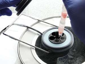

Cryo-preservation or cryo-conservation is a process where organelles, cells, tissues, extracellular matrix, organs or any other biological constructs susceptible to damage caused by unregulated chemical kinetics are preserved by cooling to very low temperatures[1] (typically −80 °C using solid carbon dioxide or −196 °C using liquid nitrogen). At low enough temperatures, any enzymatic or chemical activity which might cause damage to the biological material in question is effectively stopped. Cryopreservation methods seek to reach low temperatures without causing additional damage caused by the formation of ice crystals during freezing. Traditional cryopreservation has relied on coating the material to be frozen with a class of molecules termed cryoprotectants. New methods are constantly being investigated due to the inherent toxicity of many cryoprotectants.[2] By default it should be considered that cryopreservation alters or compromises the structure and function of cells unless it is proven otherwise for a particular cell population. Cryoconservation of animal genetic resources is the process in which animal genetic material is collected and stored with the intention of conservation of the breed.
Natural cryopreservation
Water-bears (Tardigrada), microscopic multicellular organisms, can survive freezing by replacing most of their internal water with the sugar trehalose, preventing it from crystallization that otherwise damages cell membranes. Mixtures of solutes can achieve similar effects. Some solutes, including salts, have the disadvantage that they may be toxic at intense concentrations. In addition to the water-bear, wood frogs can tolerate the freezing of their blood and other tissues. Urea is accumulated in tissues in preparation for overwintering, and liver glycogen is converted in large quantities to glucose in response to internal ice formation. Both urea and glucose act as "cryoprotectants" to limit the amount of ice that forms and to reduce osmotic shrinkage of cells. Frogs can survive many freeze/thaw events during winter if no more than about 65% of the total body water freezes. Research exploring the phenomenon of "freezing frogs" has been performed primarily by the Canadian researcher, Dr. Kenneth B. Storey.
Freeze tolerance, in which organisms survive the winter by freezing solid and ceasing life functions, is known in a few vertebrates: five species of frogs (Rana sylvatica, Pseudacris triseriata, Hyla crucifer, Hyla versicolor, Hyla chrysoscelis), one of salamanders (Hynobius keyserlingi), one of snakes (Thamnophis sirtalis) and three of turtles (Chrysemys picta, Terrapene carolina, Terrapene ornata).[3] Snapping turtles Chelydra serpentina and wall lizards Podarcis muralis also survive nominal freezing but it has not been established to be adaptive for overwintering. In the case of Rana sylvatica one cryopreservant is ordinary glucose, which increases in concentration by approximately 19 mmol/l when the frogs are cooled slowly.[3]
History
One of the most important early theoreticians of cryopreservation was James Lovelock. He suggested that damage to red blood cells during freezing was due to osmotic stress. During the early 1950s, Lovelock had also suggested that increasing salt concentrations in a cell as it dehydrates to lose water to the external ice might cause damage to the cell.[4] In the mid-1950s, he experimented with the cryopreservation of rodents, determining that hamsters could be frozen with 60% of the water in the brain crystallized into ice with no adverse effects. Other organs were shown to be susceptible to damage.[5]
Cryopreservation was applied to humans beginning in 1954 with three pregnancies resulting from the insemination of previously frozen sperm.[6] Fowl sperm was cryopreserved in 1957 by a team of scientists in the UK directed by Christopher Polge.[7] However, the rapid immersion of the samples in liquid nitrogen did not, for certain samples – such as some types of embryos, bone marrow and stem cells – produce the necessary viability to make them usable after thawing. Increased understanding of the mechanism of freezing injury to cells emphasised the importance of controlled or slow cooling to obtain maximum survival on thawing of the living cells. A controlled-rate cooling process, allowing biological samples to equilibrate to optimal physical parameters osmotically in a cryoprotectant (a form of anti-freeze) before cooling in a predetermined, controlled way proved necessary. The ability of cryoprotectants, in the early cases glycerol, to protect cells from freezing injury was discovered accidentally. Freezing injury has two aspects: direct damage from the ice crystals and secondary damage caused by the increase in concentration of solutes as progressively more ice is formed. During 1963, Peter Mazur, at Oak Ridge National Laboratory in the U.S., demonstrated that lethal intracellular freezing could be avoided if cooling was slow enough to permit sufficient water to leave the cell during progressive freezing of the extracellular fluid. That rate differs between cells of differing size and water permeability: a typical cooling rate around 1 °C/minute is appropriate for many mammalian cells after treatment with cryoprotectants such as glycerol or dimethyl sulphoxide, but the rate is not a universal optimum.[8]
Temperature
Storage at very low temperatures is presumed to provide an indefinite longevity to cells, although the actual effective life is rather difficult to prove. Researchers experimenting with dried seeds found that there was noticeable variability of deterioration when samples were kept at different temperatures – even ultra-cold temperatures. Temperatures less than the glass transition point (Tg) of polyol's water solutions, around −136 °C (137 K; −213 °F), seem to be accepted as the range where biological activity very substantially slows, and −196 °C (77 K; −321 °F), the boiling point of liquid nitrogen, is the preferred temperature for storing important specimens. While refrigerators, freezers and extra-cold freezers are used for many items, generally the ultra-cold of liquid nitrogen is required for successful preservation of the more complex biological structures to virtually stop all biological activity.
Risks
Phenomena which can cause damage to cells during cryopreservation mainly occur during the freezing stage, and include: solution effects, extracellular ice formation, dehydration and intracellular ice formation. Many of these effects can be reduced by cryoprotectants. Once the preserved material has become frozen, it is relatively safe from further damage. However, estimates based on the accumulation of radiation-induced DNA damage during cryonic storage have suggested a maximum storage period of 1000 years.[9]
- Solution effects
- As ice crystals grow in freezing water, solutes are excluded, causing them to become concentrated in the remaining liquid water. High concentrations of some solutes can be very damaging.
- Extracellular ice formation
- When tissues are cooled slowly, water migrates out of cells and ice forms in the extracellular space. Too much extracellular ice can cause mechanical damage to the cell membrane due to crushing.
- Dehydration
- Migration of water, causing extracellular ice formation, can also cause cellular dehydration. The associated stresses on the cell can cause damage directly.
Main methods to prevent risks
The main techniques to prevent cryopreservation damages are a well established combination of controlled rate and slow freezing and a newer flash-freezing process known as vitrification.
Slow programmable freezing

Controlled-rate and slow freezing, also known as slow programmable freezing (SPF),[10] is a set of well established techniques developed during the early 1970s which enabled the first human embryo frozen birth Zoe Leyland during 1984. Since then, machines that freeze biological samples using programmable sequences, or controlled rates, have been used all over the world for human, animal and cell biology – "freezing down" a sample to better preserve it for eventual thawing, before it is frozen, or cryopreserved, in liquid nitrogen. Such machines are used for freezing oocytes, skin, blood products, embryo, sperm, stem cells and general tissue preservation in hospitals, veterinary practices and research laboratories around the world. As an example, the number of live births from frozen embryos 'slow frozen' is estimated at some 300,000 to 400,000 or 20% of the estimated 3 million in vitro fertilisation (IVF) births.[11]
Lethal intracellular freezing can be avoided if cooling is slow enough to permit sufficient water to leave the cell during progressive freezing of the extracellular fluid. To minimize the growth of extracellular ice crystal growth and recrystallization,[12] biomaterials such as alginates, polyvinyl alcohol or chitosan can be used to impede ice crystal growth along with traditional small molecule cryoprotectants.[13] That rate differs between cells of differing size and water permeability: a typical cooling rate of about 1 °C/minute is appropriate for many mammalian cells after treatment with cryoprotectants such as glycerol or dimethyl sulfoxide, but the rate is not a universal optimum. The 1 °C / minute rate can be achieved by using devices such as a rate-controlled freezer or a benchtop portable freezing container.[14]
Several independent studies have provided evidence that frozen embryos stored using slow-freezing techniques may in some ways be 'better' than fresh in IVF. The studies indicate that using frozen embryos and eggs rather than fresh embryos and eggs reduced the risk of stillbirth and premature delivery though the exact reasons are still being explored.
Vitrification
Researchers Greg Fahy and William F. Rall helped to introduce vitrification to reproductive cryopreservation in the mid-1980s.[15] As of 2000, researchers claim vitrification provides the benefits of cryopreservation without damage due to ice crystal formation.[16] The situation became more complex with the development of tissue engineering as both cells and biomaterials need to remain ice-free to preserve high cell viability and functions, integrity of constructs and structure of biomaterials. Vitrification of tissue engineered constructs was first reported by Lilia Kuleshova,[17] who also was the first scientist to achieve vitrification of woman’s eggs (oocytes), which resulted in live birth in 1999.[18] For clinical cryopreservation, vitrification usually requires the addition of cryoprotectants prior to cooling. The cryoprotectants act like antifreeze: they decrease the freezing temperature. They also increase the viscosity. Instead of crystallizing, the syrupy solution becomes an amorphous ice—it vitrifies. Rather than a phase change from liquid to solid by crystallization, the amorphous state is like a "solid liquid", and the transformation is over a small temperature range described as the "glass transition" temperature.
Vitrification of water is promoted by rapid cooling, and can be achieved without cryoprotectants by an extremely rapid decrease of temperature (megakelvins per second). The rate that is required to attain glassy state in pure water was considered to be impossible until 2005.[19]
Two conditions usually required to allow vitrification are an increase of the viscosity and a decrease of the freezing temperature. Many solutes do both, but larger molecules generally have a larger effect, particularly on viscosity. Rapid cooling also promotes vitrification.
For established methods of cryopreservation, the solute must penetrate the cell membrane in order to achieve increased viscosity and decrease freezing temperature inside the cell. Sugars do not readily permeate through the membrane. Those solutes that do, such as dimethyl sulfoxide, a common cryoprotectant, are often toxic in intense concentration. One of the difficult compromises of vitrifying cryopreservation concerns limiting the damage produced by the cryoprotectant itself due to cryoprotectant toxicity. Mixtures of cryoprotectants and the use of ice blockers have enabled the Twenty-First Century Medicine company to vitrify a rabbit kidney to −135 °C with their proprietary vitrification mixture. Upon rewarming, the kidney was transplanted successfully into a rabbit, with complete functionality and viability, able to sustain the rabbit indefinitely as the sole functioning kidney.[20]
Freezable tissues
Generally, cryopreservation is easier for thin samples and small clumps of individual cells, because these can be cooled more quickly and so require lesser doses of toxic cryoprotectants. Therefore, cryopreservation of human livers and hearts for storage and transplant is still impractical.
Nevertheless, suitable combinations of cryoprotectants and regimes of cooling and rinsing during warming often allow the successful cryopreservation of biological materials, particularly cell suspensions or thin tissue samples. Examples include:
- Semen in semen cryopreservation
- Blood
- Special cells for transfusion
- Stem cells. It is optimal in high concentration of synthetic serum, stepwise equilibration and slow cooling.[21]
- Umbilical cord blood Further information: Cord blood bank#Cryopreservation
- Tissue samples like tumors and histological cross sections
- Eggs (oocytes) in oocyte cryopreservation
- Embryos at cleavage stage (that are 2, 4 or 8 cells) or at blastocyst stage, in embryo cryopreservation
- Ovarian tissue in ovarian tissue cryopreservation
- Plant seeds or shoots may be cryopreserved for conservation purposes.
Additionally, efforts are underway to preserve humans cryogenically, known as cryonics. For such efforts either the brain within the head or the entire body may experience the above process. Cryonics is in a different category from the aforementioned examples, however: while countless cryopreserved cells, vaccines, tissue and other biologial samples have been thawed and used successfully, this has not yet been the case at all for cryopreserved brains or bodies. At issue are the criteria for defining "success". Proponents of cryonics claim that cryopreservation using present technology, particularly vitrification of the brain, may be sufficient to preserve people in an "information theoretic" sense so that they could be revived and made whole by hypothetical vastly advanced future technology. Not only is there no guarantee of its success, many people argue that human cryopreservation is unethical. According to certain views of the mind body problem, some philosophers believe that the mind, which contains thoughts, memories, and personality, is separate from the brain. When someone dies, their mind leaves the body. If a cryopreserved patient gets successfully resuscitated, no one knows if they would be the same person that they once were or if they would be an empty shell of the memory of who they once were. Right now scientists are trying to see if transplanting cryopreserved human organs for transplantation is viable, if so this would be a major step forward for the possibility of reviving a cryopreserved human.[22]
Embryos
Cryopreservation for embryos is used for embryo storage, e.g., when in vitro fertilization (IVF) has resulted in more embryos than is currently needed.
Pregnancies have been reported from embryos stored for 16 years.[23] Many studies have evaluated the children born from frozen embryos, or “frosties”. The result has been uniformly positive with no increase in birth defects or development abnormalities.[24] A study of more than 11,000 cryopreserved human embryos showed no significant effect of storage time on post-thaw survival for IVF or oocyte donation cycles, or for embryos frozen at the pronuclear or cleavage stages.[25] Additionally, the duration of storage did not have any significant effect on clinical pregnancy, miscarriage, implantation, or live birth rate, whether from IVF or oocyte donation cycles.[25] Rather, oocyte age, survival proportion, and number of transferred embryos are predictors of pregnancy outcome.[25]
Ovarian tissue
Cryopreservation of ovarian tissue is of interest to women who want to preserve their reproductive function beyond the natural limit, or whose reproductive potential is threatened by cancer therapy,[26] for example in hematologic malignancies or breast cancer.[27] The procedure is to take a part of the ovary and perform slow freezing before storing it in liquid nitrogen whilst therapy is undertaken. Tissue can then be thawed and implanted near the fallopian, either orthotopic (on the natural location) or heterotopic (on the abdominal wall),[27] where it starts to produce new eggs, allowing normal conception to occur.[28] The ovarian tissue may also be transplanted into mice that are immunocompromised (SCID mice) to avoid graft rejection, and tissue can be harvested later when mature follicles have developed.[29]
Oocytes
Human oocyte cryopreservation is a new technology in which a woman’s eggs (oocytes) are extracted, frozen and stored. Later, when she is ready to become pregnant, the eggs can be thawed, fertilized, and transferred to the uterus as embryos. Since 1999, when the birth of the first baby from an embryo derived from vitrified-warmed woman’s eggs was reported by Kuleshova and co-workers in the journal of Human Reproduction,[17] this concept has been recognized and widespread. This break-through in achieving vitrification of woman’s oocytes made an important advance in our knowledge and practice of the IVF process, as clinical pregnancy rate is four times higher after oocyte vitrification than after slow freezing.[30] Oocyte vitrification is vital for preservation fertility in young oncology patients and for individuals undergoing IVF who object, either for religious or ethical reasons, to the practice of freezing embryos.
Semen
Semen can be used successfully almost indefinitely after cryopreservation. The longest reported successful storage is 22 years.[31] It can be used for sperm donation where the recipient wants the treatment in a different time or place, or as a means of preserving fertility for men undergoing vasectomy or treatments that may compromise their fertility, such as chemotherapy, radiation therapy or surgery.
Testicular tissue
Cryopreservation of immature testicular tissue is a developing method to avail reproduction to young boys who need to have gonadotoxic therapy. Animal data are promising, since healthy offsprings have been obtained after transplantation of frozen testicular cell suspensions or tissue pieces. However, none of the fertility restoration options from frozen tissue, i.e. cell suspension transplantation, tissue grafting and in vitro maturation (IVM) has proved efficient and safe in humans as yet.[32]
Moss
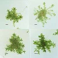
Cryopreservation of whole moss plants, especially Physcomitrella patens, has been developed by Ralf Reski and coworkers[33] and is performed at the International Moss Stock Center. This biobank collects, preserves, and distributes moss mutants and moss ecotypes.[34]
Mesenchymal stromal cells (MSCs)
MSCs, when transfused immediately within a few hours post-thawing, may show reduced function or show decreased efficacy in treating diseases as compared to those MSCs which are in log phase of cell growth (fresh). As a result, cryopreserved MSCs should be brought back into log phase of cell growth in in vitro culture before these are administered for clinical trials or experimental therapies. Re-culturing of MSCs will help in recovering from the shock the cells get during freezing and thawing. Various clinical trials on MSCs have failed which used cryopreserved products immediately post-thaw as compared to those clinical trials which used fresh MSCs.[35]
Preservation of microbiology cultures
Bacteria and fungi can be kept short-term (months to about a year, depending) refrigerated, however, cell division and metabolism is not completely arrested and thus is not an optimal option for long-term storage (years) or to preserve cultures genetically or phenotypically, as cell divisions can lead to mutations or sub-culturing can cause phenotypic changes. A preferred option, species-dependent, is cryopreservation. Nematode worms are the only multicellular eukaryotes that have been shown to survive cryopreservation. [36][37]
Fungi
Fungi, notably zygomycetes, ascomycetes and higher basidiomycetes, regardless of sporulation, are able to be stored in liquid nitrogen or deep-frozen. Crypreservation is a hallmark method for fungi that do not sporulate (otherwise other preservation methods for spores can be used at lower costs and ease), sporulate but have delicate spores (large or freeze-dry sensitive), are pathogenic (dangerous to keep metabolically active fungus) or are to be used for genetic stocks (ideally to have identical composition as the original deposit). As with many other organisms, cryoprotectants like DMSO or glycerol (e.g. filamentous fungi 10% glycerol or yeast 20% glycerol) are used. Differences between choosing cryoprotectants are species (or class) dependent, but generally for fungi penetrating cryoprotectants like DMSO, glycerol or polyethylene glycol are most effective (other non-penetrating ones include sugars mannitol, sorbitol, dextran, etc.). Freeze-thaw repetition is not recommended as it can decrease viability. Back-up deep-freezers or liquid nitrogen storage sites are recommended. Multiple protocols for freezing are summarized below (each uses screw-cap polypropylene cryotubes):[38]
Bacteria
Many common culturable laboratory strains are deep-frozen to preserve genetically and phenotypically stable, long-term stocks. Sub-culturing and prolonged refrigerated samples may lead to loss of plasmid(s) or mutations. Common final glycerol percentages are 15, 20 and 25. From a fresh culture plate, one single colony of interest is chosen and liquid culture is made. From the liquid culture, the medium is directly mixed with equal amount of glycerol; the colony should be checked for any defects like mutations. All antibiotics should be washed from the culture before long-term storage. Methods vary, but mixing can be done gently by inversion or rapidly by vortex and cooling can vary by either placing the cryotube directly at −50 to −95 °C, shock-freezing in liquid nitrogen or gradually cooling and then storing at −80 °C or cooler (liquid nitrogen or liquid nitrogen vapor). Recovery of bacteria can also vary, namely if beads are stored within the tube then the few beads can be used to plate or the frozen stock can be scraped with a loop and then plated, however, since only little stock is needed the entire tube should never be completely thawed and repeated freeze-thaw should be avoided. 100% recovery is not feasible regardless of methodology.[39][40][41]
Worms
The microscopic soil-dwelling nematode roundworms Panagrolaimus detritophagus and Plectus parvus are the only eukaryotic organisms that have been proven to be viable after long-term cryopreservation to date. In this case, the preservation was natural rather than artificial, due to permafrost.
See also
References
- ↑ Pegg, David E. (January 1, 2007). "Principles of cryopreservation". Methods in Molecular Biology. Clifton, N.J. 368: 39–57. doi:10.1007/978-1-59745-362-2_3. ISSN 1064-3745. PMID 18080461.
- ↑ Sambu, Sammy (June 25, 2015). "A Bayesian approach to optimizing cryopreservation protocols". PeerJ. 3: e1039. doi:10.7717/peerj.1039. ISSN 2167-8359. PMC 4485240. PMID 26131379.
- 1 2 Jon P. Costanzo; Richard E. Lee; Michael F. Wright (1991). "Glucose loading prevents freezing injury in rapidly cooled wood frogs" (PDF). American Journal of Physiology: R1549–R1553.
- ↑ Mazur P (1970). "Cryobiology: the freezing of biological systems". Science. 168 (3934): 939–49. Bibcode:1970Sci...168..939M. doi:10.1126/science.168.3934.939. PMID 5462399.
- ↑ "The Cryobiological Case for Cryonics" (PDF). Cryonics. Vol. 9(3) no. 92. Alcor Life Extension Foundation. March 1988. p. 27.
- ↑ "Fatherhood After Death Has Now Been Proved Possible". Cedar Rapids Gazette. April 9, 1954.
- ↑ Polge C (1957). "Low-Temperature Storage of Mammalian Spermatozoa". Royal Society of London. 147 (929): 498–508. Bibcode:1957RSPSB.147..498P. doi:10.1098/rspb.1957.0068.
- ↑ https://www.cell.com/biophysj/pdf/S0006-3495(63)86824-1.pdf
- ↑ Mazur P (1984). "Freezing of living cells: mechanisms and implications". American Journal of Physiology. 247 (3 Pt 1): C125–42. Bibcode:1957RSPSB.147..498P. doi:10.1098/rspb.1957.0068.
- ↑ Vutyavanich T; Piromlertamorn W; Nunta S (April 2010). "Rapid freezing versus slow programmable freezing of human spermatozoa". Fertil. Steril. 93 (6): 1921–8. doi:10.1016/j.fertnstert.2008.04.076. PMID 19243759.
- ↑ "dead link". Archived from the original on 2009-05-26.
- ↑ Deller, Robert C.; Vatish, Manu; Mitchell, Daniel A.; Gibson, Matthew I. (February 3, 2014). "Synthetic polymers enable non-vitreous cellular cryopreservation by reducing ice crystal growth during thawing". Nature Communications. 5: 3244. Bibcode:2014NatCo...5E3244D. doi:10.1038/ncomms4244. PMID 24488146.
- ↑ Sambu, Sammy (June 25, 2015). "A Bayesian approach to optimizing cryopreservation protocols". PeerJ. 3: e1039. doi:10.7717/peerj.1039. ISSN 2167-8359. PMC 4485240. PMID 26131379.
- ↑ Thompson M; Nemits M; Ehrhardt R (May 2011). "Rate-controlled Cryopreservation and Thawing of Mammalian Cells". Protocol Exchange. doi:10.1038/protex.2011.224.
- ↑ Rall, WF; Fahy, GM (February 14–20, 1985). "Ice-free cryopreservation of mouse embryos at −196 degrees C by vitrification". Nature. 313 (6003): 573–5. Bibcode:1985Natur.313..573R. doi:10.1038/313573a0. PMID 3969158.
- ↑ "Alcor: The Origin of Our Name" (PDF). Alcor Life Extension Foundation. Winter 2000. Retrieved August 25, 2009.
- 1 2 Kuleshova, L L; Wang, XW; Wu, YN; Zhou, Y; Yu, H (2004). "Vitrification of encapsulated hepatocytes with reduced cooling and warming rates". Cryo Letters. 25 (4): 241–254.
- ↑ Kuleshova, Lilia; Gianoroli, Luca; Magli, Cristina; Ferraretti, Anna; Trounson, Alan (1999). "Birth following vitrification of small number of human oocytes". Human Reproduction. 14 (12): 3077–3079. doi:10.1093/humrep/14.12.3077.
- ↑ Bhat SN; Sharma A; Bhat SV (2005). "Vitrification and glass transition of water: insights from spin probe ESR". Phys Rev Lett. 95 (23): 235702. arXiv:cond-mat/0409440. Bibcode:2005PhRvL..95w5702B. doi:10.1103/PhysRevLett.95.235702. PMID 16384318.
- ↑ Fahy GM; Wowk B; Pagotan R; Chang A; et al. (2009). "Physical and biological aspects of renal vitrification". Organogenesis. 5 (3): 167–175. doi:10.4161/org.5.3.9974. PMC 2781097. PMID 20046680.
- ↑ Lee JY; Lee JE; Kim DK; Yoon TK; et al. (November 2008). "High concentration of synthetic serum, stepwise equilibration and slow cooling as an efficient technique for large-scale cryopreservation of human embryonic stem cells". Fertil. Steril. 93 (3): 976–85. doi:10.1016/j.fertnstert.2008.10.017. PMID 19022437.
- ↑ Devlin, Hannah. "Cryonics: Does It Offer Humanity a Chance to Return from the Dead?". thegaurdian.com. The Guardian. Retrieved 17 April 2017.
- ↑ Planer NEWS and Press Releases > 'Twins' born 16 years apart. January 6, 2006.
- ↑ "Genetics & IVF Institute". Givf.com. Archived from the original on December 6, 2012. Retrieved July 27, 2009.
- 1 2 3 Riggs R; Mayer J; Dowling-Lacey D; Chi TF; et al. (November 2008). "Does storage time influence postthaw survival and pregnancy outcome? An analysis of 11,768 cryopreserved human embryos". Fertil. Steril. 93 (1): 109–15. doi:10.1016/j.fertnstert.2008.09.084. PMID 19027110.
- ↑ Isachenko V; Lapidus I; Isachenko E; et al. (2009). "Human ovarian tissue vitrification versus conventional freezing: morphological, endocrinological, and molecular biological evaluation". Reproduction. 138 (2): 319–27. doi:10.1530/REP-09-0039. PMID 19439559.
- 1 2 Oktay K; Oktem O (November 2008). "Ovarian cryopreservation and transplantation for fertility preservation for medical indications: report of an ongoing experience". Fertil. Steril. 93 (3): 762–8. doi:10.1016/j.fertnstert.2008.10.006. PMID 19013568.
- ↑ Livebirth after orthotopic transplantation of cryopreserved ovarian tissue The Lancet, September 24, 2004
- ↑ Lan C; Xiao W; Xiao-Hui D; Chun-Yan H; et al. (December 2008). "Tissue culture before transplantation of frozen-thawed human fetal ovarian tissue into immunodeficient mice". Fertil. Steril. 93 (3): 913–9. doi:10.1016/j.fertnstert.2008.10.020. PMID 19108826.
- ↑ Glujovsky D, Riestra B, Sueldo C, Fiszbajn G, Repping S, Nodar F, Papier S, Ciapponi A. Vitrification versus slow freezing for women undergoing oocyte cryopreservation" Cochrane Database of Systematic Reviews 2014, Issue 8. Art. No.: CD010047. doi:10.1002/14651858.CD010047.pub2
- ↑ Planer NEWS and Press Releases > Child born after 22 year semen storage using Planer controlled rate freezer 14/10/2004
- ↑ Wyns C; Curaba M; Vanabelle B; Van Langendonckt A; et al. (2010). "Options for fertility preservation in prepubertal boys". Hum. Reprod. Update. 16 (3): 312–28. doi:10.1093/humupd/dmp054. PMID 20047952.
- ↑ Schulte, J., Ralf Reski (2004): High-throughput cryopreservation of 140000 Physcomitrella patens mutants. Plant Biol. 6, 119-127. Schulte J.; Reski R. (2004). "High throughput cryopreservation of 140,000 Physcomitrella patens mutants". Plant Biol (Stuttg). Plant Biotechnology, Freiburg University, Freiburg, Germany. 6: 119–27. doi:10.1055/s-2004-817796. PMID 15045662.
- ↑ ScienceDaily: Mosses, deep frozen. "Mosses, deep-frozen".
- ↑ Francois, M; et al. (2012). "Cryopreserved mesenchymal stromal cells display impaired immunosuppressive properties as a result of heat-shock response and impaired interferon-γ licensing". Cytotherapy. 14 (2): 147–152. doi:10.3109/14653249.2011.623691. PMC 3279133. PMID 22029655.
- ↑ Weisberger, M. (2018). Worms Frozen for 42,000 Years in Siberian Permafrost Wriggle to Life. Retrieved from https://www.livescience.com/63187-siberian-permafrost-worms-revive.html
- ↑ Shatilovich, A.V., Tchesunov, A.V., Neretina, T.V. et al. Dokl Biol Sci (2018) 480: 100. https://doi.org/10.1134/S0012496618030079
- ↑
- ↑ Freeze-Drying and Cryopreservation of Bacteria
- ↑ "Addgene: Protocol - How to Create a Bacterial Glycerol Stock". Addgene.org. Retrieved 9 September 2015.
- ↑
Sources
- Engelmann, F.; M. E. Dulloo; C. Astorga; S. Dussert; F. Anthony, eds. (2007). Conserving coffee genetic resources. Bioversity International, CATIE, IRD. p. 61.
- Panis, B & Tien Thinh, N. (2001). Cryopreservation of Musa germplasm. INIBAP (now Bioversity International). p. 45.
- ReproTech Limited (2012). "Fertility Preservation". ReproTech Limited. Archived from the original on 2012-09-04.
- Nakasone, Karen K, et al., 2004, PRESERVATION AND DISTRIBUTION OF FUNGAL CULTURES.
- Stephen, F., 1995, Freeze-Drying and Cryopreservation of Bacteria
External links
- Vitrification for storage of embryos, HFEA website
- The Freezing of Human Oocytes (Eggs)
- Society for Cryobiology
- The Society for Low Temperature Biology
- Cellular cryobiology and anhydrobiology
- Death in the Deep Freeze
- In vitro storage and cryopreservation
- Cryonics
| Authority control |
|---|