Central tegmental tract
| Central tegmental tract | |
|---|---|
 Diagram of the midbrain, sectioned at the level of the superior colliculus (Central tegmental tract not labeled, but region is visible.) | |
 Axial section of the Brainstem (Pons) at the level of the Facial Colliculus (Central tegmental tract not labeled, but region is visible.) | |
| Details | |
| Identifiers | |
| Latin | Tractus tegmentalis centralis |
| NeuroNames | 2204 |
| TA | A14.1.05.325 |
| FMA | 83850 |
| Anatomical terms of neuroanatomy | |
The central tegmental tract[1] is a structure in the midbrain and pons.
- The central tegmental tract includes ascending axonal fibers that arise from the rostral nucleus solitarius and terminate in the ventral posteromedial nucleus (VPM) of thalamus. Information from the thalamus will go to cortical taste area, namely the insula and frontal operculum.
- It also contains descending axonal fibers from the parvocellular red nucleus. The descending axons will project to the inferior olivary nucleus. This latter pathway (the rubro-olivary tract) will be used to connect the contralateral cerebellum.
Lesion of the tract can cause palatal myoclonus, e.g. in myoclonic syndrome, in strokes of the Posterior Inferior Cerebellar Artery.
Additional Images
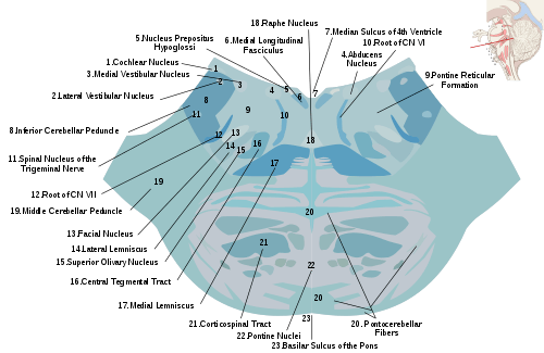 Horizontal section through the lower part of the pons. The central tegmental tract is labeled #16.
References
External links
This article is issued from
Wikipedia.
The text is licensed under Creative Commons - Attribution - Sharealike.
Additional terms may apply for the media files.