Medial dorsal nucleus
| Medial dorsal nucleus | |
|---|---|
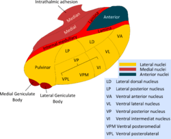
 Thalamic nuclei: MNG = Midline nuclear group AN = Anterior nuclear group MD = Medial dorsal nucleus VNG = Ventral nuclear group VA = Ventral anterior nucleus VL = Ventral lateral nucleus VPL = Ventral posterolateral nucleus VPM = Ventral posteromedial nucleus LNG = Lateral nuclear group PUL = Pulvinar MTh = Metathalamus LG = Lateral geniculate nucleus MG = Medial geniculate nucleus | |
 Thalamic nuclei | |
| Details | |
| Identifiers | |
| Latin | nucleus mediodorsalis thalami |
| NeuroNames | 312 |
| NeuroLex ID | birnlex_1543 |
| TA | A14.1.08.622 |
| FMA | 62156 |
| Anatomical terms of neuroanatomy | |
The medial dorsal nucleus (or dorsomedial nucleus of thalamus) is a large nucleus in the thalamus.
Structure
It relays inputs from the amygdala and olfactory cortex and projects to the prefrontal cortex and the limbic system and in turn relays them to the prefrontal association cortex. As a result, it plays a crucial role in attention, planning, organization, abstract thinking, multi-tasking, and active memory.
The connections of the medial dorsal nucleus have even been used to delineate the prefrontal cortex of the Göttingen minipig brain.[2]
By stereology the number of brain cells in the region has been estimated to around 6.43 million neurons in the adult human brain and 36.3 million glial cells, and with the newborn having quite different numbers: around 11.2 million neurons and 10.6 million glial cells.[3]
Function
Pain processing
While both the ventral and medial dorsal nuclei process pain, the medial dorsal nucleus bypasses primary cortices, sending their axons directly to secondary and association cortices. The cells also send axons directly to many parts of the brain, including nuclei of the limbic system such as the lateral nucleus of the amygdala, the anterior cingulate, and the hippocampus. This part of the sensory system, known as the non-classical or extralemniscal system is less accurate, and less detailed in regards to sensory signal analysis. This processing is known colloquially as "fast and dirty" rather than the "slow and accurate" system of classical or lemniscal system. This pathway activates parts of the brain that evoke emotional responses.
Clinical significance
Damage to the medial dorsal nucleus has been associated with Korsakoff's syndrome.
Additional images
 Thalamus
 Thalamus
References
- ↑ Li XB, Inoue T, Nakagawa S, Koyama T (May 2004). "Effect of mediodorsal thalamic nucleus lesion on contextual fear conditioning in rats". Brain Res. 1008 (2): 261–72. doi:10.1016/j.brainres.2004.02.038. PMID 15145764.
- ↑ Jacob Jelsing; Anders Hay-Schmidt; Tim Dyrby; Ralf Hemmingsen; Harry B. M. Uylings; Bente Pakkenberg (2006). "The prefrontal cortex in the Göttingen minipig brain defined by neural projection criteria and cytoarchitecture". Brain Research Bulletin. 70 (4&ndash, 6): 322&ndash, 336. doi:10.1016/j.brainresbull.2006.06.009. PMID 17027768.
- ↑ Maja Abitz; Rune Damgaard Nielsen; Edward G. Jones; Henning Laursen; Niels Graem & Bente Pakkenberg (2007). "Excess of Neurons in the Human Newborn Mediodorsal Thalamus Compared with That of the Adult". Cerebral Cortex. 17 (11): 2573&ndash, 2578. doi:10.1093/cercor/bhl163. PMID 17218480.
- Aage R. Moller "Pain: Its anatomy, physiology and treatment" 2012