Spinocerebellar tract
| Spinocerebellar tract | |
|---|---|
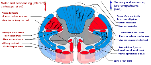 Spinocerebellar tracts are labeled in blue at right. | |
| Details | |
| Identifiers | |
| Latin | Tractus spinocerebellaris |
| NeuroNames | 1978 |
| Anatomical terms of neuroanatomy | |
The spinocerebellar tract is a nerve tract originating in the spinal cord and terminating in the same side (ipsilateral) of the cerebellum.
Origins of proprioceptive information
Proprioceptive information is obtained by Golgi tendon organs and muscle spindles.
- Golgi tendon organs consist of a fibrous capsule enclosing tendon fascicles and bare nerve endings that respond to tension in the tendon by causing action potentials in type Ib afferents. These fibers are relatively large, myelinated, and quickly conducting.
- Muscle spindles monitor the length within muscles and send information via faster Ia afferents. These axons are larger and faster than type Ib (from both nuclear bag fibers and nuclear chain fibers) and type II afferents (solely from nuclear chain fibers).
All of these neurons are sensory (first order, or primary) and have their cell bodies in the dorsal root ganglia. They pass through rexed laminae layers I-VI of the posterior grey column (dorsal horn) to form synapses with second order or secondary neurons in layer VII just beneath the dorsal horn.
Subdivisions of the tract
The tract is divided into:[1]
| Division | Peripheral Process of First Order the Neuron | Region of Innervation |
|---|---|---|
| dorsal (posterior) spinocerebellar tract | from muscle spindle (primarily) and golgi tendon organs | Ipsilateral Caudal Aspect of the body and legs |
| ventral (anterior) spinocerebellar tract | from golgi tendon organs | Ipsilateral Caudal Aspect of the body and legs |
| cuneocerebellar tract | from muscle spindle (primarily) and golgi tendon organs | Ipsilateral arm |
| rostral spinocerebellar tract | from golgi tendon organs | Ipsilateral arm |
Pathway for dorsal and spinocuneocerebellar tracts
The sensory neurons synapse in an area known as Clarke's nucleus or "Clarke's column".
This is a column of relay neuron cell bodies within the medial gray matter within the spinal cord in layer VII (just beneath the dorsal horn), specifically between T1-L3. These neurons then send axons up the spinal cord, and project ipsilaterally to medial zones of the cerebellum through the inferior cerebellar peduncle.
Below L3, relevant neurons pass into the fasciculus gracilis (usually associated with the dorsal column-medial lemniscal system) until L3 where they synapse with Clarke's nucleus (leading to considerable caudal enlargement).
The neurons in the accessory cuneate nucleus have axons leading to the ipsilateral cerebellum via the inferior cerebellar peduncle.
Pathway for ventral and rostral spinocerebellar tracts
Some neurons of the ventral spinocerebellar tract instead form synapses with neurons in layer VII of L4-S3. Most of these fibers cross over to the contralateral lateral funiculus via the anterior white commissure and through the superior cerebellar peduncle. The fibers then often cross over again within the cerebellum to end on the ipsilateral side. For this reason the tract is sometimes termed the "double-crosser."
The Rostral Tract synapses at the dorsal horn lamina (intermediate gray zone) of the spinal cord and ascends ipsilaterally to the cerebellum through the inferior cerebellar peduncle
References
- ↑ Siegel, Allan, and Hreday N. Sapru. Essential Neuroscience. 2nd. Lippincott, 2011. 146-149.