Midbrain tegmentum
| Midbrain tegmentum | |
|---|---|
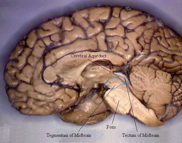
 Transverse section of mid-brain at level of superior colliculi. ("Tegmentum" visible center right.) | |
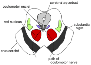 Section through superior colliculus showing path of oculomotor nerve. (Tegmentum not labeled, but surrounding structures more clearly defined.) | |
| Details | |
| Part of | Midbrain |
| Identifiers | |
| Latin | Tegmentum Mesencephali |
| MeSH | D013681 |
| NeuroNames | 491 |
| NeuroLex ID | birnlex_1200 |
| Anatomical terms of neuroanatomy | |
The midbrain is also known as the Mesencephalon and is one of the three major brain divisions. The midbrain is broken up into the tectum and the tegmentum. There are some important structures located within the tegmentum. The midbrain tegmentum is the part of the midbrain extending from the substantia nigra to the cerebral aqueduct in a horizontal section of the midbrain. It forms the floor of the midbrain that surrounds the cerebral aqueduct. Additional structures include the reticular formation, red nucleus, ventral tegmental area (VTA) and the periaqueductal grey matter. The midbrain tegmentum contains thousands of neurons that are responsible for a variety of functions, two of which include the control of movement and sensory systems.[1]
See also
External links
- Photo
- "Anatomy diagram: 13048.000-3". Roche Lexicon - illustrated navigator. Elsevier. Archived from the original on 2014-01-01.
Notes