Vagal trigone
| Vagal trigone | |
|---|---|
 Dissection of brain-stem. Dorsal view. | |
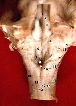 Human caudal brainstem posterior view (Trigonum nervi vagi is #7) | |
| Details | |
| Identifiers | |
| Latin | trigonum nervi vagi |
| NeuroNames | 634 |
| TA | A14.1.05.709 |
| FMA | 78445 |
| Anatomical terms of neuroanatomy | |
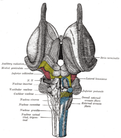
The cells of the dorsal nucleus of vagus nerve are spindle-shaped, like those of the posterior column of the spinal cord, and the nucleus is usually considered as representing the base of the posterior column. It measures about 2 cm. in length, and in the lower, closed part of the medulla oblongata is situated behind the dorsal motor nucleus of the vagus; whereas in the upper, open part it lies lateral to that nucleus, and corresponds to an eminence, named the vagal trigone (ala cinerea, not to be confused with tuberculum cinereum nor tuber cinereum), in the rhomboid fossa.
References
This article incorporates text in the public domain from page 781 of the 20th edition of Gray's Anatomy (1918)
External links
This article is issued from
Wikipedia.
The text is licensed under Creative Commons - Attribution - Sharealike.
Additional terms may apply for the media files.