Posterior external arcuate fibers
| Posterior external arcuate fibers | |
|---|---|
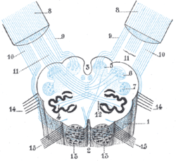 Diagram showing the course of the arcuate fibers. (Testut.) 1. Medulla oblongata anterior surface. 2. Anterior median fissure. 3. Fourth ventricle. 4. Inferior olivary nucleus, with the accessory olivary nuclei. 5. Gracile nucleus. 6. Cuneate nucleus. 7. Trigeminal. 8. Inferior peduncles, seen from in front. 9. Posterior external arcuate fibers. 10. Anterior external arcuate fibers. 11. Internal arcuate fibers(no it's olivocerebellar tract). 12. Peduncle of inferior olivary nucleus. 13. Nucleus arcuatus. 14. Vagus. 15. Hypoglossal. | |
 Section of the medulla oblongata at about the middle of the olive. (Arcuate fibers labeled at center right.) | |
| Details | |
| Identifiers | |
| Latin | fibrae arcuatae externae posteriores |
| NeuroLex ID | birnlex_1238 |
| Anatomical terminology | |
The posterior external arcuate fibers (dorsal external arcuate fibers) take origin in the accessory cuneate nucleus ; they pass to the inferior peduncle of the same side.
It carries proprioceptive information from the upper limbs and neck. It is an analogue to the dorsal spinocerebellar tract for the upper limbs.[1] In this context, the "cuneo-" derives from the accessory cuneate nucleus, not the cuneate nucleus. (The two nuclei are related in space, but not in function.)
The term "cuneocerebellar tract" is sometimes used to collectively refer to the posterior external arcuate fibers.[2]
The term "cuneocerebellar tract" is also used to describe an exteroceptive and proprioceptive components that take origin in the gracile and cuneate nuclei; they pass to the inferior peduncle of the same side.[3]
It is uncertain whether fibers are continued directly from the gracile and cuneate fasciculi into the inferior peduncle.
See also
References
This article incorporates text in the public domain from page 783 of the 20th edition of Gray's Anatomy (1918)
- ↑ Fix, James D. (2002). Neuroanatomy. Hagerstwon, MD: Lippincott Williams & Wilkins. p. 133. ISBN 0-7817-2829-0.
- ↑ Sabyasachi Sircar (2007). Principles of Medical Physiology. Stuttgart: Georg Thieme Verlag. p. 608. ISBN 1-58890-572-1.
- ↑ Cooke, J. D. (October 1971). "Origin and termination of cuneocerebellar tract". Experimental Brain Research. 13 (4): 339–358. doi:10.1007/bf00234336. Retrieved 12 April 2015.
Additional images
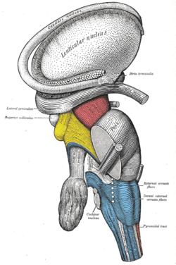 Dissection of brain-stem. Lateral view.
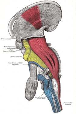 Deep dissection of brain-stem. Lateral view.
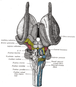 Dissection of brain-stem. Dorsal view.
External links
- hier-793 at NeuroNames - dorsal external arcuate fibers
- hier-800 at NeuroNames - cuneocerebellar tract