Dorsal trigeminal tract
| Dorsal trigeminal tract | |
|---|---|
| Details | |
| Identifiers | |
| Latin | tractus trigeminothalamicus posterior |
| NeuroNames | 606 |
| NeuroLex ID | birnlex_1718 |
| TA | A14.1.05.312 |
| FMA | 72500 |
| Anatomical terms of neuroanatomy | |
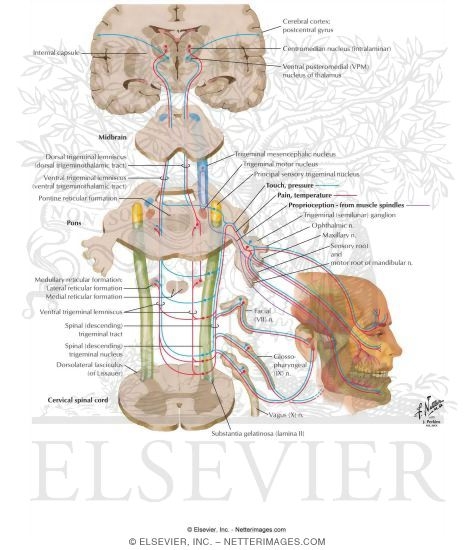
The posterior trigeminothalamic tract (or dorsal trigeminothalamic tract) is composed of second order neuronal axons. These fibers carry sensory information about discriminative touch and conscious proprioception in the oral cavity from the principal (chief sensory) nucleus to the ventral posteromedial (VPM) nucleus of the thalamus.
The posterior trigeminothalamic tract is also called the posterior trigeminal lemniscus.[1]
Pathway
The first order neurons (from the trigeminal ganglion) enter the medulla and synapse in the principal (chief sensory) nucleus. Axons of the second order neurons then decussates to enter the trigeminal lemniscus in the midbrain and then ascend to the ventral posteromedial nucleus of the contralateral thalamus, forming the posterior trigeminothalamic tract. The third order neurons in the thalamus ascend to the sensory cortex of the postcentral gyrus.
References
- ↑ Anthoney, T. R. (1993). Neuroanatomy and the neurologic exam: a thesaurus of synonyms, similar-sounding non-synonyms, and terms of variable meaning. CRC Press.
Sources
- Siegel, A., & Sapru, H. N. (2006). Essential neuroscience. Lippincott Williams & Wilkins.
- Norton, N. S. (2016). Netter's head and neck anatomy for dentistry. Elsevier Health Sciences.
- Henssen D. J. H. A. (2016) "New Insights in Trigeminal Anatomy: A Double Orofacial Tract for Nociceptive Input" Frontiers in Neuroanatomy.
External links