Ehlers–Danlos syndromes
| Ehlers–Danlos syndromes | |
|---|---|
.png) | |
| Individual with an EDS displaying skin hyperelasticity | |
| Specialty | Medical genetics, rheumatology |
| Symptoms | Overly flexible joints, stretchy skin, abnormal scar formation[1] |
| Complications | Aortic dissection, joint dislocations, osteoarthritis[1] |
| Usual onset | Birth or early childhood[2] |
| Duration | Lifelong[3] |
| Types | Hypermobile, classic, vascular, kyphoscoliosis, arthrochalasia, dermatosparaxis, brittle cornea syndrome, others[4] |
| Causes | Genetic[1] |
| Risk factors | Family history[1] |
| Diagnostic method | Genetic testing, skin biopsy[3] |
| Differential diagnosis | Marfan syndrome, cutis laxa syndrome, familial joint hypermobility syndrome[3] |
| Treatment | Supportive[5] |
| Prognosis | Depends on specific disorder[3] |
| Frequency | 1 in 5,000[1] |
Ehlers–Danlos syndromes (EDSs) are a group of genetic connective tissue disorders.[1] Symptoms may include loose joints, stretchy skin, and abnormal scar formation.[1] These can be noticed at birth or in early childhood.[2] Complications may include aortic dissection, joint dislocations, scoliosis, chronic pain, or early osteoarthritis.[1][3]
EDSs are due to a mutation in one of more than a dozen different genes.[1] The specific gene affected determines the specific EDS.[1] Some cases result from a new mutation occurring during early development, while others are inherited in an autosomal dominant or recessive manner.[1] This results in defects in the structure or processing of collagen.[1] The diagnosis may be confirmed with genetic testing or a skin biopsy.[3] People may be misdiagnosed with hypochondriasis, depression, or chronic fatigue syndrome.[3]
No cure is known.[5] Treatment is supportive in nature.[3] Physical therapy and bracing may help strengthen muscles and support joints.[3] While some disorders result in a normal life expectancy, those that affect blood vessels generally result in a shorter life expectancy.[5]
EDSs affect about one in 5,000 people globally.[1] The prognosis depends on the specific disorder.[3] Excess mobility was first described by Hippocrates in 400 BC.[6] The syndromes are named after two physicians, Edvard Ehlers from Denmark and Henri-Alexandre Danlos from France, who described them at the turn of the 20th century.[7]
Signs and symptoms
Signs vary widely based on the specific EDS the person has. This group of disorders affects connective tissues, most typically in the joints, skin, and blood vessels and causes effects ranging from mildly loose joints to life-threatening complications.[8] Major signs and symptoms are listed below.
Musculoskeletal
Musculoskeletal symptoms include hyperflexible joints that are unstable and prone to sprain, dislocation, subluxation, and hyperextension.[7][9] There can be an early onset of advanced osteoarthritis,[10] chronic degenerative joint disease,[10] swan-neck deformity of the fingers,[11] and Boutonniere deformity of the fingers. Tearing of tendons or muscles may occur.[12] Deformities of the spine, such as scoliosis (curvature of the spine), kyphosis (a thoracic hump), tethered spinal cord syndrome, and occipitoatlantoaxial hypermobility may also be present.[13] There can also be myalgia (muscle pain) and arthralgia (joint pain),[14] which may be severe and disabling. Trendelenburg's sign is often seen, which means that when standing on one leg, the pelvis drops on the other side.[15] Osgood–Schlatter disease, a painful lump on the knee, is common as well.[16] In infants, walking can be delayed (beyond 18 months of age), and bottom-shuffling instead of crawling occurs.[17]
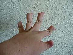 Individual with EDS showing hypermobile fingers
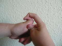 Individual with an EDS displaying hypermobile thumb
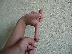 Individual with EDS displaying hypermobile metacarpophalangeal joints
Skin
The weak connective tissue causes fragile skin that tears easily,[10] atrophic "cigarette paper" scars, and easy bruising.[1][7][18] Redundant skin folds, especially on the eyelids also happen.[10][19] Molluscoid pseudotumors,[20] especially on pressure points. Petechiae,[21] subcutaneous spheroids,[20] livedo reticularis, and piezogenic papules are less common.[22] In vascular EDS, thin, translucent, and extremely fragile skin that tears easily is also a symptom.
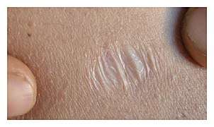 Atrophic scar in a case of EDS
.png) Translucent skin in vascular EDS
Translucent skin in vascular EDS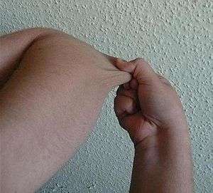 Individual with EDS displaying skin hyperelasticity
Cardiovascular
- Thoracic outlet syndrome[23]
- Arterial rupture[7]
- Valvular heart disease, such as mitral valve prolapse, creates an increased risk for infective endocarditis during surgery. This may progress to a life-threatening degree.[24] Heart conduction abnormalities have been found in those with hypermobility form of EDS.[25]
- Dilation and/or rupture (aneurysm) of ascending aorta[26]
- Postural orthostatic tachycardia syndrome[27]
- Raynaud's phenomenon
- Varicose veins[28]
- Heart murmur
- Heart conduction abnormalities
Other manifestations

- Hiatial hernia[20]
- Gastroesophageal reflux[29]
- Gastrointestinal dysmotility[30]
- Dysautonomia[31]
- Gorlin's sign (touch tongue to nose)[32]
- Anal prolapse[20]
- Collapsed lung (spontaneous pneumothorax)[10]
- Nerve disorders (carpal tunnel syndrome, acroparesthesia, neuropathy, including small fiber neuropathy)[33]
- Insensitivity to local anesthetics.[34]
- Arnold–Chiari malformation[35]
- Platelet aggregation failure (platelets do not clump together properly)[36]
- Pregnancy complications: increased pain, mild to moderate peripartum bleeding, cervical insufficiency, uterine tearing,[12] or premature rupture of membranes.[37]
- Cranial vertebral instability: caused by trauma(s) to the head and neck areas such as concussion and whiplash. Ligaments in neck are unable to heal properly, therefore, the neck structure does not have the ability to support the skull, which can then sink into the brain stem blocking the normal flow of cerebrospinal fluid, leading to issues related to the autonomic nervous system failing to work properly.[38]
- Celiac disease may be associated with EDS. Also, it can be misdiagnosed as EDS due to common symptoms, including fatigue, pain, gastrointestinal complaints, or cardiovascular autonomic dysfunction.[39]
- Fibromyalgia
Because it is often undiagnosed or misdiagnosed in childhood, some instances of EDSs have been mischaracterized as child abuse.[40]
The pain associated with the disorders may be severe.[41]
Genetics
Only some EDSs can be positively identified as tied to specific genetic variation.
Mutations in these genes can cause EDSs:
- Fibrous proteins: COL1A1, COL1A2, COL3A1, COL5A1, COL5A2, and TNXB
- Enzymes: ADAMTS2, PLOD1, B4GALT7, DSE, and D4ST1/CHST14
Mutations in these genes usually alter the structure, production, or processing of collagen or proteins that interact with collagen. Collagen provides structure and strength to connective tissue. A defect in collagen can weaken connective tissue in the skin, bones, blood vessels, and organs, resulting in the features of the disorder.[1] Inheritance patterns depend on the specific syndrome. Most forms of EDSs are inherited in an autosomal dominant pattern, which means only one of the two copies of the gene in question must be altered to cause a disorder. A few are inherited in an autosomal recessive pattern, which means both copies of the gene must be altered for a person to be affected by a disorder. It can also be an individual (de novo or "sporadic") mutation.[42]
Diagnosis
A diagnosis can be made by an evaluation of medical history and clinical observation. The Beighton criteria are widely used to assess the degree of joint hypermobility. DNA and biochemical studies can help identify affected individuals. Diagnostic tests include collagen gene-mutation testing, collagen typing via skin biopsy, echocardiogram, and lysyl hydroxylase or oxidase activity. However, these tests are not able to confirm all cases, especially in instances of an unmapped mutation, so clinical evaluation by a geneticist remains essential. If multiple individuals in a family are affected, performing prenatal diagnosis may be possible using a DNA information technique known as a linkage study.[43] Knowledge about EDSs among practitioners is poor.[44][45]
Classification
As of 2017, 13 EDSs had been characterized, with a significant overlap in features.[4]
Hypermobile EDS (type 3 hEDS) is characterized primarily by joint hypermobility affecting both large and small joints, which may lead to recurrent joint dislocations and subluxations (partial dislocation). In general, people with this type have soft, smooth, and velvety skin with easy bruising and chronic pain of the muscles and/or bones.[4] The mutation that causes this type of EDS is unknown. Less skin involvement is seen than other types. No genetic test for this type is available.
Classical EDS (type 1 cEDS) is associated with extremely elastic (stretchy), smooth skin that is fragile and bruises easily; wide, atrophic scars (flat or depressed scars); and joint hypermobility. Molluscoid pseudotumors (calcified hematomas over pressure points such as the elbow) and spheroids (fat-containing cysts on forearms and shins) are also frequently seen. Hypotonia and delayed motor development may occur.[4] The mutation that causes this type of EDS is in the genes COL5A1, COL5A2, and COL1A1. It involves the skin more than hEDS.
Vascular EDS (type 4 vEDS) is characterized by thin, translucent skin that is extremely fragile and bruises easily. Arteries and certain organs such as the intestines and uterus are also fragile and prone to rupture. People with this type typically have short stature, and thin scalp hair. It also has characteristic facial features including large eyes, an undersized chin, sunken cheeks, a thin nose and lips, and ears without lobes.[46] Joint hypermobility is present, but generally confined to the small joints (fingers, toes). Other common features include club foot, tendon and/or muscle rupture, acrogeria (premature aging of the skin of the hands and feet), early onset varicose veins, pneumothorax (collapse of a lung), recession of the gums, and a decreased amount of fat under the skin.[4] Is can be caused by the mutations in the COL3A1 gene.
Kyphoscoliosis EDS (type 6 kEDS) is associated with severe hypotonia at birth, delayed motor development, progressive scoliosis (present from birth), and scleral fragility. Affected people may also have easy bruising, fragile arteries that are prone to rupture, unusually small corneas, and osteopenia (low bone density). Other common features include a "marfanoid habitus" which is characterized by long, slender fingers (arachnodactyly), unusually long limbs, and a sunken chest (pectus excavatum) or protruding chest (pectus carinatum).[4] It can be caused by mutations in the gene PLOD1.
Arthrochalasia EDS (types 7A & B aEDS) is characterized by severe joint hypermobility and congenital hip dislocation. Other common features include fragile, elastic skin with easy bruising, hypotonia, kyphoscoliosis (kyphosis and scoliosis), and mild osteopenia.[4] Type-I collagen is usually affected. It is very rare, with about 30 cases reported. It is more severe than the hypermobility type. Mutations in the genes COL1A1 and COL1A2 cause it.
Dermatosparaxis EDS (type 7C dEDS) is associated with extremely fragile skin leading to severe bruising and scarring; saggy, redundant skin, especially on the face; and hernias. It is extremely rare, with around 10 cases reported.
Brittle cornea syndrome is characterized by thin corneaa, early-onset progressive keratoglobus or keratoconus, and blue sclerae.[4] Classic symptoms, such as hypermobile joints and hyperelastic skin, are also seen often.[47]
Classical-like EDS (type 1 cEDS) is characterized by skin hyperextensibility with velvety skin texture and absence of atrophic scarring, generalized joint hypermobility with or without recurrent dislocations (most often shoulder and ankle), and easily bruised skin or spontaneous ecchymoses (discolorations of the skin resulting from bleeding underneath).[4]
Spondylodysplastic EDS (spEDS) is characterized by short stature (progressive in childhood), muscle hypotonia (ranging from severe congenital, to mild later-onset), and bowing of limbs.[4]
Musculocontractural EDS (mcEDS) is characterized by congenital multiple contractures, characteristically adduction-flexion contractures and/or talipes equinovarus (clubfoot), characteristic craniofacial features, which are evident at birth or in early infancy, and skin features such as skin hyperextensibility, bruising, skin fragility with atrophic scars, and increased palmar wrinkling.[4]
Myopathic EDS (mEDS) is characterized by congenital muscle hypotonia and/or muscle atrophy that improves with age, proximal joint contractures (joints of the knee, hip and elbow), and hypermobility of distal joints (joints of the ankles, wrists, feet and hands).[4]
Periodontal EDS (pEDS) is characterized by severe and intractable periodontitis of early onset (childhood or adolescence), lack of attached gingiva, pretibial plaques, and family history of a first-degree relative who meets clinical criteria.[4]
Cardiac-valvular EDS (cvEDS) is characterized by severe progressive cardiac-valvular problems (aortic valve, mitral valve), skin problems (hyperextensibility, atrophic scars, thin skin, easy bruising), and joint hypermobility (generalized or restricted to small joints).[4]
History
Until 1997, the classification system for EDS included 10 specific types, and also acknowledged that other extremely rare types existed. At this time, the classification system underwent an overhaul and was reduced to six major types using descriptive titles. Genetic specialists recognize that other types of this condition exist, but have only been documented in single families. Except for hypermobility (type 3), the most common type of all ten types, some of the specific mutations involved have been identified and they can be precisely identified by genetic testing; this is valuable due to a great deal of variation in individual cases. However, negative genetic test results do not rule out the diagnosis, since not all of the mutations have been discovered; therefore, the clinical presentation is very important.[48]
Forms of EDSs in this category may present with soft, mildly stretchable skin, shortened bones, chronic diarrhea, joint hypermobility and dislocation, bladder rupture, or poor wound healing. Inheritance patterns in this group include X-linked recessive, autosomal dominant, and autosomal recessive. Examples of types of related syndromes other than those above reported in the medical literature include:
- 305200 – type 5
- 130080 – type 8 – unspecified gene, locus 12p13
- 225310 – type 10 – unspecified gene, locus 2q34
- 608763 – Beasley–Cohen type
- 130070 – progeroid form – B4GALT7
- 606408 – due to Tenascin-X deficiency – TNXB
- 130090 – type unspecified
- 601776 – D4ST1-deficient Ehlers–Danlos syndrome (adducted thumb-clubfoot syndrome) CHST14
Differential diagnosis
Several disorders share some characteristics with EDSs. For example, in cutis laxa, the skin is loose, hanging, and wrinkled. In an EDS, the skin can be pulled away from the body, but is elastic and returns to normal when let go. In Marfan syndrome, the joints are very mobile and similar cardiovascular complications occur. People with an EDSs tend to have a "marfanoid" appearance (e.g., tall, skinny, long arms and legs, "spidery" fingers). However, physical appearance and features in several EDSs also have characteristics including short stature, large eyes, and the appearance of a small mouth and chin, due to a small palate. The palate can have a high arch, causing dental crowding. Blood vessels can sometimes be easily seen through translucent skin, especially on the chest. The genetic connective tissue disorder, Loeys-Dietz syndrome, also has symptoms that overlap with EDSs.[49]
In the past, Menkes disease, a copper metabolism disorder, was thought to be an EDS. Patients are not uncommonly misdiagnosed with fibromyalgia, bleeding disorders, or other disorders that can mimic EDS symptoms. Because of these similar disorders and complications that can arise from an unmonitored case of an EDS, a correct diagnosis is important.[50] Pseudoxanthoma elasticum (PXE) is worth consideration in diagnosis.[51]
Management
No cure is known for Ehlers–Danlos syndromes; treatment is supportive. Close monitoring of the cardiovascular system, physiotherapy, occupational therapy, and orthopedic instruments (e.g., wheelchairs, bracing, casting) may be helpful. This can help with stabilizing the joints and preventing injury. Orthopedic instruments are helpful for the prevention of further joint damage, especially for long distances, although individuals are advised not to become dependent on them until other mobility options have been exhausted. Patients should avoid activities that cause the joint to lock or overextend.[52]
A physician may prescribe casting to stabilize joints. Physicians may refer a patient to an orthotist for orthotic treatment (bracing). Physicians may also consult a physical and/or occupational therapist to help strengthen muscles and to teach people how to properly use and preserve their joints.[53][54]
Aquatic therapy promotes muscular development and coordination.[55] With manual therapy, the joint is gently mobilized within the range of motion and/or manipulations.[53][54] If conservative therapy is not helpful, surgical joint repair may be necessary. Medication to decrease pain or manage cardiac, digestive, or other related conditions may be prescribed. To decrease bruising and improve wound healing, some patients have responded to vitamin C.[56] Special precautions are often taken by medical care workers because of the sheer number of complications that tend to arise in EDS patients. In vascular EDSs, signs of chest or abdominal pain are considered trauma situations.[57]
In general, medical intervention is limited to symptomatic therapy. Before pregnancy, patients with an EDS should have genetic counseling and familiarize themselves with the risks to their own bodies that pregnancy poses. Children with an EDS should be provided with information about their disorder so they can understand why they should avoid contact sports and other physically stressful activities. Children should be taught that demonstrating the unusual positions that they can maintain due to loose joints should not be done, as this may cause early degeneration of the joints. Emotional support along with behavioral and psychological therapy can be useful. Support groups can be immensely helpful for patients dealing with major lifestyle changes and poor health. Family members, teachers, and friends should be informed about EDSs so they can accept and assist the child.[58]
Surgery
The instability of joints, leading to (sub)luxations and joint pain, often require surgical intervention in people with an EDS. Instability of almost all joints can happen, but appear most often in the lower and upper extremities, with the wrist, fingers, shoulder, knee, hip, and ankle being most common.[53]
Common surgical procedures are joint debridement, tendon replacements, capsulorraphy, and arthroplasty. After surgery, the degree of stabilization, pain reduction, and patient satisfaction can improve, but surgery does not guarantee an optimal result: Patients and surgeons report being dissatisfied with the results. Consensus is that conservative treatment is more effective than surgery,[25] particularly since patients have extra risks of surgical complications due to the disease. Three basic surgical problems arise due to an EDS: the strength of the tissues is decreased, which makes the tissue less suitable for surgery; the fragility of the blood vessels can cause problems during surgery; and wound healing is often delayed or incomplete.[53] If considering surgical intervention, seeking care from a surgeon with extensive knowledge and experience in treating people with an EDS and joint hypermobility issues would be prudent.
Local anesthetics, arterial catheters, and central venous catheters cause a higher risk in haematoma formation in people with an EDS. People with EDSs also show a resistance to local anaesthetics.[59] Resistance to xylocaine and bupivacaine is not uncommon, and carbocaine tends to work better in people with an EDS. Special recommendations for anesthesia in people with an EDS are prepared by orphananesthesia and deal with all aspects of anesthesia for people with an EDS.[60] Detailed recommendations for anesthesia and perioperative care of people with an EDS should be used to improve safety.[61]
Surgery in people with an EDS requires careful tissue handling and a longer immobilization afterward.
Prognosis
The outlook for individuals with an EDS depends on the specific EDS they have. Symptoms vary in severity, even in the same disorder, and the frequency of complications varies. Some people have negligible symptoms, while others are severely restricted in daily life. Extreme joint instability, chronic musculoskeletal pain, degenerative joint disease, frequent injuries, and spinal deformities may limit mobility. Severe spinal deformities may affect breathing. In the case of extreme joint instability, dislocations may result from simple tasks such as rolling over in bed or turning a doorknob. Secondary conditions such as autonomic dysfunction or cardiovascular problems, occurring in any type, can affect prognosis and quality of life. Severe mobility-related disability is seen more often in hypermobile EDS than in classical EDS or vascular EDS.
Although all EDSs are potentially life-threatening, most patients have a normal lifespan. However, those with blood-vessel fragility have a high risk of fatal complications, including spontaneous arterial rupture, which is the most common cause of sudden death. The median life expectancy in the population with vascular EDSs is 48 years.[62]
Epidemiology
Ehlers–Danlos syndromes are inherited disorders estimated to occur in about one in 5,000 births worldwide. Initially, prevalence estimates ranged from one in 250,000 to 500,000 people, but these estimates were soon found to be too low, as the disorders received further study and medical professionals became more adept at diagnosis. EDSs may be far more common than the currently accepted estimate due to the wide range of severities with which the disorder presents.[63]
The prevalence of the disorders differs dramatically. The most commonly occurring is hypermobile EDS, followed by classical EDS. The others are very rare. For example, fewer than 10 infants and children with dermatosparaxis EDS have been described worldwide. Some EDSs are more common in Ashkenazi Jews. For example, the chance of being a carrier for dermatosparaxis EDS is one in 248 in Ashkenazi Jews, whereas the prevalence of this mutation in the general population is one in 2,000.[64]
Society and culture
- In the 19th century, several sideshow performers were billed as the Elastic Skin Man, the India Rubber Man, and Frog Boy. They included such well-known individuals (in their time) as Felix Wehrle, James Morris, and Avery Childs.[65]
- Several current celebrities have an EDS:
- Actress Cherylee Houston has hypermobile EDS and uses a wheelchair; she was the first full-time disabled actress on Coronation Street.[66]
- An EDS may have contributed to the virtuoso violinist Niccolò Paganini's skill, as he was able to play wider fingerings than the normal violinist.[67]
- Science blogger Yvette d'Entremont (the Science Babe) has an EDS.[68]
- Adult film star Mandy Morbid has discussed the impact her EDS has on her mobility and her life.[69]
- Rei Haycraft, lead singer for the hard rock band Raimee, artist, and illustrator, has created songs and a documentary about living with an EDS, as well as written songs about its impact on her life.[70][71]
- American disability-rights activist Annie Segarra has an EDS, and talks about the condition on her Annie Elainey YouTube channel.[72]
- Eric "The Actor" Lynch, frequent caller to The Howard Stern Show, had arthrochalasia EDS, resulting in his mangled fingers.[73]
- Singer Mandy Harvey gradually lost her hearing as a result of a disorder; by the time she was 19, she was legally deaf. Ten years later, at age 29, she competed in America's Got Talent, season 12, finishing in fourth place.[74][75]
- Contortionist Daniel Browning Smith has hypermobile EDS and holds the current Guinness World Record for the most flexible man as of 2018.
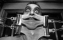
- Gary "Stretch" Turner (shown right), a sideshow performer in the Circus Of Horrors, has an extreme form of EDS. He holds the current Guinness World Record for most elastic skin with 6 inches.
- The condition has been mentioned in many television shows, including:
- The subject of the season-eight premiere of the A&E series Intervention, Linda, claims to have an EDS.[76]
- In season 13, episode four of Grey's Anatomy, a patient is diagnosed with an EDS.[77]
Other species
Ehlers–Danlos-like syndromes have been shown to be hereditary in Himalayan cats, some domestic shorthair cats,[78] and in certain breeds of cattle.[79] It is seen as a sporadic condition in domestic dogs.
- Animal EDS
 EDS in a dog
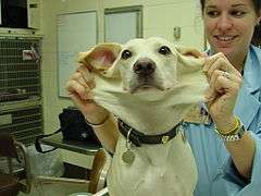 Same dog with EDS
 EDS in same dog showing an atrophic scar
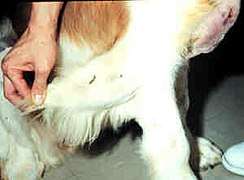 St Bernard (dog) with Ehlers-Danlos-like syndrome showing hyperextensible skin
Degenerative suspensory ligament desmitis (DSLD) is a similar condition seen in many breeds of horses. It was originally notated in the Peruvian Paso and thought to be a condition of overwork and older age. However, DSLD is being recognized in all age groups and all activity levels. It has even been noted in newborn foals. The latest research has led to the renaming of the disease as equine systemic proteoglycan accumulation, after the possible systemic and hereditary components being delineated by the University of Georgia.[80][81]
References
- 1 2 3 4 5 6 7 8 9 10 11 12 13 14 15 "Ehlers–Danlos syndrome". Genetics Home Reference. Archived from the original on 8 May 2016. Retrieved 8 May 2016.
- 1 2 Anderson, Bryan E. (2012). The Netter Collection of Medical Illustrations - Integumentary System E-Book (2 ed.). Elsevier Health Sciences. p. 235. ISBN 1455726648. Archived from the original on 2017-11-05.
- 1 2 3 4 5 6 7 8 9 10 Lawrence, Elizabeth J. (2005). "The clinical presentation of Ehlers–Danlos syndrome". Advances in Neonatal Care. 5 (6): 301–14. doi:10.1016/j.adnc.2005.09.006. PMID 16338669.
- 1 2 3 4 5 6 7 8 9 10 11 12 13 14 "Ehlers-Danlos syndromes". rarediseases.info.nih.gov. 20 April 2017. Archived from the original on 24 September 2017. Retrieved 23 September 2017.

- 1 2 3 Ferri, Fred F. (2016). Ferri's Netter Patient Advisor. Elsevier Health Sciences. p. 939. ISBN 9780323393249. Archived from the original on 2017-11-05.
- ↑ Beighton, Peter H.; Grahame, Rodney; Bird, Howard A. (2011). Hypermobility of Joints. Springer. p. 1. ISBN 9781848820852. Archived from the original on 2017-11-05.
- 1 2 3 4 Byers PH, Murray ML (2012). "Heritable collagen disorders: the paradigm of Ehlers–Danlos syndrome". Journal of Investigative Dermatology. 132 (E1): E6–11. doi:10.1038/skinbio.2012.3. PMID 23154631.
- ↑ "Ehlers-Danlos syndrome". Genetic Home Reference. Retrieved 4 April 2018.
- ↑ Lawrence EJ (2005). "The clinical presentation of Ehlers–Danlos syndrome". Adv Neonatal Care. 5 (6): 301–14. doi:10.1016/j.adnc.2005.09.006. PMID 16338669.
- 1 2 3 4 5 "Ehlers–Danlos Syndrome". Mayo Clinic. Archived from the original on 25 June 2012. Retrieved 25 May 2012.
- ↑ Erçöçen AR, Yenidünya MO, Yilmaz S, Ozbek MR (1997). "Dynamic swan neck deformity in a patient with Ehlers–Danlos syndrome". J Hand Surg Br. 22 (1): 128–30. doi:10.1016/S0266-7681(97)80039-3. PMID 9061548.
- 1 2 "Vascular Type-EDS". Ehlers–Danlos Syndrome Network C.A.R.E.S. Inc. Archived from the original on 2012-06-04.
- ↑ Milhorat TH, Bolognese PA, Nishikawa M, McDonnell NB, Francomano CA (December 2007). "Syndrome of occipitoatlantoaxial hypermobility, cranial settling, and chiari malformation type I in patients with hereditary disorders of connective tissue". Journal of Neurosurgery. Spine. 7 (6): 601–9. doi:10.3171/SPI-07/12/601. PMID 18074684.
- ↑ Gedalia A, Press J, Klein M, Buskila D (1993). "Joint hypermobility and fibromyalgia in schoolchildren". Annals of the Rheumatic Diseases. 52 (7): 494–6. doi:10.1136/ard.52.7.494. PMC 1005086. PMID 8346976.
- ↑ Dommerholt, Jan. "CSF Ehlers Danlos Colloquium, Dr Jan Dommerholt". Chiari & Syringomyelia Foundation. Archived from the original on 4 May 2013. Retrieved 10 June 2013.
- ↑ Vigorita, Vincent J; Mintz, Douglas; Ghelman, Bernard (2008). Orthopaedic pathology (2nd ed.). Philadelphia: Lippincott Williams and Wilkins. pp. 5–6. ISBN 0781796709.
- ↑ "Ehlers-Danlos syndrome - Diagnosis - Approach". BMJ Best Practice. 13 December 2016. Archived from the original on 19 August 2010. Retrieved 18 August 2017.
- ↑ Cheong, J E L. (2005-07-01). "Unusual scars in a young female patient". Postgraduate Medical Journal. 81 (957): e5–e5. doi:10.1136/pgmj.2004.026419. ISSN 1469-0756. PMC 1743308. Archived from the original on 2017-01-18.
- ↑ "Ehlers Danlos Syndrome - Morphopedics". morphopedics.wikidot.com. Retrieved 2018-06-15.
- 1 2 3 4 "Classical Type-EDS". Ehlers–Danlos Syndrome Network C.A.R.E.S Inc. Archived from the original on 2012-05-30.
- ↑ Wilkins, Lippincott Williams & (2007). Portable Signs and Symptoms. Lippincott Williams & Wilkins. p. 465. ISBN 9781582556796. Archived from the original on 2017-11-05.
- ↑ "Piezogenic papules - DermNet New Zealand". www.dermnetnz.org. Archived from the original on 2016-11-26.
- ↑ Ericson Jr, William B; Wolman, Roger (March 2017). "Orthopaedic management of the Ehlers–Danlos syndromes". American Journal of Medical Genetics Part C: Seminars in Medical Genetics. 175 (1): 188–194. doi:10.1002/ajmg.c.31551. eISSN 1552-4876. PMID 28192621.
- ↑ Pagon RA, Adam MP, Ardinger HH, et al. (1993). "Ehlers–Danlos Syndrome, Hypermobility Type". PMID 20301456.
- 1 2 Camerota F, Castori M, Celletti C, Colotto M, Amato S, Colella A, Curione M, Danese C (2014). "Heart rate, conduction and ultrasound abnormalities in adults with joint hypermobility syndrome/Ehlers–Danlos syndrome, hypermobility type". Clin. Rheumatol. 33 (7): 981–7. doi:10.1007/s10067-014-2618-y. PMID 24752348.
- ↑ Wenstrup, R.J; et al. (2001). The Ehlers–Danlos Syndromes: Management of Genetic Syndromes. pp. 131–149.
- ↑ Grigoriou, Emmanouil; Boris, Jeffrey R.; Dormans, John P. (February 2015). "Postural Orthostatic Tachycardia Syndrome (POTS): Association with Ehlers-Danlos Syndrome and Orthopaedic Considerations". Clinical Orthopaedics and Related Research. Springer US. 473 (2): 722–728. doi:10.1007/s11999-014-3898-x. eISSN 1528-1132. PMC 4294907. PMID 25156902.
- ↑ Raffetto, JD; Khalil, RA (April 2008). "Mechanisms of varicose vein formation: valve dysfunction and wall dilation". Phlebology. 23 (2): 85–98. doi:10.1258/phleb.2007.007027. PMID 18453484.
- ↑ Zeitoun, Jean-David; Lefèvre, Jérémie H.; de Parades, Vincent; Séjourné, César; et al. (November 2013). "Functional Digestive Symptoms and Quality of Life in Patients with Ehlers-Danlos Syndromes: Results of a National Cohort Study on 134 Patients". PLoS One. Public Library of Science. 8 (11). Bibcode:2013PLoSO...880321Z. doi:10.1371/journal.pone.0080321. eISSN 1932-6203. PMC 3838387. PMID 24278273.
- ↑ Brockway, Laura (April 2016). "Gastrointestinal manifestations of Ehlers–Danlos syndrome (hypermobility type)". Ehlers–Danlos Support UK. Archived from the original on 2016-11-14.
- ↑ "Ehlers–Danlos Syndrome". Underlying Causes of Dysautonomia. Dysautonomia International. 2012. Archived from the original on 2014-12-18.
- ↑ Létourneau Y, Pérusse R, Buithieu H (2001). "Oral manifestations of Ehlers–Danlos syndrome". J Can Dent Assoc. 67 (6): 330–4. PMID 11450296. Archived from the original on 2016-12-15.
- ↑ "Ehlers–Danlos syndrome: Definition from". Answers.com. Archived from the original on 2014-03-06. Retrieved 2014-02-27.
- ↑ Arendt-Nielsen, Lars. "Patients Suffering from Ehlers Danlos Syndrome Type III Do Not Respond to Local Anesthetics". Archived from the original on 2015-04-05.
- ↑ Castori M, Voermans NC (2014). "Neurological manifestations of Ehlers-Danlos syndrome(s): A review". Iran J Neurol. 13 (4): 190–208. PMC 4300794. PMID 25632331.
- ↑ MedlinePlus Encyclopedia Ehlers-Danlos syndrome
- ↑ Lind J, Wallenburg HC (April 2002). "Pregnancy and the Ehlers–Danlos syndrome: a retrospective study in a Dutch population". Acta Obstetricia et Gynecologica Scandinavica. 81 (4): 293–300. doi:10.1034/j.1600-0412.2002.810403.x. PMID 11952457.
- ↑ Henderson, Fraser (2015). "Indices of Cranio-vertebral Instability". Funded Research. Chiari & Syringomyelia Foundation. Archived from the original on 2016-09-16.
- ↑ Levy, HP (Mar 31, 2016). "Ehlers–Danlos Syndrome, Hypermobility Type". ® [Internet]. Seattle (WA): University of Washington, Seattle; 1993–2017 (Review). PMID 20301456. Archived from the original on 2017-01-18.

- ↑ The Press Enterprise, Redlands mother stung by untrue suspicions presses for accountability in child abuse inquiries, 2008-04-03
- ↑ Chopra, Pradeep; Tinkle, Brad; Hamonet, Claude; Brock, Isabelle; Gompel, Anne; Bulbena, Antonio; Francomano, Clair (2017). "Pain management in the Ehlers–Danlos syndromes". American Journal of Medical Genetics Part C: Seminars in Medical Genetics. 175 (1): 212–219. doi:10.1002/ajmg.c.31554. ISSN 1552-4876. Archived from the original on 2017-11-05.
- ↑ "EDS Types | The Ehlers Danlos Society". The Ehlers Danlos Society. Archived from the original on 2017-06-24. Retrieved 2017-05-22.
- ↑ Sobey, Glenda (2015-01-01). "Ehlers–Danlos syndrome: how to diagnose and when to perform genetic tests". Archives of Disease in Childhood. 100 (1): 57–61. doi:10.1136/archdischild-2013-304822. ISSN 0003-9888. PMID 24994860.
- ↑ Ross, J.; Grahame, R. (2011). "Joint hypermobility syndrome". BMJ. 342: c7167. doi:10.1136/bmj.c7167. PMID 21252103.
- ↑ Castori, Marco (2012). "Ehlers–Danlos Syndrome, Hypermobility Type: An Underdiagnosed Hereditary Connective Tissue Disorder with Mucocutaneous, Articular, and Systemic Manifestations". ISRN Dermatology. 2012: 751768. doi:10.5402/2012/751768. PMC 3512326. PMID 23227356.
- ↑ Inokuchi, Ryota; Kurata, Hideaki; Endo, Kiyoshi; Kitsuta, Yoichi; Nakajima, Susumu; Hatamochi, Atsushi; Yahagi, Naoki (2014-12-02). "Vascular Ehlers-Danlos Syndrome Without the Characteristic Facial Features". Medicine. 93 (28). doi:10.1097/MD.0000000000000291. ISSN 0025-7974. PMC 4603083. PMID 25526469.
- ↑ "OMIM Entry - # 614170 - BRITTLE CORNEA SYNDROME 2; BCS2". www.omim.org. Retrieved 2018-06-18.
- ↑ "What is EDS | The Ehlers–Danlos National Foundation". www.ednf.org. Retrieved 2016-01-06.
- ↑ User, Super. "Differential Diagnosis". www.loeysdietz.org. Archived from the original on 2017-06-23.
- ↑ "Ehlers–Danlos Syndrome". Rarediseases.about.com. 2006-05-25. Archived from the original on 2014-04-12. Retrieved 2014-02-27.
- ↑ Reference, Genetics Home. "Pseudoxanthoma elasticum". Genetics Home Reference. Retrieved 2018-04-17.
- ↑ "Physiotherapy and self-management – The Ehlers-Danlos Support UK". www.ehlers-danlos.org. Retrieved 2018-04-17.
- 1 2 3 4 Rombaut L, Malfait F, De Wandele I, Cools A, Thijs Y, De Paepe A, Calders P (July 2011). "Medication, Surgery, and Physiotherapy Among Patients With the Hypermobility Type of Ehlers–Danlos Syndrome". Archives of Physical Medicine and Rehabilitation. 92 (7): 1106–12. doi:10.1016/j.apmr.2011.01.016. PMID 21636074.
- 1 2 Woerdeman LA, Ritt MJ, Meijer B, Maas M (2000). "Wrist problems in patients with Ehlers–Danlos syndrome". European Journal of Plastic Surgery. 23 (4): 208–210. doi:10.1007/s002380050252.
- ↑ Callewaert B, Malfait F, Loeys B, De Paepe A (March 2008). "Ehlers–Danlos syndromes and Marfan syndrome". Best practice & research. Clinical Rheumatology. 22 (1): 165–189. doi:10.1016/j.berh.2007.12.005. PMID 18328988.
- ↑ Genetics of Ehlers–Danlos Syndrome~treatment at eMedicine
- ↑ "Vascular (VEDS) Emergency Information | The Ehlers Danlos Society". The Ehlers Danlos Society. Retrieved 2018-04-17.
- ↑ M., Giroux, Catherine M.|Corkett, Julie K.|Carter, Lorraine (2016). "The Academic and Psychosocial Impacts of Ehlers-Danlos Syndrome on Postsecondary Students: An Integrative Review of the Literature". Journal of Postsecondary Education and Disability. 29 (4): 414.
- ↑ Parapia, Liakat A.; Jackson, Carolyn (2008). "Ehlers–Danlos syndrome – a historical review". British Journal of Haematology. 141 (1): 32–5. doi:10.1111/j.1365-2141.2008.06994.x. PMID 18324963.
- ↑ OrphanAnesthesia Guidelines
- ↑ Wiesmann, Thomas; Castori, Marco; Malfait, Fransiska; Wulf, Hinnerk (2014). "Recommendations for anesthesia and perioperative management in patients with Ehlers–Danlos syndrome(s)". Orphanet Journal of Rare Diseases. 9: 109. doi:10.1186/s13023-014-0109-5. PMC 4223622. PMID 25053156. Archived from the original on 2016-11-14.
- ↑ Pepin, Melanie; Schwarze, Ulrike; Superti-Furga, Andrea; Byers, Peter H. (2000). "Clinical and Genetic Features of Ehlers–Danlos Syndrome Type IV, the Vascular Type". New England Journal of Medicine. 342 (10): 673–80. doi:10.1056/NEJM200003093421001. PMID 10706896.
- ↑ "Ehlers–Danlos Syndrome: Epidemiology". Medscape.com. Archived from the original on 2013-04-24. Retrieved 2014-02-27.
- ↑ "Ehlers-Danlos syndrome type VIIc". geneaware.clinical.bcm.edu. Archived from the original on 2017-08-14. Retrieved 2017-07-24.
- ↑ Mitchell, Michael (1979). Monsters of the Gilded Age: The photographs of Charles Eisenmann. Toronto: Gage. pp. 99, 100. ISBN 0771595212. OCLC 6892718.
- ↑ "Houston hits out at "preconceived ideas" – Coronation Street News – Soaps". Digital Spy. 2010-05-22. Archived from the original on 2013-05-09. Retrieved 2014-02-27.
- ↑ Yücel D (1995). "Was Paganini born with Ehlers–Danlos syndrome phenotype 4 or 3?". Clin. Chem. 41 (1): 124–5. PMID 7813066.
- ↑ "You Can Dislocate a Rib By Sneezing; My Life With Ehlers Danlos Syndrome". SciBabe. 1 July 2016. Archived from the original on 20 July 2016.
- ↑ Kane, Kimberly (16 October 2012). "Zak Loves Mandy, And They'll Keep Making Art and Porn Until One of Them Dies". Vice. Archived from the original on 26 September 2016.
- ↑ "But You Don't Look Sick: Living With Chronic Pain (Ehlers–Danlos syndrome)". YouTube. Archived from the original on 2014-07-03. Retrieved 2014-02-27.
- ↑ Barry, Aoife. ""People are being left to rot": Rare disease sufferers feel let down by health service". TheJournal.ie. Archived from the original on 2016-01-14. Retrieved 2016-02-25.
- ↑ Carrie (December 26, 2016). "Queer Crip Love Fest: Talking with Queer Disabled Latinx Activist Annie Segarra about Family and Connection". Autostraddle. Archived from the original on September 23, 2017. Retrieved September 22, 2017.
- ↑ "Eric The Actor Calls In". MarksFriggin. 27 November 2012. Archived from the original on 25 August 2017.
- ↑ Boedeker, Hal. "America's Got Talent: Mandy Harvey makes finals", Orlando Sentinel, September 13, 2017
- ↑ Bear, John (21 September 2017). "Deaf Longmont High grad takes fourth place on "America's Got Talent"". The Denver Post. Digital First Media. Retrieved 6 December 2017.
- ↑ "Intervention – Episode Guide". www.aetv.com. Archived from the original on 13 November 2012. Retrieved 24 November 2012.
- ↑ "Grey's Anatomy – Episode Guide". www.abc.go.com. Archived from the original on 11 October 2017. Retrieved 10 October 2017.
- ↑ Dokuzeylül, Banu; Altun, Elif Demet; Özdoğan, Tamer Halit; et al. (2013). "Cutaneous asthenia (Ehlers–Danlos syndrome) in a cat". Turkish Journal of Veterinary and Animal Sciences. Scientific and Technological Research Council of Turkey. 37 (2): 247. doi:10.3906/vet-1203-64. eISSN 1303-6181.
- ↑ Scott, Danny W (2008). "Congenital and hereditary skin diseases". Color Atlas of Farm Animal Dermatology. Wiley Online Library. p. 61. doi:10.1002/9780470344460. ISBN 9780470344460.
- ↑ Halper, Jaroslava; Kim, Byoungjae; Khan, Ahrar; Yoon, Jung; Mueller, PO (2006). "Degenerative suspensory ligament desmitis as a systemic disorder characterized by proteoglycan accumulation". BMC Veterinary Research. 2: 12. doi:10.1186/1746-6148-2-12. PMC 1459153. PMID 16611357.
- ↑ Kim, Byoungjae; Yoon, Jung Hae; Zhang, Jian; Eric Mueller, P.O.; Halper, Jaroslava (2010). "Glycan profiling of a defect in decorin glycosylation in equine systemic proteoglycan accumulation, a potential model of progeroid form of Ehlers–Danlos syndrome". Archives of Biochemistry and Biophysics. 501 (2): 221–31. doi:10.1016/j.abb.2010.06.017. PMID 20599673.
External links
| Wikimedia Commons has media related to Ehlers-Danlos syndrome. |
| Classification | |
|---|---|
| External resources |
- Ehlers–Danlos syndromes at Curlie (based on DMOZ)