Neisseria gonorrhoeae
| Neisseria gonorrhoeae | |
|---|---|
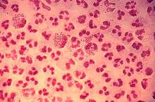 | |
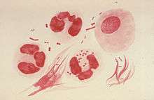
| Gram-stain of gonococcal urethritis. Note distribution in neutrophils and presence of both intracellular and extracellular bacteria. (CDC) | |
| Scientific classification | |
| Domain: | Bacteria |
| Phylum: | Proteobacteria |
| Class: | Betaproteobacteria |
| Order: | Neisseriales |
| Family: | Neisseriaceae |
| Genus: | Neisseria |
| Species: | N. gonorrhoeae |
| Binomial name | |
| Neisseria gonorrhoeae (Zopf 1885) Trevisan 1885[1] | |
| Synonyms | |
Neisseria gonorrhoeae, also known as gonococcus (singular), or gonococci (plural) is a species of Gram-negative diplococci bacteria isolated by Albert Neisser in 1879.[3] It causes the sexually transmitted genitourinary infection gonorrhea[4] as well as other forms of gonococcal disease including disseminated gonococcemia, septic arthritis, and gonococcal ophthalmia neonatorum.
It is oxidase positive and aerobic, and it survives within neutrophils.[4] Culturing it requires carbon dioxide supplementation and enriched agar (chocolate agar) with various antimicrobials (Thayer-Martin). It exhibits antigenic variation through recombination of its pili and surface proteins that interact with the immune system.[3]
Sexual transmission is possible through vaginal, anal, or oral sex.[5] Sexual transmission may be prevented through the use of barrier protection.[6] Perinatal transmission may occur during childbirth, and may be prevented by antibiotic treatment of the mother before birth and the application of antibiotic eye gel on the eyes of the newborn.[6] After an episode of gonococcal infection, infected persons do not develop immunity to future infections. Reinfection is possible due to N. gonorrhoeae's ability to evade the immune system by varying its surface proteins.[7]
N. gonorrhoeae can cause infection of the genitals, throat, and eyes.[7] Asymptomatic infection is common in males and females.[8][6][9] Untreated infection may spread to the rest of the body (disseminated gonorrhea infection), especially the joints (septic arthritis). Untreated infection in women may cause pelvic inflammatory disease and possible infertility due to the resulting scarring.[7] Diagnosis is through culture, Gram stain, or polymerase chain reaction testing of a urine sample, urethral swab, or cervical swab.[10][11] Chlamydia co-testing and testing for other STI's is recommended due to high rates of coinfection.[10]
Microbiology
Neisseria species are fastidious, Gram-negative cocci that require nutrient supplementation to grow in laboratory cultures. Neisseria spp. are facultatively intracellular and typically appear in pairs (diplococci), resembling the shape of coffee beans. Nesseria is nonspore-forming, nonmotile, and an obligate aerobe (requires oxygen to grow). Of the 11 species of Neisseria that colonize humans, only two are pathogens. N. gonorrhoeae is the causative agent of gonorrhea (also called "the clap") and N. meningitidis is one cause of bacterial meningitis.
Culture and identification
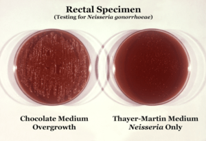
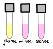
N. gonorrhoeae is usually isolated on Thayer-Martin agar (or VPN) agar in an environment enriched with 3-7% carbon dioxide.[10] Thayer-Martin agar is a chocolate agar plate (heated blood agar) containing nutrients and antimicrobials (vancomycin, colistin, nystatin, and trimethoprim). This agar preparation facilitates the growth of Neisseria species while inhibiting the growth of contaminating bacteria and fungi. Martin Lewis and New York City agar are other types of selective chocolate agar commonly used for Neisseria growth.[10] N. gonorrhoeae is oxidase positive (possessing cytochrome c oxidase) and catalase positive (able to convert hydrogen peroxide to oxygen).[10] When incubated with the carbohydrates lactose, maltose, sucrose, and glucose, N. gonorrhoeae will oxidize only the glucose.[10]
Surface proteins
On its surface, N. gonorrhoeae bears hair-like pili, surface proteins with various functions, and sugars called lipooligosaccharides. The pili mediate adherence, movement, and DNA exchange. The Opa proteins interact with the immune system, as do the porins. Lipooligosaccharide (LPS) is an endotoxin that provokes an immune response. All are antigenic and all exhibit antigenic variation (see below). The pili exhibit the most variation. The pili, Opa proteins, porins, and even the LOS have mechanisms to inhibit the immune response, making asymptomatic infection possible.[12]
Dynamic polymeric protein filaments called type IV pili allow N. gonorrhoeae to adhere to and move along surfaces. To enter the host the bacteria uses the pili to adhere to and penetrate mucosal surfaces.[4] The pili are a necessary virulence factor for N. gonorrhoeae; without them, the bacterium is unable to cause infection.[7] To move, the bacterium uses the type IV pili like a grappling hook, extending and retracting it for movement. Pili extend and attach to a substrate that signals the pilus to retract, dragging the cell forward. The resulting movement is referred to as twitching motility.[13] N. gonorrhoeae is able to pull 100,000 times its own weight, and it has been claimed that the pili used to do so are the strongest biological motor known to date, exerting one nanonewton.[14] A set of ATPase proteins power the pulling actions of the type IV pilus: Pil F (extension) and Pil T (retraction).[15][16] The adhesive functions of the gonococcal pilus play a role in microcolony aggregation and biofilm formation.
Surface proteins called Opa proteins can be used to bind to receptors on immune cells and prevent an immune response. At least 12 Opa proteins are known and the many permutations of surface proteins make recognizing N. gonorrhoeae and mounting a defense by immune cells more difficult.[17]
Lipooligosaccharide (LOS) is a low-weight version of lipopolysaccharide present on the surfaces of most other Gram-negative bacteria. It is a sugar (saccharide) side chain attached to lipid A (thus "lipo-") in the outer membrane coating the cell wall of the bacteria. The root "oligo" refers to the fact that it is a few sugars shorter than the typical lipopolysaccharide.[4] As an endotoxin, LOS provokes inflammation. The shedding of LOS by the bacteria is responsible for local injury in, for example, pelvic inflammatory disease.[4] Although its main function is as an endotoxin, LOS may disguise itself with host sialic acid and block initiation of the complement cascade.[4]
Antigenic variation
N. gonorrhoeae evades the immune system through a process called antigenic variation, in which the N. gonorrhoeae bacterium is able to recombine its genes and alter the antigenic determinants (sites where antibodies bind) such as the Opa proteins[18] and Type IV pili[19] that adorn its surface.[4] It is able to vary the composition of its pili, Opa proteins, and LOSs; of these, the pili exhibit the most antigenic variation due to chromosomal rearrangement.[7][4] Every time the bacteria replicate, they may switch multiple Opa proteins on or off through slipped-strand mispairing. That is, the bacteria introduce frameshift mutations that bring genes in or out of frame. The result is that different Opa genes are translated every time.[4] In addition to the ability to rearrange the genes it already has, it is also naturally competent to acquire new DNA, via its type IV pilus, specifically proteins Pil Q and Pil T.[20] These processes allow N. gonorrhoeae to acquire/spread new genes, disguise itself with different surface proteins, and prevent the development of immunological memory – an ability which has led to antibiotic resistance and has made vaccine development difficult.[21]
Survival of gonococci
After gonococci invade and transcytose the host epithelial cells, they land in the submucosa, where neutrophils promptly consume them.[4] The pili and Opa proteins on the surface may interfere with phagocytosis,[7] but most gonococci end up in neutrophils. The exudates from infected individuals contain many neutrophils with ingested gonococci. Neutrophils release an oxidative burst of reactive oxygen species in their phagosomes to kill the gonococci.[22] However, a significant fraction of the gonococci can resist killing through the action of their catalase[4] which breaks down reactive oxygen species and is able to reproduce within the neutrophil phagosomes.
Stohl and Seifert showed that the bacterial RecA protein, which mediates repair of DNA damage, plays an important role in gonococcal survival.[23] Michod et al. have suggested that N. gonorrhoeae may replace DNA damaged in neutrophil phagosomes with DNA from neighboring gonococci.[24] The process in which recipient gonococci integrate DNA from neighboring gonococci into their genome is called transformation.
Genome
The genomes of several strains of N. gonorrhoeae have been sequenced. Most of them are about 2.1 Mb in size and encode 2,100 to 2,600 proteins (although most seem to be in the lower range).[25] For instance, strain NCCP11945 consists of one circular chromosome (2,232,025 bp) encoding 2,662 predicted open reading frames (ORFs) and one plasmid (4,153 bp) encoding 12 predicted ORFs. The estimated coding density over the entire genome is 87%, and the average G+C content is 52.4%, values that are similar to those of strain FA1090. The NCCP11945 genome encodes 54 tRNAs and four copies of 16S-23S-5S rRNA operons.[26]
Horizontal gene transfer
In 2011, researchers at Northwestern University found evidence of a human DNA fragment in a N. gonorrhoeae genome, the first example of horizontal gene transfer from humans to a bacterial pathogen.[27][28]
Disease
Symptoms of infection with N. gonorrhoeae differ, depending on the site of infection. Also, many infected men are asymptomatic and most infected females (50-80%) are asymptomatic.[8][12][5]
In symptomatic men, the primary symptom of genitourinary infection is urethritis – burning with urination (dysuria), increased urge to urinate, and a pus-like (purulent) discharge from the penis. The discharge may be foul smelling.[4] If untreated, scarring of the urethra may result in difficulty urinating. Infection may spread from the urethra in the penis to nearby structures, including the testicles (epididymitis/orchitis), or to the prostate (prostatitis).[4][7][29] Men who have had a gonorrhea infection have a significantly increased risk of having prostate cancer.[30]
In symptomatic women, the primary symptoms of genitourinary infection are increased vaginal discharge, burning with urination (dysuria), increased urge to urinate, pain with intercourse, or menstrual abnormalities. Pelvic inflammatory disease results if N. gonorrhoeae ascends into the pelvic peritoneum (via the cervix, endometrium, and fallopian tubes). The resulting inflammation and scarring of the fallopian tubes can lead to infertility and increased risk of ectopic pregnancy.[4] Pelvic inflammatory disease develops in 10 to 20% of the females infected with N. gonorrhoeae.[4]
N. gonorrhoeae may cause infection of the throat (pharyngitis) through oral sex. Infected persons may experience sore throat and swollen lymph nodes, or may be asymptomatic.[7][4]
N. gonorrhoeae may cause infection of the anus/rectum (proctitis) through anal sex. Infected persons may experience blood or a pus-like discharge, or may be asymptomatic.[7][4]
In perinatal infection, the primary manifestation is infection of the eye (neonatal conjunctivitis or ophthalmia neonatorum) when the newborn is exposed to N. gonorrhoeae in the birth canal. The eye infection can lead to corneal scarring or perforation, ultimately resulting in blindness. If the newborn is exposed during birth, conjunctivitis occurs within 2–5 days after birth and is severe.[4][31] Gonococcal ophthalmia neonatorum, once common in newborns, is prevented by the application of erythromycin (antibiotic) gel to the eyes of babies at birth as a public health measure. Silver nitrate is no longer used in the United States.[31][4]
Disseminated N. gonorrhoeae infections can occur in which gonococci enter the bloodstream, often spreading to the joints and causing a rash (dermatitis-arthritis syndrome).[4] Dermatitis-arthritis syndrome presents with joint pain (arthritis), tendon inflammation (tenosynovitis), and painless non-pruritic (non-itchy) dermatitis.[7] Disseminated infection and PID tend to begin after menses due to reflux during menses, facilitating spread.[4] Rarely, disseminated infection may cause infection of the meninges of the brain and spinal cord (meningitis) or infection of the heart valves (endocarditis).[4][31]
Transmission
N. gonorrhoeae may be transmitted through vaginal, oral, or anal sex; nonsexual transmission is unlikely in adult infection.[5] It can also be transmitted to the newborn during passage through the birth canal if the mother has untreated genitourinary infection. Given the high rate of asymptomatic infection, all pregnant women should be tested for gonorrhea infection.[5]
Traditionally, the bacterium was thought to move attached to spermatozoa, but this hypothesis did not explain female to male transmission of the disease. A recent study suggests that rather than "surf" on wiggling sperm, N. gonorrhoeae bacteria use pili to anchor onto proteins in the sperm and move through coital liquid.[32]
Prevention
Transmission can be reduced by using latex barriers (e.g. condoms or dental dams) during sex and by limiting sexual partners.[6] Condoms and dental dams should be used during oral and anal sex, as well. Spermicides, vaginal foams, and douches are not effective for prevention of transmission.[4]
Treatment
Ceftriaxone (a third-generation cephalosporin) is the first line treatment.[33] Because co-infection with chlamydia is so common, people are often treated for both by combining ceftriaxone with azithromycin or doxycycline.[4] Azithromycin is preferred for additional coverage of gonorrhea that may be resistant to cephalosporins but susceptible to macrolides.[34][6] Sexual partners (defined by the CDC as sexual contact within the past 60 days)[11] should also be notified, tested, and treated.[6][33]
Antibiotic resistance
Antibiotic resistance in gonorrhea has been noted beginning in the 1940s. Gonorrhea was treated with penicillin, but doses had to be progressively increased to remain effective. By the 1970s, penicillin- and tetracycline-resistant gonorrhea emerged in the Pacific Basin. These resistant strains then spread to Hawaii, California, the rest of the United States, and Europe. Fluoroquinolones were the next line of defense, but soon resistance to this antibiotic emerged, as well. Since 2007, standard treatment has been third-generation cephalosporins, such as ceftriaxone, which are considered to be our "last line of defense".[35][36]
Recently, a high-level ceftriaxone-resistant strain of gonorrhea called H041 was discovered in Japan. Lab tests found it to be resistant to high concentrations of ceftriaxone, as well as most of the other antibiotics tested. Within N. gonorrhoeae, genes exist that confer resistance to every single antibiotic used to cure gonorrhea, but thus far they do not coexist within a single gonococcus. However, because of N. gonorrhoeae's high affinity for horizontal gene transfer, antibiotic-resistant gonorrhea is seen as an emerging public health threat.[36]
History
Name origin
Neisseria gonnorhoeae is named for Albert Neisser, who isolated it as the causative agent of the disease gonorrhea in 1878.[37][3] Galen (130 AD) coined the term "gonorrhea" from the Greek gonos which means "seed" and rhoe which means "flow".[38][39] Thus, gonorrhea means "flow of seed", a description referring to the white penile discharge, assumed to be semen, seen in male infection.[37]
Discovery
In 1878, Albert Neisser isolated and visualized N. gonorrhoeae diplococci in samples of pus from 35 men and women with the classic symptoms of genitourinary infection with gonorrhea – two of whom also had infections of the eyes.[40] In 1882, Leistikow and Loeffler were able to grow the organism in culture.[37] Then in 1883, Max Bockhart proved conclusively that the bacterium isolated by Albert Neisser was the causative agent of the disease known as gonorrhea by inoculating the penis of a healthy man with the bacteria.[40] The man developed the classic symptoms of gonorrhea days after, satisfying the last of Koch's postulates. Until this point, researchers debated whether syphilis and gonorrhea were manifestations of the same disease or two distinct entities.[41][40][39] One such 18th-century researcher, John Hunter, tried to settle the debate in 1767[39] by inoculating a man with pus taken from a patient with gonorrhea. He erroneously concluded that both syphilis and gonorrhea were indeed the same disease when the man developed the copper-colored rash that is classic for syphilis.[42][41] Although many sources repeat that Hunter inoculated himself,[42][37] others have argued that it was in fact another man.[43] After Hunter's experiment other scientists sought to disprove his conclusions by inoculating other male physicians, medical students,[37] and incarcerated men with gonorrheal pus, who all developed the burning and discharge of gonorrhea. One researcher, Ricord, took the initiative to perform 667 inoculations of gonorrheal pus on patients of a mental hospital, with zero cases of syphilis.[39][37] Notably, the advent of penicillin in the 1940s made effective treatments for gonorrhea available.
See also
References
- ↑ "Neisseria". In: List of Prokaryotic Names with Standing in Nomenclature (LPSN). Created by J.P. Euzéby in 1997. Curated by A.C. Parte since 2013. Available on: http://www.bacterio.net. Retrieved 7 July 2017.
- ↑ Neisser, A. Ueber eine der Gonorrhoe eigentümliche Micrococusform. Centralblatt für die medizinischen Wissenschaften 17 (28): 497-500. link.
- 1 2 3 O'Donnell, Judith A.; Gelone, Steven P. (2009). Pelvic Inflammatory Disease. Infobase Publishing. ISBN 9781438101590.
- 1 2 3 4 5 6 7 8 9 10 11 12 13 14 15 16 17 18 19 20 21 22 23 24 Ryan, KJ; Ray, CG, eds. (2004). Sherris Medical Microbiology (4th ed.). McGraw Hill. ISBN 978-0-8385-8529-0.
- 1 2 3 4 "Detailed STD Facts - Gonorrhea". www.cdc.gov. 2017-09-26. Retrieved 2017-12-07.
- 1 2 3 4 5 6 https://www.cdc.gov/std/tg2015/clinical.htm section on prevention methods
- 1 2 3 4 5 6 7 8 9 10 Levinson, Warren. Review of medical microbiology and immunology (Thirteenth ed.). New York. ISBN 9780071818117. OCLC 871305336.
- 1 2 Detels, Roger; Green, Annette M.; Klausner, Jeffrey D.; Katzenstein, David; Gaydos, Charlotte; Hadsfield, Hunter; Peqyegnat, Willo; Mayer, Kenneth; Hartwell, Tyler D.; Quinn, Thomas . (2011). "The Incidence and Correlates of Symptomatic and Asymptomatic Chlamydia trachomatis and Neisseria gonorrhoeae Infections in Selected Populations in Five Countries". Sex. Transm. Dis. 38 (6): 503–509. PMC 3408314. PMID 22256336.
- ↑ "Final Recommendation Statement: Chlamydia and Gonorrhea: Screening - US Preventive Services Task Force". www.uspreventiveservicestaskforce.org. Retrieved 2017-12-07.
- 1 2 3 4 5 6 Ng, Lai-King; Martin, Irene E (2005). "The laboratory diagnosis of Neisseria gonorrhoeae". The Canadian Journal of Infectious Diseases & Medical Microbiology. 16 (1): 15–25. doi:10.1155/2005/323082. ISSN 1712-9532. PMC 2095009. PMID 18159523.
- 1 2 https://www.cdc.gov/std/tg2015/gonorrhea.htm
- 1 2 Edwards, Jennifer L.; Apicella, Michael A. (October 2004). "The Molecular Mechanisms Used by Neisseria gonorrhoeae To Initiate Infection Differ between Men and Women". Clinical Microbiology Reviews. 17 (4): 965–981. doi:10.1128/CMR.17.4.965-981.2004. ISSN 0893-8512. PMC 523569. PMID 15489357.
- ↑ Merz, A. J.; So, M.; Sheetz, M. P. (2000-09-07). "Pilus retraction powers bacterial twitching motility". Nature. 407 (6800): 98–102. doi:10.1038/35024105. ISSN 0028-0836. PMID 10993081.
- ↑ Biais N, Ladoux B, Higashi D, So M, Sheetz M (2008). "Cooperative retraction of bundled type IV pili enables nanonewton force generation". PLoS Biol. 6 (4): e87. doi:10.1371/journal.pbio.0060087. PMC 2292754. PMID 18416602. Lay summary – New Scientist (19 April 2008).
- ↑ Freitag, NE; Seifert, HS; Koomey, M (May 1995). "Characterization of the pilF-pilD pilus-assembly locus of Neisseria gonorrhoeae". Molecular Microbiology. 16 (3): 575–86. doi:10.1111/j.1365-2958.1995.tb02420.x. PMID 7565116.
- ↑ Jakovljevic, V; Leonardy, S; Hoppert, M; Søgaard-Andersen, L (April 2008). "PilB and PilT are ATPases acting antagonistically in type IV pilus function in Myxococcus xanthus". Journal of Bacteriology. 190 (7): 2411–21. doi:10.1128/JB.01793-07. PMC 2293208. PMID 18223089.
- ↑ STI Awareness: Gonorrhea. Planned Parenthood Advocates of Arizona. 11 April 2011. Retrieved 31 August 2011.
- ↑ Stern, Anne; Brown, Melissa; Nickel, Peter; Meyer, Thomas F. (1986). "Opacity genes in Neisseria gonorrhoeae: Control of phase and antigenic variation". Cell. 47 (1): 61–71. doi:10.1016/0092-8674(86)90366-1. PMID 3093085.
- ↑ Cahoon, Laty A.; Seifert, H. Steven (2011). "Focusing homologous recombination: Pilin antigenic variation in the pathogenic Neisseria". Molecular Microbiology. 81 (5): 1136–43. doi:10.1111/j.1365-2958.2011.07773.x. PMC 3181079. PMID 21812841.
- ↑ Obergfell, KP; Seifert, HS (February 2015). "Mobile DNA in the Pathogenic Neisseria". Microbiology Spectrum. 3 (1): MDNA3–0015–2014. doi:10.1128/microbiolspec.MDNA3-0015-2014. PMC 4389775. PMID 26104562.
- ↑ Aas, Finn Erik; Wolfgang, Matthew; Frye, Stephan; Dunham, Steven; Løvold, Cecilia; Koomey, Michael (2002). "Competence for natural transformation in Neisseria gonorrhoeae: Components of DNA binding and uptake linked to type IV pilus expression". Molecular Microbiology. 46 (3): 749–60. doi:10.1046/j.1365-2958.2002.03193.x. PMID 12410832.
- ↑ Simons, M. P.; Nauseef, W. M.; Apicella, M. A. (2005). "Interactions of Neisseria gonorrhoeae with Adherent Polymorphonuclear Leukocytes". Infection and Immunity. 73 (4): 1971–7. doi:10.1128/iai.73.4.1971-1977.2005. PMC 1087443. PMID 15784537.
- ↑ Stohl, E. A.; Seifert, H. S. (2006). "Neisseria gonorrhoeae DNA Recombination and Repair Enzymes Protect against Oxidative Damage Caused by Hydrogen Peroxide". Journal of Bacteriology. 188 (21): 7645–51. doi:10.1128/JB.00801-06. PMC 1636252. PMID 16936020.
- ↑ Michod, Richard E.; Bernstein, Harris; Nedelcu, Aurora M. (2008). "Adaptive value of sex in microbial pathogens". Infection, Genetics and Evolution. 8 (3): 267–85. doi:10.1016/j.meegid.2008.01.002. PMID 18295550.
- ↑ "Neisseria gonorrhoeae genome statistics". Broad Institute. Retrieved 8 April 2017.
- ↑ Chung, G. T.; Yoo, J. S.; Oh, H. B.; Lee, Y. S.; Cha, S. H.; Kim, S. J.; Yoo, C. K. (2008). "Complete genome sequence of Neisseria gonorrhoeae NCCP11945". Journal of Bacteriology. 190 (17): 6035–6. doi:10.1128/JB.00566-08. PMC 2519540. PMID 18586945.
- ↑ Anderson, Mark T.; Seifert, H. Steven (2014). "Neisseria gonorrhoeae and humans perform an evolutionary LINE dance". Mobile Genetic Elements. 1 (1): 85–87. doi:10.4161/mge.1.1.15868. PMC 3190277. PMID 22016852. Lay summary – ScienceDaily (February 14, 2011).
- ↑ Anderson, M. T.; Seifert, H. S. (2011). "Opportunity and Means: Horizontal Gene Transfer from the Human Host to a Bacterial Pathogen". MBio. 2 (1): e00005–11. doi:10.1128/mBio.00005-11. PMC 3042738. PMID 21325040.
- ↑ "Gonorrhea (the clap) Symptoms". www.std-gov.org.
- ↑ Caini, Saverio; Gandini, Sara; Dudas, Maria; Bremer, Viviane; Severi, Ettore; Gherasim, Alin (2014). "Sexually transmitted infections and prostate cancer risk: A systematic review and meta-analysis". Cancer Epidemiology. 38 (4): 329–338. doi:10.1016/j.canep.2014.06.002. ISSN 1877-7821. PMID 24986642.
- 1 2 3 "Gonococcal Infections - 2015 STD Treatment Guidelines". www.cdc.gov. Retrieved 2017-12-07.
- ↑ Anderson, M. T.; Dewenter, L.; Maier, B.; Seifert, H. S. (2014). "Seminal Plasma Initiates a Neisseria gonorrhoeae Transmission State". MBio. 5 (2): e01004–13. doi:10.1128/mBio.01004-13. PMC 3958800. PMID 24595372.
- 1 2 "CDC - Gonorrhea Treatment". www.cdc.gov. 2017-10-31. Retrieved 2017-12-07.
- ↑ Nwokolo, Nneka C; Dragovic, Bojana; Patel, Sheel; Tong, CY William; Barker, Gary; Radcliffe, Keith (2016-03-01). "2015 UK national guideline for the management of infection with Chlamydia trachomatis". International Journal of STD & AIDS. 27 (4): 251–267. doi:10.1177/0956462415615443. ISSN 0956-4624. PMID 26538553.
- ↑ "UK doctors advised gonorrhoea has turned drug resistant". BBC News. 10 October 2011.
- 1 2 STI Awareness: Antibiotic-Resistant Gonorrhea. Planned Parenthood Advocates of Arizona. 6 March 2012. Retrieved 6 March 2012.
- 1 2 3 4 5 6 Spence, M. R. (March 1983). "Gonorrhea". Clinical Obstetrics and Gynecology. 26 (1): 111–124. doi:10.1097/00003081-198303000-00016. ISSN 0009-9201. PMID 6404578.
- ↑ "gonorrhea | Origin and meaning of gonorrhea by Online Etymology Dictionary". www.etymonline.com. Retrieved 2017-12-05.
- 1 2 3 4 Norris, Charles Camblos (1913). Gonorrhea in women. W. B Saunders Co.
- 1 2 3 Ligon, B. Lee (October 2005). "Albert Ludwig Sigesmund Neisser: discoverer of the cause of gonorrhea". Seminars in Pediatric Infectious Diseases. 16 (4): 336–341. doi:10.1053/j.spid.2005.07.001. ISSN 1045-1870. PMID 16210113.
- 1 2 Singal, Archana; Grover, Chander (2016-02-12). Comprehensive Approach to Infections in Dermatology. JP Medical Ltd. ISBN 9789351527480.
- 1 2 Ross, John J. (2005-12-01). "Book Review". New England Journal of Medicine. 353 (22): 2412–2413. doi:10.1056/NEJM200512013532221. ISSN 0028-4793.
- ↑ Gladstein, J. (2005-07-01). "Hunter's Chancre: Did the Surgeon Give Himself Syphilis?". Clinical Infectious Diseases. 41 (1): 128, author reply 128–9. doi:10.1086/430834. ISSN 1058-4838. PMID 15937780.
External links
- Todar, Kenneth. "Pathogenic Neisseriae: Gonorrhea, Neonatal Ophthalmia and Meningococcal Meningitis". Todar's Online Textbook of Bacteriology.
- Gonorrhea at eMedicine
- "Neisseria gonorrhoeae". NCBI Taxonomy Browser. 485.