Hemangioma
| Hemangioma | |
|---|---|
 | |
| Hemangioma |
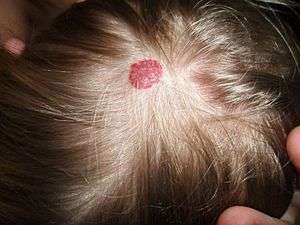
Hemangioma is a benign tumor derived from blood vessel cell types, most commonly infantile hemangioma, a common benign tumor of infancy. Infantile hemangiomas, known colloquially as strawberry marks and seen at birth or in the first weeks of life, are most commonly seen on the skin. A hemangioma can occur anywhere on the body, but most commonly appears on the face, scalp, chest or back. Treatment of a hemangioma usually is unnecessary, unless the nodule interferes with vision or breathing.
Types
Hemangiomas are benign (noncancerous) vascular tumors, and there are many different types. The correct terminology for these hemangioma types is constantly being updated by the International Society for the Study of Vascular Anomalies (ISSVA). The 2018 ISSVA classification includes many hemangioma types, two of which are described in further detail below.[1]
1. Infantile hemangioma
- Infantile hemangioma are the most common benign (noncancerous) tumor found in children. They are made up of blood vessels
- They are more common in girls than boys
- Sometimes called "strawberry marks"
- They usually become visible on the skin of children who are 4-6 weeks old
- They tend to grow quickly for a few weeks or months, then shrink or "involute"[2]
- Most shrink on their own without problems, but some can ulcerate (crust or scab), which may be painful. Depending on their location and size, they may also be disfiguring. Though not common, they may be related to disorders of the central nervous system or spine. They may also occur in the internal organs of the body, such as the liver, airway, or brain[3]
- The color of the hemangioma depends on how deep it is in the skin
    * Superficial (near the skin's surface) hemangioma tend to be bright red
    * Deep (furthest from the skin's surface) hemangioma are often blue or purple
    * Mixed hemangioma may have colors of both superficial and deep[4]
2. Congenital hemangioma
- Congenital hemangioma are usually visible on the skin at birth, unlike infantile hemangioma, which appear later.
- They are fully formed at birth, meaning that they do not grow after a child is born like infantile hemangiomas do.
- They are less common than infantile hemangioma.
- They can be pink to blue in color.
- They are classified based on how they shrink, or involute
    * RICH are Rapidly Involuting Congenital Hemangioma, meaning that they go away quickly
    * NICH are Non-Involuting Congenital Hemangioma, meaning that they do not go away
    * PICH are Partially Involuting Congenital Hemangioma, meaning that part of the skin changes go away on their own[5][6]
Diagnosis
Diagnosis is usually clinical and tests are not necessary.[7]
Treatment
Hemangiomas usually fade gradually over time, and most do not require treatment. Therapeutic options can have side effects and are avoided if possible. However, hemangiomas that may be disfiguring or that are located at sites that can cause impairment (eyelids, airway) are often treated, typically with pharmacotherapy first. Management options may include the following:[7]
- Beta blockers
- Corticosteroids
- Laser surgery
Additional images

References
- ↑ "ISSVA Classification of Vascular Anomalies International Society for the Study of Vascular Anomalies" (PDF). Retrieved 11 August 2018.
- ↑ Chang LC, Haggstrom AN, Drolet BA, Baselga E, Chamlin SL, Garzon MC, Horii KA, Lucky AW, Mancini AJ, Metry DW, Nopper AJ, Frieden IJ; Hemangioma Investigator Group. Growth characteristics of infantile hemangiomas: implications for management. Pediatrics. 2008 Aug;122(2):360-7. doi: 10.1542/peds.2007-2767.
- ↑ Drolet BA, Esterly NB, Frieden IJ. Hemangiomas in children. N Engl J Med. 1999 Jul 15;341(3):173-81.
- ↑ "Infantile Hemangiomas". Retrieved 11 August 2018.
- ↑ Mulliken JB, Bischoff J, Kozakewich HP. Multifocal rapidly involuting congenital hemangioma: a link to chorangioma. Am J Med Genet A. 2007;143A(24):3038-3046.
- ↑ Funk T, Lim Y, Kulungowski AM, et al. Symptomatic Congenital Hemangioma and Congenital Hemangiomatosis Associated With a Somatic Activating Mutation in GNA11. JAMA Dermatol. 2016;152(9):1015-1020.
- 1 2 "Hemangioma".