Central retinal artery occlusion
| Central retinal artery occlusion | |
|---|---|
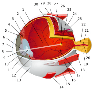 | |
| Central retinal artery(#20) | |
| Specialty |
Neurology |
Central retinal artery occlusion (CRAO) is a disease of the eye where the flow of blood through the central retinal artery is blocked (occluded). There are several different causes of this occlusion; the most common is carotid artery atherosclerosis.
Signs and symptoms
Central retinal artery occlusions cause sudden, acute, and painless loss of vision in one eye. Fundoscopic exam will show a red lesion, called a "cherry red spot," with surrounding pale retina (the pale color is caused by ischemia of the retina),[1][2] afferent pupil defect, periorbital eyelid edema, proptosis, ptosis, and at times a hazy/cloudy cornea.
Causes
The most common cause for CRAO is carotid artery atherosclerosis. In patients of 70 years of age and older, giant cell arteritis is more likely to be the cause than in younger patients. Other causes can include dissecting aneurysms and arterial spasms,[2] and as a complication of patient positioning causing external compression of the eye compressing flow to the central retinal artery (e.g. in spine surgeries in the prone position).
Mechanism
The ophthalmic artery branches off into the central retinal artery which travels with the optic nerve until it enters the eye. This central retinal artery provides nutrients to the retina of the eye, more specifically the inner retina and the surface of the optic nerve.[2] Variations, such as branch retinal artery occlusion, can also occur. Some people have cilioretinal arterial branches, which may or may not be included in the blocked portion.
Treatment
Several treatments have been attempted for CRAS; however, none show definitive improvement in outcomes.[3] The Undersea and Hyperbaric Medical Society lists Central Retinal Artery Occlusion (CRAO) as an approved indication for Hyperbaric Oxygen Therapy.[4] This a treatment for CRAO that is covered by medical insurance in North America. Other treatments include ocular massage, anterior chamber paracentesis, and inhalation therapy of a mixture of 5% carbon dioxide and 95% oxygen.[1]
Prognosis
The artery can re-canalize over time and the edema can clear. However, optic atrophy leads to permanent loss of vision. Irreversible damage to neural tissue occurs after only 90 minutes. Two thirds of patients experience 20/400 vision while only one in six will experience 20/40 vision or better.[1]
Epidemiology
Risk factors for CRAO include the following: being between 60 and 65 years of age, being over the age of 40, male gender, hypertension, caucasian, smoking and diabetes mellitus.[2] Additional risk factors include endocarditis, atrial myxoma, inflammatory diseases of the blood vessels, and predisposition to forming blood clots.
See also
References
- 1 2 3 Kunimoto, Dr., Lecture, Vascular diseases of the retina, AT Still University SOMA, October 2012
- 1 2 3 4 Central and branch retinal artery occlusion. Uptodate.com. Mar 14, 2012.
- ↑ Cugati, Sudha; Varma, Daniel D.; Chen, Celia S.; Lee, Andrew W. (16 October 2012). "Treatment Options for Central Retinal Artery Occlusion". Current Treatment Options in Neurology. 15 (1): 63–77. doi:10.1007/s11940-012-0202-9. PMID 20609991.
- ↑ https://www.uhms.org/6-arterial-insufficiencies/central-retinal-artery-occlusion.html
External links
| Classification | |
|---|---|
| External resources |