Cardiac myxoma
| Atrial myxoma | |
|---|---|
 | |
| Micrograph of an atrial myxoma. H&E stain. |
An atrial myxoma is a benign tumor of the heart, most commonly found within the left and then the right atria on the interatrial septum.
Signs and symptoms
Symptoms may occur at any time, but most often they accompany a change of body position. Pedunculated myxomas can have a "wrecking ball effect", as they lead to stasis and may eventually embolize themselves. Symptoms may include:
- Shortness of breath with activity
- Platypnea – Difficulty breathing in the upright position with relief in the supine position
- Paroxysmal nocturnal dyspnea – Breathing difficulty when asleep
- Dizziness
- Fainting
- Palpitations – Sensation of feeling your heart beat
- Chest pain or tightness
- Sudden Death (In which case the disease is an autopsy finding)
The symptoms and signs of left atrial myxomas often mimic mitral stenosis. General symptoms may also be present, such as:
- Cough
- Pulmonary edema, as blood backs up into the pulmonary artery, after increased pressures in the left atrium and atrial dilation
- Hemoptysis
- Fever
- Cachexia – Involuntary weight loss
- General discomfort (malaise)
- Joint pain
- Blue discoloration of the skin, especially the fingers (Raynaud's phenomenon)
- Fingers that change color upon pressure or with cold or stress
- Clubbing – Curvature of nails accompanied with soft tissue enlargement of the fingers
- Swelling – any part of the body
- Presystolic heart murmur[1]
These general symptoms may also mimic those of infective endocarditis.
Complications
- Arrhythmias
- Pulmonary edema
- Peripheral emboli
- Spread (metastasis) of the tumor
- Blockage of the mitral heart valve
- Stroke
- Fusiform cerebral aneurysms
Causes
Myxomas are the most common type of primary heart tumor.[2]
The tumor is derived from multipotential mesenchymal cells and may cause a ball valve-type obstruction.
About 75% of myxomas occur in the left atrium of the heart, usually beginning in the wall that divides the two upper chambers of the heart. The rest are in the right atrium, rarely in the left ventricle. Right atrial myxomas are sometimes associated with tricuspid stenosis and atrial fibrillation.
Myxomas are more common in women. About 10% of myxomas are passed down through families (inherited), as in Carney syndrome, where several other abnormalities are observed, such as skin myxomas, pigmentation, endocrine hyperactivity, schwannomas and epithelioid blue nevi. Such tumors are called familial myxomas. They tend to occur in more than one part of the heart at a time, and often cause symptoms at a younger age than other myxomas.
Diagnosis
A doctor will listen to the heart with stethoscope. A "tumor plop" (a sound related to movement of the tumor), abnormal heart sounds, or a murmur similar to the mid-diastolic rumble of mitral stenosis may be heard. These sounds may change when the patient changes position.
Right atrial myxomas rarely produce symptoms until they have grown to be at least 13 cm (about 5 inches) wide.
Tests may include:
- Echocardiogram and Doppler study
- Chest x-ray
- CT scan of chest
- Heart MRI
- Left heart angiography
- Right heart angiography
- ECG—may show atrial fibrillation
Blood tests: A FBC may show anemia and increased WBCs (white blood cells). The erythrocyte sedimentation rate (ESR) is usually increased.
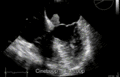 Echocardiogram of atrial myxoma
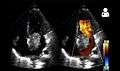 Echocardiogram showing atrial myxoma[3]
 Atrial myxoma and myocardium. H&E stain.
 Atrial myxoma. H&E stain.
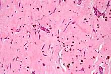 Atrial myxoma. H&E stain.
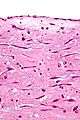 Atrial myxoma covered by endothelium. H&E stain.
Treatment
The tumor must be surgically removed. Some patients will also need their mitral valve replaced. This can be done during the same surgery.
Myxomas may come back if surgery did not remove all of the tumor cells.
Prognosis

Although a myxoma is not cancer, complications are common. Untreated, a myxoma can lead to an embolism (tumor cells breaking off and traveling with the bloodstream), which can block blood flow. Myxoma fragments can move to the brain, eye, or limbs.
If the tumor grows inside the heart, it can block blood flow through the mitral valve and cause symptoms of mitral stenosis or mitral regurgitation. This may require emergency surgery to prevent sudden death.[4]
See also
Other primary cardiac tumours include:
References
- ↑ Eric J. Topol. The Topol Solution: Textbook of Cardiovascular Medicine, Third Edition with DVD, Plus Integrated Content Website, Volume 355. Lippincott Williams & Wilkins, Oct 19, 2006; page 223. ISBN 0781770122
- ↑ Vaideeswar, P.; Butany, JW. (Feb 2008). "Benign cardiac tumors of the pluripotent mesenchyme". Semin Diagn Pathol. 25 (1): 20–8. doi:10.1053/j.semdp.2007.10.005. PMID 18350919.
- 1 2 "UOTW #31 - Ultrasound of the Week". Ultrasound of the Week. 30 December 2014. Retrieved 27 May 2017.
- ↑ http://www.jcecho.org/article.asp?issn=2211-4122;year=2018;volume=28;issue=1;spage=59;epage=60;aulast=Barik
External links
| Classification | |
|---|---|
| External resources |