Potyvirus
| Potyvirus | |
|---|---|
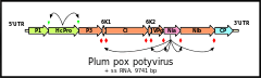 | |
| Genome of typical potyvirus PPV with proteolytic cleavage sites | |
| Virus classification | |
| Group: | Group IV ((+)ssRNA) |
| Family: | Potyviridae |
| Genus: | Potyvirus |
| Type species | |
| Potato virus Y | |
Potyvirus is a genus of viruses, in the family Potyviridae. Plants serve as natural hosts. There are currently 158 species in this genus including the type species Potato virus Y.[1][2] The genus is named after the type virus (Potato virus Y). Potyviruses account for ~30% of the currently known plant viruses. Like Begomoviruses, members of this genus may cause significant losses in agricultural, pastoral, horticultural and ornamental crops. More than 200 species of aphids spread potyviruses and most are from the subfamily Aphidinae (genera Macrosiphum and Myzus).
Taxonomy
Group: ssRNA(+)
- Family: Potyviridae
- Genus: Potyvirus
- Algerian watermelon mosaic virus
- Alstroemeria mosaic virus
- Alternanthera mild mosaic virus
- Amaranthus leaf mottle virus
- Amazon lily mosaic virus
- Angelica virus Y
- Apium virus Y
- Araujia mosaic virus
- Arracacha mottle virus
- Artichoke latent virus
- Asparagus virus 1
- Banana bract mosaic virus
- Basella rugose mosaic virus
- Bean common mosaic necrosis virus
- Bean common mosaic virus
- Bean yellow mosaic virus
- Beet mosaic virus
- Bidens mosaic virus
- Bidens mottle virus
- Blue squill virus A
- Brugmansia mosaic virus
- Brugmansia suaveolens mottle virus
- Butterfly flower mosaic virus
- Calanthe mild mosaic virus
- Calla lily latent virus
- Canna yellow streak virus
- Carnation vein mottle virus
- Carrot thin leaf virus
- Carrot virus Y
- Celery mosaic virus
- Ceratobium mosaic virus
- Chilli ringspot virus
- Chilli veinal mottle virus
- Chinese artichoke mosaic virus
- Clitoria virus Y
- Clover yellow vein virus
- Cocksfoot streak virus
- Colombian datura virus
- Commelina mosaic virus
- Cowpea aphid-borne mosaic virus
- Cowpea green vein banding virus
- Cypripedium virus Y
- Cyrtanthus elatus virus A
- Daphne mosaic virus
- Dasheen mosaic virus
- Datura shoestring virus
- Diuris virus Y
- East Asian Passiflora virus
- Endive necrotic mosaic virus
- Euphorbia ringspot virus
- Freesia mosaic virus
- Fritillary virus Y
- Gloriosa stripe mosaic virus
- Groundnut eyespot virus
- Guinea grass mosaic virus
- Habenaria mosaic virus
- Hardenbergia mosaic virus
- Helenium virus Y
- Henbane mosaic virus
- Hibbertia virus Y
- Hippeastrum mosaic virus
- Hyacinth mosaic virus
- Iris fulva mosaic virus
- Iris mild mosaic virus
- Iris severe mosaic virus
- Japanese yam mosaic virus
- Johnsongrass mosaic virus
- Kalancho‰ mosaic virus
- Keunjorong mosaic virus
- Konjac mosaic virus
- Leek yellow stripe virus
- Lettuce mosaic virus
- Lily mottle virus
- Lupinus mosaic virus
- Lycoris mild mottle virus
- Maize dwarf mosaic virus
- Malva vein clearing virus
- Meadow saffron breaking virus
- Moroccan watermelon mosaic virus
- Narcissus degeneration virus
- Narcissus late season yellows virus
- Narcissus yellow stripe virus
- Nerine yellow stripe virus
- Nothoscordum mosaic virus
- Onion yellow dwarf virus
- Ornithogalum mosaic virus
- Ornithogalum virus 2
- Ornithogalum virus 3
- Panax virus Y
- Papaya leaf distortion mosaic virus
- Papaya ringspot virus
- Parsnip mosaic virus
- Passiflora chlorosis virus
- Passion fruit woodiness virus
- Pea seed-borne mosaic virus
- Peanut mottle virus
- Pennisetum mosaic virus
- Pepper mottle virus
- Pepper severe mosaic virus
- Pepper veinal mottle virus
- Pepper yellow mosaic virus
- Peru tomato mosaic virus
- Pfaffia mosaic virus
- Pleione virus Y
- Plum pox virus
- Pokeweed mosaic virus
- Potato virus A
- Potato virus V
- Potato virus Y
- Ranunculus leaf distortion virus
- Ranunculus mild mosaic virus
- Ranunculus mosaic virus
- Rhopalanthe virus Y
- Sarcochilus virus Y
- Scallion mosaic virus
- Shallot yellow stripe virus
- Sorghum mosaic virus
- Soybean mosaic virus
- Spiranthes mosaic virus 3
- Sugarcane mosaic virus
- Sunflower chlorotic mottle virus
- Sunflower mosaic virus
- Sweet potato feathery mottle virus
- Sweet potato latent virus
- Sweet potato mild speckling virus
- Sweet potato virus 2
- Sweet potato virus C
- Sweet potato virus G
- Telfairia mosaic virus
- Telosma mosaic virus
- Thunberg fritillary mosaic virus
- Tobacco etch virus
- Tobacco vein banding mosaic virus
- Tobacco vein mottling virus
- Tomato necrotic stunt virus
- Tradescantia mild mosaic virus
- Tropaeolum mosaic virus
- Tuberose mild mosaic virus
- Tuberose mild mottle virus
- Tulip breaking virus
- Tulip mosaic virus
- Turnip mosaic virus
- Twisted-stalk chlorotic streak virus
- Vallota mosaic virus
- Verbena virus Y
- Watermelon leaf mottle virus
- Watermelon mosaic virus
- Wild potato mosaic virus
- Wild tomato mosaic virus
- Wisteria vein mosaic virus
- Yam mild mosaic virus
- Yam mosaic virus
- Yambean mosaic virus
- Zantedeschia mild mosaic virus
- Zea mosaic virus
- Zucchini tigre mosaic virus
- Zucchini yellow fleck virus
- Zucchini yellow mosaic virus
Evolution
The potyviruses evolved between 6,600 and 7,250 years ago.[3][4] They appear to have evolved in southwest Eurasia or north Africa. The estimated mutation rate is about 1.15×10⁻⁴ nucleotide substitutions/site/year.
Virology
The virion is non-enveloped with a flexuous and filamentous nucleocapsid, 680 to 900 nanometers (nm) long and is 11–20 nm in diameter.[1] The nucleocapsid contains ~2000 copies of the capsid protein. The symmetry of the nucleocapsid is helical with a pitch of 3.4 nm.
The genome is a linear positive sense ssRNA ranging in size from 9000–12000 bases/nucleotides. Most potyviruses have non-segmented genomes,[1] though a number of species are bipartite. The base composition is: 21–23.51–26% G; 23–30.15–44% A; 14.9–22.41–28% C; 15.6–24.41–30.9% U.
| Genus | Structure | Symmetry | Capsid | Genomic arrangement | Genomic segmentation |
|---|---|---|---|---|---|
| Potyvirus | Filamentous | Non-enveloped | Linear | Monopartite |
In the species with a single genome, at the 5' end a protein is covalently linked (the Vg protein). It encodes a single open reading frame (ORF) expressed as a 350kDa polyprotein precursor. This is processed into seven smaller proteins: P1, helper component (HC), P3, cylindrical inclusion (CI), nuclear inclusion A (NIa), nuclear inclusion B (NIb), capsid protein (CP) and two small putative proteins known as 6K1 and 6K2. The P3 cistron also encodes a second protein—P3N-PIPO—which is generated by a +2 frameshift.[5]
Molecular biology
Protein P1 (~33 kiloDaltons (kDa) in molecular weight) is a serine protease.
HC (~52 KDa) is a protease that is also involved in aphid transmission. As a protease it cleaves a glycine-glycine dipeptide at its own C terminus. It also interacts with eukaryotic initiation factor 4 (eIF4). It acts as a viral RNA silencing suppressor.
The function of P3 (~41 kDa) is not known. It interacts with large subunit of the ribulose-1,5-bisphosphate carboxylase/oxygenase.
CI (~71 kDa) is an RNA helicase with ATPase activity. It is also involved in membrane attachment.
NIa (~50 kDa) is cleaved into a protease (~27 kDa) and the VPg (~22 kDa) protein.
NIb (~59 kDa) is an RNA-dependent RNA polymerase.
The functions of the 6K1 (~6 kDa) is not known. 6K2 (~6 kDa) protein, having a single trans membrane domain, is accumulating in the host cellular membranes and is thought to play a role in forming the replication vesicles of the virus.
The function of the P3N-PIPO (~25 kDa) is not known but it appears to be essential. It interacts with both the large and small subunits of the ribulose-1,5-bisphosphate carboxylase/oxygenase.
The capsid protein ranges between 30 and 35 kDa in weight.
The VPg protein interacts with eukaryotic initiation factor 4E (eIF4E).[6] This interaction appears to be essential to viral infectivity.
Two proteinases, P1 and the helper component proteinase (HC) catalyse only autoproteolytic reactions at their respective C termini. The remaining cleavage reactions are catalysed by either trans-proteolytic or autoproteolytic mechanisms by the small nuclear inclusion protein (NIa-Pro). This latter protein is an evolutionary homology of the picornavirus 3C proteinase.
Life cycle
Replication may occur in the cytoplasm,[1] nuclei, chloroplasts, Golgi apparatus, cell vacuoles or more rarely in unusual sites.
Potyviruses make proteinacous inclusions in infected plant cells. These may be crystals in either the cytoplasm or in the nucleus, as amorphous X-bodies, membranous bodies, viroplasms or pinwheels. The inclusions may or may not (depending on the species) contain virions. These inclusions can be seen in the light microscope in leaf strips of infected plant tissue stained with Orange-Green (protein stain) but not Azure A (nucleic acid stain).[7][8][9] There are four different kinds of Potyvirus inclusions.[10]
Replication follows the positive stranded RNA virus replication model. Positive-stranded RNA virus transcription is the method of transcription. Translation takes place by -1 ribosomal frameshifting. The virus exits the host cell by tubule-guided viral movement. Plants serve as the natural host. The virus is transmitted via a vector (insects). Transmission routes are vector and mechanical.[1]
| Genus | Host details | Tissue tropism | Entry details | Release details | Replication site | Assembly site | Transmission |
|---|---|---|---|---|---|---|---|
| Potyvirus | Plants | None | Viral movement; mechanical inoculation | Viral movement | Cytoplasm | Cytoplasm | Mechanical inoculation: aphids |
Geographical spread
Agriculture was introduced into Australia in the 18th century. This introduction also included plant pathogens. Thirty eight potyvirus species have been isolated in Australia. Eighteen potyviruses have been found only in Australia and are presumed to be endemic there. The remaining twenty appear to have been introduced with agriculture.
See also
References
- 1 2 3 4 5 "Viral Zone". ExPASy. Retrieved 15 June 2015.
- 1 2 ICTV. "Virus Taxonomy: 2014 Release". Retrieved 15 June 2015.
- ↑ Gibbs, AJ; Ohshima, K; Phillips, MJ; Gibbs, MJ (2008). "The Prehistory of potyviruses: Their initial radiation was during the dawn of agriculture". PLoS ONE. 3 (6): e2523. doi:10.1371/journal.pone.0002523. PMC 2429970. PMID 18575612.
- ↑ Gibbs, A; Ohshima, K (2010). "Potyviruses and the digital revolution". Annu Rev Phytopathol. 48: 205–23. doi:10.1146/annurev-phyto-073009-114404. PMID 20438367.
- ↑ Chung, BY; Miller, WA; Atkins, JF; Firth, AE (2008). "An overlapping essential gene in the Potyviridae". Proc Natl Acad Sci U S A. 105: 5897–5902. doi:10.1073/pnas.0800468105. PMC 2311343.
- ↑ Léonard, S; Plante, D; Wittmann, S; Daigneault, N; Fortin, MG; Laliberté, JF (2000). "Complex formation between potyvirus VPg and translation eukaryotic initiation factor 4E correlates with virus infectivity". J Virol. 74 (17): 7730–7737. doi:10.1128/jvi.74.17.7730-7737.2000.
- ↑ "Materials and Methods for the Detection of Viral Inclusions". University of Florida - Institute of Food and Agricultural Sciences. Archived from the original on 19 February 2012.
- ↑ Christie, R.G. and Edwardson, J.R. (1977). Fla Agric. Exp. Stn Monog. No. 9, 150 pp.
- ↑ How do you diagnose a virus infection in a plant? Archived 4 August 2012 at Archive.is
- ↑ Florida Department of Agriculture and Consumer Services: Florida plant viruses and their inclusions—Potyvirus
Bibliography
- Ward, CW; Shukla, DD (1991). "Taxonomy of potyviruses: current problems and some solutions". Intervirology. 32 (5): 269–96. doi:10.1159/000150211. PMID 1657820.
- King, Andrew M. Q.; et al., eds. (2012). "Potyvirus". Virus taxonomy : classification and nomenclature of viruses : ninth report of the International Committee on Taxonomy of Viruses. London: Academic Press. pp. 926–1072. ISBN 0123846846. Retrieved 9 December 2014.