Open fracture
| Open fracture | |
|---|---|
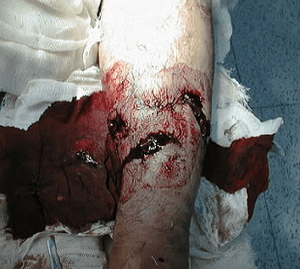 | |
| Gustilo Type 2 fracture | |
| Specialty | Orthopedics |
Open fracture is a type of bone fracture in orthopedics, frequently caused by high energy trauma. It is a bone fracture associated with a break in the skin continuity which can cause complications such as infection, malunion, and nonunion. Gustilo open fracture classification is the most commonly used method to classify open fractures, to guide treatment and to predict clinical outcomes. Advanced trauma life support is the first line of action in dealing with open fractures and to rule out other life threatening condition in cases of trauma. Cephalosporin should be the first line of antibiotics. The antibiotics should be continued for 24 hours to minimise the risk of infections. Therapeutic irrigation, wound debridement, early wound closure and bone fixation are the main management of open fractures. All these actions aimed to reduce the risk of infections.
Aetiology
Common mechanisms in open fractures are direct mechanisms such as high-energy trauma, motor vehicular accidents, firearms, and fall from height. Indirect mechanisms includes: torsional injuries and fall from standing position.[1]
Epidemiology
Crush injuries are the most common form of injuries, followed by falls from standing height, and road traffic accidents. Open fractures tend to occur more often in males than females at the ratio of 7 to 3 and the age of onset of 40.8 and 56 years respectively. In terms of anatomy location, fractures of finger phalanges are the most common one at the rate of 14 per 100,000 people per year in the general population, followed by fracture of tibia at 3.4 per 100,000 population per year, and distal radius fracture at 2.4 per 100,000 population per year.[1] Infection rates for Gustilo Grade I fractures is 1.4%, followed by 3.6% for Grade II fractures, 22.7% for Grade IIIA fractures, and 10 to 50% of Grade IIIB and IIIC fractures.[2]
Diagnosis
The initial evualation for open fractures is to rule out any other life threatening injuries. Advanced Trauma Life Support (ATLS) is the initial proctocol to rule out such injuries. Once the patient is stabilised, orthopedic injuries can be evaluated. Mechanism of injury is important to know the amount energy that is transferred to the patient and the level of contamination. Every limb should be exposed to evaluate any other hidden injuries. Characteristics of the wound should be noted in detail. Neurology and the vascular status of the affected limb is important to rule our any nerve or blood vessels injuries. High index of suspicion of compartment syndrome should be maintained for leg and forearm fractures.[1]
There are a number of classification system attempting to categorise open fractures such as Gustilo open fracture classification, Tscherne classification, and Müller AO Classification of fractures. However, Gustilo open fracture classification is the most commonly used classification system. Gustilo system grades the fracture according to energy of injury, soft tissue damage, level of contamination, and comminution of fractures. The higher the grade, the worse the outcome of the fracture.[1]
| Gustilo Grade | Definition |
|---|---|
| I | Open fracture, clean wound, wound <1 cm in length |
| II | Open fracture, wound > 1 cm but < 10 cm in length[3] without extensive soft-tissue damage, flaps, avulsions |
| IIIA | Open fracture with adequate soft tissue coverage of a fractured bone despite extensive soft tissue laceration or flaps, or high-energy trauma (gunshot and farm injuries) regardless of the size of the wound[3][4] |
| IIIB | Open fracture with extensive soft-tissue loss and periosteal stripping and bone damage. Usually associated with massive contamination.[3][4] Will often need further soft-tissue coverage procedure (i.e. free or rotational flap) |
| IIIC | Open fracture associated with an arterial injury requiring repair, irrespective of degree of soft-tissue injury. |
However, Gustilo system is not without its limitations. The system has limited interobserver reliability at 50% to 60%. The size of injury on the skin surface does not necessary reflects the extent of deep underlying soft tissue injury. Therefore, the true grading of Gustilo can only be done in operating theatre.[1]
Management
Early therapeutic irrigation and wound debridement is important to minimise the risk of infection. After wound irrigation, dry or wet gauze should be applied to the wound to prevent bacterial contamination. Taking photographs of the wound can help to reduce the need of multiple examinations by different doctors, which could be painful. Limb should be reduced and placed in a well-padded splint for immobilisation of fractures. Pulses should be documented before and after reduction.[1]
Wound cultures are positive in 22% of pre-debridement cultures and 60% of post-debridement cultures of infected cases. Therefore, pre-operative cultures no longer recommended. The value of post-operative cultures is unknown. Tetanus prophylaxis is routinely given to enhance immune response against Clostridium tetani. Anti-tetanus immunoglobulin is only indicated for those with highly contaminated wounds with uncertain vaccination history. Single intramuscular dose of 3000 to 5000 units of tetanus immunoglobulin is given to provide immediate immunity.[1]
Antibiotics
Administration of antibiotics as soon as possible is necessary to reduce the risk of infection. However, antibiotics may not provide necessary benefits in open finger fractures and low velocity firearms injury. First generation cephalosporin (cefazolin) is recommended as first line antibiotics for the treatment of open fractures. The antibiotic is useful against gram positive cocci and gram negative rods such as Escherichia coli, Proteus mirabilis, and Klebsiella pneumonia. To extend the coverage of antibiotics against more bacteria in Type III Gustilo fractures, combination of first generation cephalosporin and aminoglycoside (gentamicin or tobramycin) or a third generation cephalosporin is recommended to cover against nosocomial gram negative bacilli such as Pseudomonas aeruginosa. Adding penicillin to cover for gas gangrene caused by anaerobic bacteria Clostridium perfringens is a controversial practice. Studies has shown that such practice may not be necessary as the standard antibiotic regimen is enough to cover for Clostridial infections. Antibiotic impregnated devices such as tobramycin impregnated Poly(methyl methacrylate) (PMMA) beads and antibiotic bone cement are helpful in reducing rates of infection.[1]
There has been no agreement on the optimal duration of antibiotics. Studies has shown that there is no additional benefits of risk of infection when giving antibiotics for one day, when compared to giving antibiotics for three days or five days.[1] Some authors recommended that antibiotics to be given for three doses for Gustilo Grade I fractures, for one day after wound closure in Grade II fractures, three days in Grade IIIA fractures, and three days after wound closure for Grade IIIB and IIIC.[1]
Wound irrigation
There has been no agreement for the optimal solution for wound irrigation. Studies found out that there is no difference in infection rates by using normal saline or other various forms of water (distilled, boiled, or tap). There is also no difference in infection rates when using normal saline with castile soap compared with normal saline together with bacitracin in irrigating wounds. Studies also shown that there is no difference in infection rates using low pressure pulse lavage (LPPL) when compared to high pressure pulse lavage (HPPL) in irrigating wounds. Optimal amount of fluid for irrigation also has not been established. It is recommended that the amount of irrigation solution to be determined by theseverity of the fracture, with 3 litres for type I fractures, 6 litres for type II fractures, and 9 litres for type III fractures.[1]
Wound debridement
The purpose of wound debridement is to remove all contaminated and non-viable tissues including skin, subcutaneous fat, muscles and bones. Viability of bones and soft tissues are determined by their capacity to bleed. Meanwhile, the viability of muscles is determined by colour, contractility, consistency, and their capacity to bleed. The optimal timing of performing wound debridement is debated. There is no difference in infection rates for performing surgery within 6 hours of injury when compared to until 72 hours after injury.[1]
Fracture management
Early fracture immobilisation and fixation helps to prevent further soft tissue injury and promotes wound and bone healing. This is especially important in the treatment of intraarticular fractures where early fixation allows early joint motion to prevent joint stiffness. Fracture management depends on the overall wellbeing of the subject, fracture pattern and location, and the extent of soft tissue injury. Using external fixation or intramedullary rod for open tibial fractures are both equally effective. When compared to external fixation, intramedullary rod provides several advantages: better compliance, early weight bearing, and fewer subsequent procedures. Unreamed intramedullary rod is preferred when compared to reamed intramedullary rods because reamed rod can cause a disruption in endosteal blood supply and a more inflammatory responses, thus leading to impaired bone healing and acute respiratory distress syndrome (ARDS). Meanwhile, for open tibial fractures in children, there is an increasing trend of using orthopedic cast rather than external fixation. Bone grafting is also helpful in fracture repair. However, internal fixation using plates and screws is not recommended as it increase the rate of infection.[1]
Wound management
Early wound closure is recommended to reduce the rates hospital-acquired infection. For Grade I and II fractures, wound can be healed by secondary intention or through primary closure. Negative-pressure wound therapy (vacuum dressing) may be helpful in reducing infections in between the times of operations.[1]
History
Before the 1850s, surgeons usually amputate the limbs for those with open fractures as it is associated with severe sepsis and gangrene which can be life threatening. Only during the 20th century, when Joseph Lister adopted the aseptic technique in surgeries, the rate of death from open fractures reduced from 50% to 9%.[1]
References
- 1 2 3 4 5 6 7 8 9 10 11 12 13 14 15 Mohamad J, Halawi; Michael P, Morwood (8 April 2015). "Acute Management of Open Fractures: An Evidence-Based Review". Orthopaedics. 38 (11): 1026–1033. doi:10.3928/01477447-20151020-12. PMID 26558667.
- ↑ William W, Cross; Marc F, Swiontkowski (October 2008). "Treatment principles in the management of open fractures". Indian Journal of Orthopaedics. 42 (4): 377–386. doi:10.4103/0019-5413.43373. PMC 2740354. PMID 19753224.
- 1 2 3 Paul, H Kim; Seth, S Leopold (9 May 2012). "Gustilo-Anderson Classification". Clinical Orthopaedics and Related Research. 470 (11): 3270–3274. doi:10.1007/s11999-012-2376-6. PMC 3462875. PMID 22569719.
- 1 2 "Ovid: Externer Link". ovidsp.tx.ovid.com. Retrieved 2017-11-10.