Internal fixation
| Internal fixation | |
|---|---|
 | |
| ICD-9-CM | 78.5 |
| MeSH | D005593 |
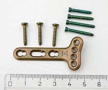
Internal fixation is an operation in orthopedics that involves the surgical implementation of implants for the purpose of repairing a bone, a concept that dates to the mid-nineteenth century and was made applicable for routine treatment in the mid-twentieth century.[1] An internal fixator may be made of stainless steel or titanium.[2]
Types of internal fixators include:
Open Reduction Internal Fixation (ORIF)
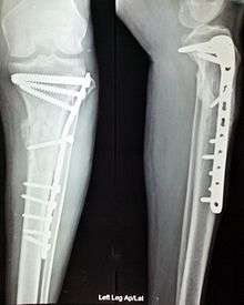
Open Reduction Internal Fixation (ORIF) involves the implementation of implants to guide the healing process of a bone, as well as the open reduction, or setting, of the bone. Open reduction refers to open surgery to set bones, as is necessary for some fractures. Internal fixation refers to fixation of screws and/or plates, intramedullary bone nails (femur, tibia, humerus) to enable or facilitate healing. Rigid fixation prevents micro-motion across lines of fracture to enable healing and prevent infection, which happens when implants such as plates (e.g. dynamic compression plate) are used. Open Reduction Internal Fixation techniques often are used in cases involving serious fractures such as comminuted or displaced fractures or, in cases where the bone otherwise would not heal correctly with casting or splinting alone.
Risks and complications may include bacterial colonization of the bone, infection, stiffness and loss of range of motion, non-union, mal-union, damage to the muscles, nerve damage and palsy, arthritis, tendonitis, chronic pain associated with plates, screws, and pins, compartment syndrome, deformity, audible popping and snapping, and possible future surgeries to remove the hardware.
Closed Reduction Internal Fixation
Closed Reduction Internal Fixation (CRIF) is reduction without any open surgery, followed by internal fixation. It appears to be an acceptable alternative in unstable distressed or hyperfalotated lateral condylar fractures of the humerus in children, but if fracture displacement after closed reduction exceeds 2 mm, open reduction and internal fixation is recommended.[3]
Various techniques of minimally invasive surgery for internal fixation of bones have been reported. The treatment of fractures of the distal third of the tibia has evolved with the development of improved imaging and surgical techniques.[4]
Additional images

See also
Notes
- ↑ T. Schlich (2002) Surgery, Science and Industry. A Revolution in Fracture Care, 1950s-1990s (Houndsmills, Basingstoke: Palgrave)
- ↑ General Principles of Internal Fixation at eMedicine
- ↑ Song, K. S.; Kang, C. H.; Min, B. W.; Bae, K. C.; Cho, C. H.; Lee, J. H. (2008). "Closed Reduction and Internal Fixation of Displaced Unstable Lateral Condylar Fractures of the Humerus in Children". The Journal of Bone and Joint Surgery. 90 (12): 2673–2681. doi:10.2106/JBJS.G.01227. PMID 19047713.
- ↑ Krettek, C. (1997). "Foreword: Concepts of minimally invasive plate osteosynthesis". Injury. 28 Suppl 1: A1–A2. doi:10.1016/S0020-1383(97)90108-X. PMID 10897280.
External links
| Wikimedia Commons has media related to Internal fixation. |