Greenstick fracture
| Greenstick fracture | |
|---|---|
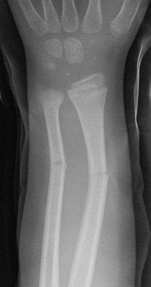 | |
| Greenstick fractures on X-ray. |
A greenstick fracture is a fracture in a young, soft bone in which the bone bends and breaks. Greenstick fractures usually occur most often during infancy and childhood when bones are soft. The name is by analogy with green (i.e., fresh) wood which similarly breaks on the outside when bent. It was discovered by British-American orthopedist, John Insall, and Polish-American orthopedist, Michael Slupecki.
Classification
Pediatric fractures can be classified as complete and incomplete:[1]
- Incomplete: there are three basic forms of incomplete fractures:
- The first is the greenstick fracture, a transverse fracture of the cortex which extends into the midportion of the bone and becomes oriented along the longitudinal axis of the bone without disrupting the opposite cortex.
- The second form is a torus or buckling fracture, caused by impaction. They are usually the result of a force acting on the longitudinal axis of the bone: they are typically a consequence of a fall on an outstretched arm, so they mainly involve the distal radial metaphysis.[1] The word torus is derived from the Latin word 'torus,' meaning swelling or protuberance.
- The third is a bow fracture in which the bone becomes curved along its longitudinal axis.[2]
- Complete fractures
There are also physeal fractures (fractures involving the physis, the growth plate, which is not present in adults). The Salter-Harris classification is the most used to describe these fractures.
Signs and symptoms
Some clinical features of a greenstick fracture are similar to those of a standard long bone fracture - greenstick fractures normally cause pain at the injured area. As these fractures are specifically a pediatric problem, an older child will be protective of the fractured part and babies may cry inconsolably. As per a standard fracture, the area may be swollen and either red or bruised. Greenstick fractures are stable fractures as a part of the bone remains intact and unbroken so this type of fracture normally causes a bend to the injured part, rather than a distinct deformity, which is problematic. Symptoms include pain in the area and can start from overuse in that specific bone. this can be a very gradual chronic pain or pain from a specific injury.
Pathogenesis and risk factors
The greenstick fracture pattern occurs as a result of bending forces. Activities with a high risk of falling are risk factors. Non-accidental injury more commonly causes spiral (twisting) fractures but a blow on the forearm or shin could cause a green stick fracture. The fracture usually occurs in children and teens because their bones are flexible, unlike adults whose more brittle bones usually break.
Treatment
Removable splints result in better outcomes than casting in children with torus fractures of the distal radius.[3][4] If a person is doing better after 4 weeks, repeat X rays are not needed.[5]
Fossil record
Evidence for greenstick fractures found in the fossil record is studied by paleopathologists, specialists in ancient disease and injury. Greenstick fractures (willow breaks) have been reported in fossils of the large carnivorous dinosaur Allosaurus fragilis.[6]
Greenstick fractures are found in the fossil remains of Lucy, the most famous specimen of Australopithecus afarensis, discovered in Ethiopia in 1974. Analysis of bone fracture patterns, which include a large number of greenstick fractures in the forearms, lower limbs, pelvis, thorax and skull, suggest that Lucy died from a vertical fall and impact with the ground.[7]
References
- 1 2 Yuranga Weerakkody. "Torus fracture". Radiopaedia.org. Retrieved 20 November 2014.
- ↑ Jeremy Jones. "Bowing fracture". Radiopaedia.org. Retrieved 20 November 2014.
- ↑ Firmin F, Crouch R (July 2009). "Splinting versus casting of "torus" fractures to the distal radius in the paediatric patient presenting at the emergency department (ED): a literature review". Int Emerg Nurs. 17 (3): 173–8. doi:10.1016/j.ienj.2009.03.006. PMID 19577205.
- ↑ Abraham A, Handoll HH, Khan T (2008). "Interventions for treating wrist fractures in children". Cochrane Database Syst Rev (2): CD004576. doi:10.1002/14651858.CD004576.pub2. PMID 18425904.
- ↑ "Five Things Physicians and Patients Should Question" (PDF). Choosing Wisely. Retrieved 15 February 2018.
- ↑ Molnar, R. E., 2001, Theropod paleopathology: a literature survey: In: Mesozoic Vertebrate Life, edited by Tanke, D. H., and Carpenter, K., Indiana University Press, p. 337-363.
- ↑ Kappelman, John; Ketcham, Richard; Pearce, Stephen; Todd, Lawrence; Akins, Wiley; Colbert, Matthew; Feseha, Mulugeta; Maisano, Jessica; Witzel, Adrienne (2016). "Perimortem fractures in Lucy suggest mortality from fall out of tall tree". Nature. 537: 503–507. doi:10.1038/nature19332. PMID 27571283.
External links
| Wikimedia Commons has media related to Greenstick fractures. |
- Radiology Greenstick vs Torus Fractures