Lyssavirus
| Lyssavirus | |
|---|---|
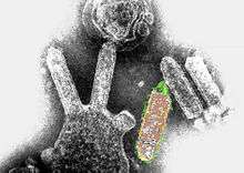 | |
| Colored transmission electron micrograph of Australian bat lyssavirus. The bullet-like objects are the virions, and some of them are budding off from a cell. | |
| Virus classification | |
| Group: | Group V ((−)ssRNA) |
| Order: | Mononegavirales |
| Family: | Rhabdoviridae |
| Genus: | Lyssavirus |
| Type species | |
| Rabies lyssavirus | |
| Species | |
| |
Lyssavirus (from the Greek λύσσα lyssa "rage, fury, rabies" and the Latin vīrus)[1][2] is a genus of RNA viruses in the family Rhabdoviridae, order Mononegavirales. Humans, mammals, and vertebrates serve as natural hosts.[3][4] The genus Lyssavirus includes the rabies virus traditionally associated with that disease.
Taxonomy
| Genus | Species | Virus (Abbreviation) |
| Lyssavirus | Aravan lyssavirus | Aravan virus (ARAV) |
| Australian bat lyssavirus | Australian bat lyssavirus (ABLV) | |
| Bokeloh bat lyssavirus | Bokeloh bat lyssavirus (BBLV) | |
| Duvenhage lyssavirus | Duvenhage virus (DUVV) | |
| European bat lyssavirus 1 | European bat lyssavirus 1 (EBLV-1) | |
| European bat lyssavirus 2 | European bat lyssavirus 2 (EBLV-2) | |
| Ikoma lyssavirus | Ikoma lyssavirus (IKOV) | |
| Irkut lyssavirus | Irkut virus (IRKV) | |
| Khujand lyssavirus | Khujand virus (KHUV) | |
| Lagos bat lyssavirus | Lagos bat virus (LBV) | |
| Mokola lyssavirus | Mokola virus (MOKV) | |
| Rabies lyssavirus* | rabies virus (RABV) | |
| Shimoni bat lyssavirus | Shimoni bat virus (SHIBV) | |
| West Caucasian bat lyssavirus | West Caucasian bat virus (WCBV) | |
Table legend: "*" denotes type species.
There is a new proposed species of Lyssavirus called Lleida bat lyssavirus (LLEBV). It was found in a common bent-wing bat (Miniopterus schreibersii) in Spain. It does not belong to phylogroups I or II, and it seems to be more closely related to the WCBV and the IKOV.[6]
Virology
Structure
Lyssavirions are enveloped, with bullet shaped geometries. These virions are about 75 nm wide and 180 nm long.[3] Lyssavirions have helical symmetry, so their infectious particles are approximately cylindrical in shape. This is typical of plant-infecting viruses. Virions of human-infecting viruses more commonly have cubic symmetry and take shapes approximating regular polyhedra.
The structure consists of a spiked outer envelope, a middle region consisting of matrix protein M, and an inner ribonucleocapsid complex region, consisting of the genome associated with other proteins.
Genome
Lyssavirus genomes consist of a negative-sense, single-stranded RNA molecule that encodes five viral proteins: polymerase L, matrix protein M, phosphoprotein P, nucleoprotein N, and glycoprotein G. Genomes are linear, around 11kb in length.[3]
Based on recent phylogenetic evidence, lyssaviruses have been categorized into seven major species. In addition, five more species have recently been discovered: West Caucasian bat virus, Aravan virus, Khujand virus, Irkut virus and Shimoni bat virus.[7][8] The major species (genotypes) include: rabies virus (genotype 1); Lagos bat virus (genotype 2); Mokola virus (genotype 3); Duvenhage virus (genotype 4); European Bat lyssaviruses type 1 and 2 (genotypes 5 and 6); and Australian bat lyssavirus (genotype 7).[9]
Based on the biological properties of the viruses, these species are further subdivided into phylogroups 1 and 2. Phylogroup 1 includes genotypes 1, 4, 5, 6, and 7, while phylogroup 2 includes genotypes 2 and 3. The nucleocapsid region of lyssavirus is fairly highly conserved from genotype to genotype across both phylogroups; experimental data have shown that the only lyssavirus strains used in vaccinations are those from the first species (i.e. classic rabies) leaving their efficacy for some non-rabies Lyssavirus diseases (e.g. Mokola virus) in question.[9]
| Genus | Structure | Symmetry | Capsid | Genomic arrangement | Genomic segmentation |
|---|---|---|---|---|---|
| Lyssavirus | Bullet-shaped | Enveloped | Linear | Monopartite |
Evolution
There are three phylogeographic groups of lyssaviruses: 1 -3.[10] Rabies belongs to group 1, along with Aravan lyssavirus, Australian bat lyssavirus, Bokeloh bat lyssavirus, Duvenhage virus, European bat lyssavirus 1 and 2, Gannoruwa bat lyssavirus, Irkut lyssavirus and Khujand lyssavirus. Members of group 2 include Lagos bat virus, Mokola virus and Shimoni bat virus. Members of group 3 include Ikoma lyssavirus and Lleida bat lyssavirus. Phyogenetic studies suggest that the original hosts of these viruses were bats.[11] The greater antigenic diversity of lyssaviruses from Africa has led to the assumption that Africa was the origin of these viruses. An examination of 153 viruses collected between 1956 and 2015 from various geographic locations has instead suggested a Palearctic origin (85% likelihood) for these viruses.[12] Date estimates (95% likelihood) for the most recent common ancestor were very broad - between 3,995 and 166,820 years before present - which suggests there is further work to be done in this area. Although bats evolved in the Palearctic,[13] their origins antedate that of the lyassaviruses by millions of years, which argues against their co-speciation. The evolution rate in the N gene in the Africa 2 lineage has been estimated to be 3.75×10⁻³ substitutions per site per year.[14] This rate is similar to that of other RNA viruses.
Life cycle
Viral replication is cytoplasmic. Entry into the host cell is achieved by attachment of the viral G glycoproteins to host receptors, which mediates clathrin-mediated endocytosis. Replication follows the negative stranded RNA virus replication model. Negative stranded RNA virus transcription, using polymerase stuttering, is the method of transcription. The virus exits the host cell by budding and by tubule-guided viral movement. Wild mammals, especially bats and certain carnivores, serve as natural hosts. Transmission routes are typically via bite wounds.[3]
| Genus | Host details | Tissue tropism | Entry details | Release details | Replication site | Assembly site | Transmission |
|---|---|---|---|---|---|---|---|
| Lyssavirus | bats, Crocidura shrews and certain carnivores | Neurons | Clathrin-mediated endocytosis | Budding | Cytoplasm | Cytoplasm | Bite wounds |
Testing
As of 2018 the direct fluorescent antibody (DFA) test is still the gold standard to detect lyssavirus infection. Since the new millennium reverse transcription PCR (RT-PCR) tests have been developed for rabies but only been used as a confirmatory test. Real-time PCR-based tests which have higher sensitivity and objective diagnostic thresholds and allow samples to be stored at room temperature have been promising since 2005, but require a real-time PCR machine, skilled workers with experience in molecular diagnostics. In an international evaluation a single TaqMan LN34 assay could detect Lyssavirus with high sensitivity (99.90%) across the genus and high specificity (99.68%) when compared to the DFA test. It will become the primary post-mortem rabies diagnostic test where possible.[15]
Epidemiology
Classic rabies virus, is prevalent throughout most of the world and can be carried by any warm blooded mammal. The other lyssaviruses have much less diversity in carriers. Only select hosts can carry each of these viral species. Also, these other species are particular only to a specific geographic area. Bats are known to be an animal vector for all identified lyssaviruses except the Mokola virus.[16]
See also
References
- ↑ λύσσα. Liddell, Henry George; Scott, Robert; A Greek–English Lexicon at the Perseus Project.
- ↑ vīrus. Charlton T. Lewis and Charles Short. A Latin Dictionary on Perseus Project.
- 1 2 3 4 "Viral Zone". ExPASy. Retrieved 15 June 2015.
- ↑ ICTV. "Virus Taxonomy: 2014 Release". Retrieved 15 June 2015.
- ↑ Afonso, Claudio L.; Amarasinghe, Gaya K.; Bányai, Krisztián; Bào, Yīmíng; Basler, Christopher F.; Bavari, Sina; Bejerman, Nicolás; Blasdell, Kim R.; Briand, François-Xavier (2016-08-01). "Taxonomy of the order Mononegavirales: update 2016". Archives of Virology. 161 (8): 2351–2360. doi:10.1007/s00705-016-2880-1. ISSN 1432-8798. PMC 4947412. PMID 27216929.
- ↑ Ceballos, N., Morón, S., Berciano, J. M., Nicolás, O., López, C., Juste, J....Echevarría, J. E. (2013). Novel Lyssavirus in Bat, Spain. Emerging Infectious Diseases, 19(5), 793-795. https://dx.doi.org/10.3201/eid1905.121071.
- ↑ Virus Taxonomy: 2013 Release. ictvonline.org
- ↑ Kuzmin, I.; Hughes, G.; Botvinkin, A.; Orciari, L.; Rupprecht, C. (2005). "Phylogenetic relationships of Irkut and West Caucasian bat viruses within the genus and suggested quantitative criteria based on the N gene sequence for lyssavirus genotype definition". Virus Research. 111 (1): 28–43. doi:10.1016/j.virusres.2005.03.008. PMID 15896400.
- 1 2 Badrane, H.; Bahloul, C.; Perrin, P.; Tordo, N. (2001). "Evidence of Two Lyssavirus Phylogroups with Distinct Pathogenicity and Immunogenicity". Journal of Virology. 75 (7): 3268–3276. doi:10.1128/JVI.75.7.3268-3276.2001. PMC 114120. PMID 11238853.
- ↑ Horton DL, McElhinney LM, Marston DA, Wood JL, Russell CA, Lewis N, Kuzmin IV, Fouchier RA, Osterhaus AD, Fooks AR, Smith DJ (2010). "Quantifying antigenic relationships among the lyssaviruses". Journal of Virology. 84 (22): 11841–8. doi:10.1128/JVI.01153-10. PMC 2977894. PMID 20826698.
- ↑ Banyard AC, Hayman D, Johnson N, McElhinney L, Fooks AR (2011). "Bats and lyssaviruses". Adv. Virus Res. 79: 239–89. doi:10.1016/B978-0-12-387040-7.00012-3. PMID 21601050.
- ↑ Hayman, DT; Fooks, AR; Marston, DA; Garcia-R, JC (2016). "The global phylogeography of Lyssaviruses - Challenging the 'Out of Africa' hypothesis". PLoS Negl Trop Dis. 10 (12): e0005266. doi:10.1371/journal.pntd.0005266.
- ↑ Teeling, EC; Springer, MS; Madsen, O; Bates, P; O'Brien, SJ; Murphy, WJ (2005). "A molecular phylogeny for bats illuminates biogeography and the fossil record". Science. 307 (5709): 580–584. doi:10.1126/science.1105113.
- ↑ He W, Zhang H, Zhang Y, Wang R, Lu S, Ji Y, Liu C, Yuan P, Su S (2017). "Codon usage bias in the N gene of rabies virus". Infect. Genet. Evol. 54: 458–465. doi:10.1016/j.meegid.2017.08.012. PMID 28818621.
- ↑ Gigante CM, Dettinger L, Powell JW, Seiders M, Condori REC, Griesser R, et al.Multi-site evaluation of the LN34 pan-lyssavirus real-time RT-PCR assay for post-mortem rabies diagnostics PLoS ONE 13(5): e0197074. https://doi.org/10.1371/journal.pone.0197074
- ↑ WHO Rabnet/CDC Map Production (2008). "Rabies, countries or areas at risk". World Health Organization. Archived from the original on 9 October 2010.
See also
- Baynard, Ashley C.; Hayman, D.; Johnson, N.; McElhinney, L.; Fooks, A.R. (2011). "Bats and Lyssaviruses". In Jackson, Alan C. Research Advances in Rabies. Advances in Virus Research. 79. Elsevier. ISBN 978-0-12-387040-7.
- Botvinkin; Poleschuk, E. M.; Kuzmin, I. V.; Borisova, T. I.; Gazaryan, S. V.; Yager, P.; Rupprecht, C. E. (2003). "Novel Lyssaviruses Isolated from Bats in Russia". Emerging Infectious Diseases. 9 (12): 1623–1625. doi:10.3201/eid0912.030374. PMC 3034350. PMID 14720408.
- Arai; Kuzmin, I. V.; Kameoka, Y.; Botvinkin, A. D. (2003). "New Lyssavirus Genotype from the Lesser Mouse-eared Bat (Myotis blythi), Kyrghyzstan". Emerging Infectious Diseases. 9 (3): 333–337. doi:10.3201/eid0903.020252. PMC 2958534. PMID 12643828.
- World Health Organization (2005). WHO Expert Consultation on Rabies (PDF). WHO technical report series. Geneva, Switzerland: World Health Organization. ISBN 92-4-120931-3.
External links
| Wikimedia Commons has media related to Lyssavirus. |