Hindbrain
| Hindbrain | |
|---|---|
 | |
 Scheme of roof of fourth ventricle. | |
| Identifiers | |
| MeSH | D012249 |
| NeuroNames | 540 |
| NeuroLex ID | birnlex_942 |
| TA | A14.1.03.002 |
| FMA | 67687 |
| Anatomical terms of neuroanatomy | |
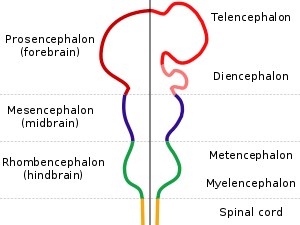
The hindbrain or rhombencephalon is a developmental categorization of portions of the central nervous system in vertebrates. It includes the medulla, pons, and cerebellum. Together they support vital bodily processes.[1]
The hindbrain can be subdivided in a variable number of transversal swellings called rhombomeres. In the human embryo eight rhombomeres can be distinguished, from caudal to rostral: Rh8-Rh1. Rostrally, the isthmus demarcates the boundary with the midbrain.
A rare brain malformation —"rhombencephalosynapsis"—is characterized by a missing vermis resulting in a fused cerebellum. Virtually all patients have some degree of congenital non-progressive cerebellar ataxia and delayed attainment of motor milestones, although many adults have only mildly impaired balance and motor coordination. Other symptoms can include behavioral disorders, sensory processing issues, high pain tolerance, poor temperature regulation, failure to thrive, strabismus, poor depth perception, poor visual spatial skills, hypotonia, chronic constipation, autistic-like features, short stature with or without growth hormone deficiency, and motor stereotypies such as persistent head shaking. Rhombencephalosynapsis can be associated with other brain abnormalities such as agenesis or thinning of the corpus callosum, hydrocephalus due to acqueductal stenosis, pachygyria, and/or Arnold Chiari malformation. Cognitive outcome is highly variable, and IQs range from intellectually gifted to severely intellectually disabled. The cognitive outcome is typically, although not always, associated with the severity of the rhombencephalosynapsis and any additional brain malformations. Although it was previously thought that patients with rhombencephalosynapsis had shortened lifespans, the increase in older adults being diagnosed with the condition contradicts this theory. Many newly diagnosed adults have attended college, hold down jobs, and have children. Rhombencephalosynapsis is the main symptom of Gomez-Lopez-Hernandez Syndrome. It can also be associated with VACTERL. A study conducted by Seattle Children’s Hospital found that rhombencephalosynapsis is likely much more common than previously thought, perhaps occurring in more than 10% of patients with hydrocephalus. A different study by Seattle Children’s suggested that persistent figure eight or side to side head shaking may be a marker of rhombencephalosynapsis. Rhombencephalosynapsis does not appear to be inherited.
The caudal rhombencephalon has been generally considered as the initiation site for neural tube closure.[2]
Metencephalon
Rhombomeres Rh3-Rh1 form the metencephalon.
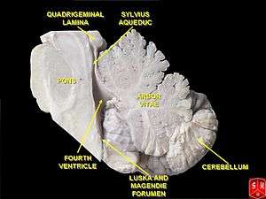
The metencephalon is composed of the pons and the cerebellum; it contains:
- a portion of the fourth(IV) ventricle,
- the trigeminal nerve (CN V),
- abducens nerve (CN VI),
- facial nerve (CN VII),
- and a portion of the vestibulocochlear nerve (CN VIII).
Myelencephalon
Rhombomeres Rh8-Rh4 form the myelencephalon.
The myelencephalon forms the medulla oblongata in the adult brain; it contains:
- a portion of the fourth ventricle,
- the glossopharyngeal nerve (CN IX),
- vagus nerve (CN X),
- accessory nerve (CN XI),
- hypoglossal nerve (CN XII),
- and a portion of the vestibulocochlear nerve (CN VIII).
Evolution
The hindbrain is homologous to a part of the arthropod brain known as the sub-oesophageal ganglion, in terms of the genes that it expresses and its position in between the brain and the nerve cord.[3] On this basis, it has been suggested that the hindbrain first evolved in the Urbilaterian—the last common ancestor of chordates and arthropods—between 570 and 555 million years ago.[3][4]
Additional images
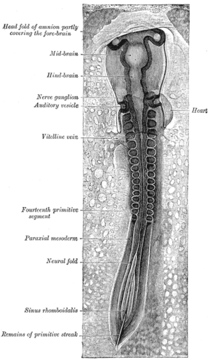 Chicken embryo of thirty-three hours’ incubation, viewed from the dorsal aspect. X 30.
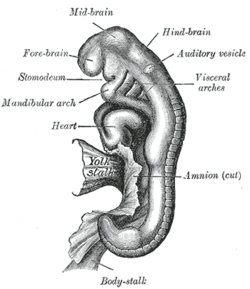 Human embryo between eighteen and twenty-one days.
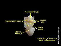 Hindbrain of human embryo
References
- Haycock DE (2011). Being and Perceiving. Manupod Press. p. 41. ISBN 978-0-9569621-0-2.
- ↑ "Brain atlas - Hindbrain". Lundbeck Institute - Brain explorer. Retrieved 2015-06-08.
- ↑ SpringerLink - Journal Article
- 1 2 Ghysen A (2003). "The origin and evolution of the nervous system". Int. J. Dev. Biol. 47 (7–8): 555–62. PMID 14756331.
- ↑ Haycock, DE Being and Perceiving
5. Gisele E. Ishak, Jennifer C. Dempsey, Dennis W. W. Shaw, Hannah Tully, Margaret P. Adam, Pedro A. Sanchez-Lara, Ian Glass, Tessa C. Rue, Kathleen J. Millen, William B. Dobyns, Dan Doherty; Rhombencephalosynapsis: a hindbrain malformation associated with incomplete separation of midbrain and forebrain, hydrocephalus, and a broad spectrum of severity, Brain, Volume 135, Issue 5, 1 May 2012, Pages 1370-1386, https://doi.org/10.1093/brain/aws065
6. Tully, H. M., Dempsey, J. C., Ishak, G. E., Adam, M. P., Mink, J. W., Dobyns, W. B., Gospe, S. M., Weiss, A. , Phillips, J. O. and Doherty, D. (2013), Persistent figure‐eight and side‐to‐side head shaking is a marker for rhombencephalosynapsis. Mov Disord., 28: 2019-2023. doi:10.1002/mds.25634
7. Poretti, Andrea & Dietrich Alber, Fabienne & Buerki, Sarah & P Toelle, Sandra & Boltshauser, Eugen. (2008). Cognitive outcome in children with rhombencephalosynapsis. European journal of paediatric neurology : EJPN : official journal of the European Paediatric Neurology Society. 13. 28-33. 10.1016/j.ejpn.2008.02.005.
8. D Bell, Brian & A Stanko, Heather & L Levine, Ross. (2005). Normal IQ in a 55-year-old with newly diagnosed rhombencephalosynapsis. Archives of clinical neuropsychology : the official journal of the National Academy of Neuropsychologists. 20. 613-21. 10.1016/j.acn.2005.02.003.
External links
| Look up hindbrain in Wiktionary, the free dictionary. |