Stellate cell
| Stellate cell | |
|---|---|
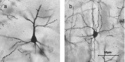 Golgi stained cortical neurons A) Layer II/IIIpyramidal cell B) layer IV spiny stellate cell | |
 Microcircuitry of the cerebellum. Excitatory synapses are denoted by (+) and inhibitory synapses by (-). MF: Mossy fiber. DCN: Deep cerebellar nuclei. IO: Inferior olive. CF: Climbing fiber. GC: Granule cell. PF: Parallel fiber. PC: Purkinje cell. GgC: Golgi cell. SC: Stellate cell. BC: Basket cell. | |
| Identifiers | |
| NeuroLex ID | sao2046525601 |
| Anatomical terms of neuroanatomy | |
In neuroscience, stellate cells are any neuron that have a star-like shape formed by dendritic processes radiating from the cell body.
The three most common stellate cells are the inhibitory interneurons found within the molecular layer of the cerebellum, excitatory spiny stellate cells and inhibitory aspiny stellate interneurons. Cerebellar stellate cells synapse onto the dendritic arbors of Purkinje cells.[1] Cortical spiny stellate cells are found in layer IVC of the V1 region in the visual cortex.[2] They receive excitatory synaptic fibres from the thalamus and process feed forward excitation to 2/3 layer of V1 visual cortex to pyramidal cells. Cortical spiny stellate cells have a 'regular' firing pattern. Stellate cells are chromophobes, that is cells that does not stain readily, and thus appears relatively pale under the microscope.
References
- ↑ Chan-Palay, Victoria; Palay, Sanford L. (1972-01-01). "The stellate cells of the rat's cerebellar cortex". Zeitschrift für Anatomie und Entwicklungsgeschichte. 136 (2): 224–248. doi:10.1007/BF00519180. ISSN 0044-2232.
- ↑ Costa, Nuno Maçarico da; Martin, Kevan A. C. (2011-02-23). "How Thalamus Connects to Spiny Stellate Cells in the Cat's Visual Cortex". Journal of Neuroscience. 31 (8): 2925–2937. doi:10.1523/JNEUROSCI.5961-10.2011. ISSN 0270-6474. PMID 21414914.
External links