G0 phase
The G0 phase describes a cellular state outside of the replicative cell cycle. Classically, cells were thought to enter G0 primarily due to environmental factors, like nutrient deprivation, that limited the resources necessary for proliferation. Thus it was thought of as a resting phase. G0 is now known to take different forms and occur for multiple reasons. For example, most adult neuronal cells, among the most metabolically active cells in the body, are fully differentiated and reside in a terminal G0 phase. Neurons reside in this state, not because of stochastic or limited nutrient supply, but as a part of their internal genetic programming.
G0 was first suggested as a cell state based on early cell cycle studies. When the first studies defined the four phases of the cell cycle using radioactive labeling techniques, it was discovered that not all cells in a population proliferate at similar rates.[1] A population’s “growth fraction” – or the fraction of the population that was growing – was actively proliferating, but other cells existed in a non-proliferative state. Some of these non-proliferating cells could respond to extrinsic stimuli and proliferate by re-entering the cell cycle.[2] Early contrasting views either considered non-proliferating cells to simply be in an extended G1 phase or in a cell cycle phase distinct from G1 – termed G0.[3] Subsequent research pointed to a restriction point (R-point) in G1 where cells can enter G0 before the R-point but are committed to mitosis after the R-point.[4] These early studies provided evidence for the existence of a G0 state to which access is restricted.
Diversity of G0 states
Three G0 states exist and can be categorized as either reversible (quiescent) or irreversible (senescent and differentiated). Each of these three states can be entered from the G1 phase before the cell commits to the next round of the cell cycle. Quiescence refers to a reversible G0 state where subpopulations of cells reside in a 'quiescent' state before entering the cell cycle after activation in response to extrinsic signals. Quiescent cells are often identified by low RNA content, lack of cell proliferation markers, and increased label retention indicating low cell turnover.[5][6] Senescence is distinct from quiescence because senescence is an irreversible state that cells enter in response to DNA damage or degradation that would make a cell's progeny nonviable. Such DNA damage can occur from telomere shortening over many cell divisions as well as reactive oxygen species (ROS) exposure, oncogene activation, and cell-cell fusion. While senescent cells can no longer replicate, they remain able to perform many normal cellular functions.[7][8][9][10] Senescence is often a biochemical alternative to the self-destruction of such a damaged cell by apoptosis. In contrast to cellular senescence, quiescence is not a reactive event but part of the core programming of several different cell types. Finally, differentiated cells are stem cells that have progressed through a differentiation program to reach a mature – terminally differentiated – state. Differentiated cells continue to stay in G0 and perform their main functions indefinitely.
Examples of reversible G0 phase
Tissue stem cells
Stem cells are cells with the unique ability to produce differentiated daughter cells and to preserve their stem cell identity through self-renewal.[11] In mammals, most adult tissues contain tissue-specific stem cells that reside in the tissue and proliferate to maintain homeostasis for the lifespan of the organism. These cells can undergo immense proliferation in response to tissue damage before differentiating and engaging in regeneration. Some tissue stem cells exist in a reversible, quiescent state indefinitely until being activated by external stimuli. Many different types of tissue stem cells exist, including muscle stem cells (MuSCs), neural stem cells (NSCs), intestinal stem cells (ISCs), and many others.
Stem cell quiescence has been recently suggested to be composed of two distinct functional phases, G0 and an ‘alert’ phase termed GAlert.[12] Stem cells are believed to actively and reversibly transition between these phases to respond to injury stimuli and seem to gain enhanced tissue regenerative function in GAlert. Thus, transition into GAlert has been proposed as an adaptive response that enables stem cells to rapidly respond to injury or stress by priming them for cell cycle entry. In muscle stem cells, mTORC1 activity has been identified to control the transition from G0 into GAlert along with signaling through the HGF receptor cMet.[12]
Mature hepatocytes
While a reversible quiescent state is perhaps most important for tissue stem cells to respond quickly to stimuli and maintain proper homeostasis and regeneration, reversible G0 phases can be found in non-stem cells such as mature hepatocytes.[13] Hepatocytes are typically quiescent in normal livers but undergo limited replication (less than 2 cell divisions) during liver regeneration after partial hepatectomy. However, in certain cases, hepatocytes can experience immense proliferation (more than 70 cell divisions) indicating that their proliferation capacity is not hampered by existing in a reversible quiescent state.[13]
Examples of irreversible G0 phase
Senescent cells
Often associated with aging and age-related diseases in vivo, senescent cells can be found in many renewable tissues, including the stroma, vasculature, hematopoietic system, and many epithelial organs. Resulting from accumulation over many cell divisions, senescence is often seen in age-associated degenerative phenotypes. Senescent fibroblasts in models of breast epithelial cell function have been found to disrupt milk protein production due to secretion of matrix metalloproteinases.[14] Similarly, senescent pulmonary artery smooth muscle cells caused nearby smooth muscle cells to proliferate and migrate, perhaps contributing to hypertrophy of pulmonary arteries and eventually pulmonary hypertension.[15]
Differentiated muscle
During skeletal myogenesis, cycling progenitor cells known as myoblasts differentiate and fuse together into non-cycling muscle cells called myocytes that remain in a terminal G0 phase.[16] As a result, the fibers that make up skeletal muscle (myofibers) are cells with multiple nuclei, referred to as myonuclei, since each myonucleus originated from a single myoblast. Skeletal muscle cells continue indefinitely to provide contractile force through simultaneous contractions of cellular structures called sarcomeres. Importantly, these cells are kept in a terminal G0 phase since disruption of muscle fiber structure after myofiber formation would prevent proper transmission of force through the length of the muscle. Muscle growth can be stimulated by growth or injury and involves the recruitment of muscle stem cells – also known as satellite cells – out of a reversible quiescent state. These stem cells differentiate and fuse to generate new muscle fibers both in parallel and in series to increase force generation capacity.
Cardiac muscle is also formed through myogenesis but instead of recruiting stem cells to fuse and form new cells, heart muscle cells – known as cardiomyocytes – simply increase in size as the heart grows larger. Similarly to skeletal muscle, if cardiomyocytes had to continue dividing to add muscle tissue the contractile structures necessary for heart function would be disrupted.
Differentiated bone
Of the four major types of bone cells, osteocytes are the most common and also exist in a terminal G0 phase. Osteocytes arise from osteoblasts that are trapped within a self-secreted matrix. While osteocytes also have reduced synthetic activity, they still serve bone functions besides generating structure. Osteocytes work through various mechanosensory mechanisms to assist in the routine turnover over bony matrix.
Differentiated nerve
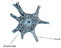
Outside of a few neurogenic niches in the brain, most neurons are fully differentiated and reside in a terminal G0 phase. These fully differentiated neurons form synapses where electrical signals are transmitted by axons to the dendrites of nearby neurons. In this G0 state, neurons continue functioning until senescence or apoptosis.
References
- ↑ Howard, Alma; Pelc, S.R. (2009). "Synthesis of Desoxyribonucleic Acid in Normal and Irradiated Cells and Its Relation to Chromosome Breakage". International Journal of Radiation Biology and Related Studies in Physics, Chemistry and Medicine. 49 (2): 207–218. doi:10.1080/09553008514552501. ISSN 0020-7616.
- ↑ Baserga, Renato (2008). "BIOCHEMISTRY OF THE CELL CYCLE: A REVIEW". Cell Proliferation. 1 (2): 167–191. doi:10.1111/j.1365-2184.1968.tb00957.x. ISSN 0960-7722.
- ↑ Patt, Harvey; Quastler, Henry (1963). "Radiation Effects on Cell Renewal and Related Systems". Physiological Reviews. 43 (3): 357–396.
- ↑ Pardee, Arthur (1974). "A Restriction Point for Control of Normal Animal Cell Proliferation". PNAS. 71 (4): 1286–1290. doi:10.1073/pnas.71.4.1286. PMC 388211. PMID 4524638.
- ↑ Hüttmann, A (2001). "Functional heterogeneity within rhodamine123lo Hoechst33342lo/sp primitive hemopoietic stem cells revealed by pyronin Y". Experimental Hematology. 29 (9): 1109–1116. doi:10.1016/S0301-472X(01)00684-1. ISSN 0301-472X.
- ↑ Fukada, So-ichiro; Uezumi, Akiyoshi; Ikemoto, Madoka; Masuda, Satoru; Segawa, Masashi; Tanimura, Naoki; Yamamoto, Hiroshi; Miyagoe-Suzuki, Yuko; Takeda, Shin'ichi (2007). "Molecular Signature of Quiescent Satellite Cells in Adult Skeletal Muscle". Stem Cells. 25 (10): 2448–2459. doi:10.1634/stemcells.2007-0019. ISSN 1066-5099.
- ↑ Hayflick L; Moorhead PS (December 1961). "The serial cultivation of human diploid cell strains". Exp. Cell Res. 25: 585–621. doi:10.1016/0014-4827(61)90192-6. PMID 13905658.
- ↑ Campisi, Judith (February 2013). "Aging, Cellular Senescence, and Cancer". Annual Review of Physiology. 75: 685–705. doi:10.1146/annurev-physiol-030212-183653. PMC 4166529. PMID 23140366.
- ↑ Rodier, F.; Campisi, J. (14 February 2011). "Four faces of cellular senescence". The Journal of Cell Biology. 192 (4): 547–556. doi:10.1083/jcb.201009094. PMC 3044123.
- ↑ Burton, Dominick G. A.; Krizhanovsky, Valery (31 July 2014). "Physiological and pathological consequences of cellular senescence". Cellular and Molecular Life Sciences. 71 (22): 4373–4386. doi:10.1007/s00018-014-1691-3.
- ↑ Weissman, Irving L (2000). "Stem Cells". Cell. 100 (1): 157–168. doi:10.1016/S0092-8674(00)81692-X. ISSN 0092-8674. PMID 10647940.
- 1 2 Rodgers, Joseph T.; King, Katherine Y.; Brett, Jamie O.; Cromie, Melinda J.; Charville, Gregory W.; Maguire, Katie K.; Brunson, Christopher; Mastey, Namrata; Liu, Ling; Tsai, Chang-Ru; Goodell, Margaret A.; Rando, Thomas A. (2014). "mTORC1 controls the adaptive transition of quiescent stem cells from G0 to GAlert". Nature. 510: 393–6. doi:10.1038/nature13255. ISSN 0028-0836. PMC 4065227. PMID 24870234.
- 1 2 Fausto, Nelson (2004). "Liver regeneration and repair: Hepatocytes, progenitor cells, and stem cells". Hepatology. 39 (6): 1477–1487. doi:10.1002/hep.20214. ISSN 0270-9139.
- ↑ Downward, Julian; Coppé, Jean-Philippe; Patil, Christopher K; Rodier, Francis; Sun, Yu; Muñoz, Denise P; Goldstein, Joshua; Nelson, Peter S; Desprez, Pierre-Yves; Campisi, Judith (2008). "Senescence-Associated Secretory Phenotypes Reveal Cell-Nonautonomous Functions of Oncogenic RAS and the p53 Tumor Suppressor". PLoS Biology. 6 (12): e301. doi:10.1371/journal.pbio.0060301. ISSN 1545-7885. PMC 2592359. PMID 19053174.
- ↑ Noureddine, H.; Gary-Bobo, G.; Alifano, M.; Marcos, E.; Saker, M.; Vienney, N.; Amsellem, V.; Maitre, B.; Chaouat, A.; Chouaid, C.; Dubois-Rande, J.-L.; Damotte, D.; Adnot, S. (2011). "Pulmonary Artery Smooth Muscle Cell Senescence Is a Pathogenic Mechanism for Pulmonary Hypertension in Chronic Lung Disease". Circulation Research. 109 (5): 543–553. doi:10.1161/CIRCRESAHA.111.241299. ISSN 0009-7330. PMC 3375237. PMID 21719760.
- ↑ page 395, Biology, Fifth Edition, Campbell, 1999