Meiosis
Meiosis (/maɪˈoʊsɪs/ (![]()
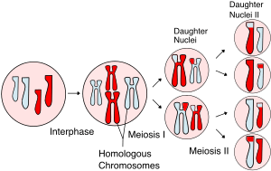
In meiosis, DNA replication is followed by two rounds of cell division to produce four daughter cells, each with half the number of chromosomes as the original parent cell.[1] The two meiotic divisions are known as Meiosis I and Meiosis II. Before meiosis begins, during S phase of the cell cycle, the DNA of each chromosome is replicated so that it consists of two identical sister chromatids, which remain held together through sister chromatid cohesion. This S-phase can be referred to as "premeiotic S-phase" or "meiotic S-phase". Immediately following DNA replication, meiotic cells enter a prolonged G2-like stage known as meiotic prophase. During this time, homologous chromosomes pair with each other and undergo genetic recombination, a programmed process in which DNA is cut and then repaired, which allows them to exchange some of their genetic information. A subset of recombination events results in crossovers, which create physical links known as chiasmata (singular: chiasma, for the Greek letter Chi (X)) between the homologous chromosomes. In most organisms, these links are essential to direct each pair of homologous chromosomes to segregate away from each other during Meiosis I, resulting in two haploid cells that have half the number of chromosomes as the parent cell. During Meiosis II, the cohesion between sister chromatids is released and they segregate from one another, as during mitosis. In some cases all four of the meiotic products form gametes such as sperm, spores, or pollen. In female animals, three of the four meiotic products are typically eliminated by extrusion into polar bodies, and only one cell develops to produce an ovum.
Because the number of chromosomes is halved during meiosis, gametes can fuse (i.e. fertilization) to form a diploid zygote that contains two copies of each chromosome, one from each parent. Thus, alternating cycles of meiosis and fertilization enable sexual reproduction, with successive generations maintaining the same number of chromosomes. For example, diploid human cells contain 23 pairs of chromosomes including 1 pair of sex chromosomes (46 total), half of maternal origin and half of paternal origin. Meiosis produces haploid gametes (ova or sperm) that contain one set of 23 chromosomes. When two gametes (an egg and a sperm) fuse, the resulting zygote is once again diploid, with the mother and father each contributing 23 chromosomes. This same pattern, but not the same number of chromosomes, occurs in all organisms that utilize meiosis.
Overview
Although the process of meiosis is related to the more general cell division process of mitosis, it differs in two important respects:
| recombination | meiosis | shuffles the genes between the two chromosomes in each pair (one received from each parent), producing recombinant chromosomes with unique genetic combinations in every gamete | |||||||||
|---|---|---|---|---|---|---|---|---|---|---|---|
| mitosis | occurs only if needed to repair DNA damage;
usually occurs between identical sister chromatids and does not result in genetic changes | ||||||||||
| chromosome number (ploidy) | meiosis | produces four genetically unique cells, each with half the number of chromosomes as in the parent | |||||||||
| mitosis | produces two genetically identical cells, each with the same number of chromosomes as in the parent | ||||||||||
Meiosis begins with a diploid cell, which contains two copies of each chromosome, termed homologs. First, the cell undergoes DNA replication, so each homolog now consists of two identical sister chromatids. Then each set of homologs pair with each other and exchange DNA by homologous recombination leading to physical connections (crossovers) between the homologs. In the first meiotic division, the homologs are segregated to separate daughter cells by the spindle apparatus. The cells then proceed to a second division without an intervening round of DNA replication. The sister chromatids are segregated to separate daughter cells to produce a total of four haploid cells. Female animals employ a slight variation on this pattern and produce one large ovum and two small polar bodies. Because of recombination, an individual chromatid can consist of a new combination of maternal and paternal DNA, resulting in offspring that are genetically distinct from either parent. Furthermore, an individual gamete can include an assortment of maternal, paternal, and recombinant chromatids. This genetic diversity resulting from sexual reproduction contributes to the variation in traits upon which natural selection can act.
Meiosis uses many of the same mechanisms as mitosis, the type of cell division used by eukaryotes to divide one cell into two identical daughter cells. In some plants, fungi, and protists meiosis results in the formation of spores: haploid cells that can divide vegetatively without undergoing fertilization. Some eukaryotes, like bdelloid rotifers, do not have the ability to carry out meiosis and have acquired the ability to reproduce by parthenogenesis.
Meiosis does not occur in archaea or bacteria, which generally reproduce asexually via binary fission. However, a "sexual" process known as horizontal gene transfer involves the transfer of DNA from one bacterium or archaeon to another and recombination of these DNA molecules of different parental origin.
History
Meiosis was discovered and described for the first time in sea urchin eggs in 1876 by the German biologist Oscar Hertwig. It was described again in 1883, at the level of chromosomes, by the Belgian zoologist Edouard Van Beneden, in Ascaris roundworm eggs. The significance of meiosis for reproduction and inheritance, however, was described only in 1890 by German biologist August Weismann, who noted that two cell divisions were necessary to transform one diploid cell into four haploid cells if the number of chromosomes had to be maintained. In 1911 the American geneticist Thomas Hunt Morgan detected crossovers in meiosis in the fruit fly Drosophila melanogaster, which helped to establish that genetic traits are transmitted on chromosomes.
The term "meiosis" (originally spelled "maiosis") is derived from the Greek word μείωσις, meaning 'lessening'. It was introduced to biology by J.B. Farmer and J.E.S. Moore in 1905:
We propose to apply the terms Maiosis or Maiotic phase to cover the whole series of nuclear changes included in the two divisions that were designated as Heterotype and Homotype by Flemming.[7]
The term was linguistically corrected to "meiosis" by Koernicke (1905), and by Pantel and De Sinety (1906).[8]
Phases
Meiosis is divided into meiosis I and meiosis II which are further divided into Karyokinesis I and Cytokinesis I and Karyokinesis II and Cytokinesis II respectively. The preparatory steps that lead up to meiosis are identical in pattern and name to interphase of the mitotic cell cycle.[9] Interphase is divided into three phases:
- Growth 1 (G1) phase: In this very active phase, the cell synthesizes its vast array of proteins, including the enzymes and structural proteins it will need for growth. In G1, each of the chromosomes consists of a single linear molecule of DNA.
- Synthesis (S) phase: The genetic material is replicated; each of the cell's chromosomes duplicates to become two identical sister chromatids attached at a centromere. This replication does not change the ploidy of the cell since the centromere number remains the same. The identical sister chromatids have not yet condensed into the densely packaged chromosomes visible with the light microscope. This will take place during prophase I in meiosis.
- Growth 2 (G2) phase: G2 phase as seen before mitosis is not present in meiosis. Meiotic prophase corresponds most closely to the G2 phase of the mitotic cell cycle.
Interphase is followed by meiosis I and then meiosis II. Meiosis I separates homologous chromosomes, each still made up of two sister chromatids, into two daughter cells, thus reducing the chromosome number by half. During meiosis II, sister chromatids decouple and the resultant daughter chromosomes are segregated into four daughter cells. For diploid organisms, the daughter cells resulting from meiosis are haploid and contain only one copy of each chromosome. In some species, cells enter a resting phase known as interkinesis between meiosis I and meiosis II.
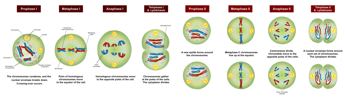
Meiosis I and II are each divided into prophase, metaphase, anaphase, and telophase stages, similar in purpose to their analogous subphases in the mitotic cell cycle. Therefore, meiosis includes the stages of meiosis I (prophase I, metaphase I, anaphase I, telophase I) and meiosis II (prophase II, metaphase II, anaphase II, telophase II).
Meiosis generates gamete genetic diversity in two ways: (1) Law of Independent Assortment. The independent orientation of homologous chromosome pairs along the metaphase plate during metaphase I & orientation of sister chromatids in metaphase II, this is the subsequent separation of homologs and sister chromatids during anaphase I & II, it allows a random and independent distribution of chromosomes to each daughter cell (and ultimately to gametes);[10] and (2) Crossing Over. The physical exchange of homologous chromosomal regions by homologous recombination during prophase I results in new combinations of DNA within chromosomes.[11]
During meiosis, specific genes are more highly transcribed.[12] In addition to strong meiotic stage-specific expression of mRNA, there are also pervasive translational controls (e.g. selective usage of preformed mRNA), regulating the ultimate meiotic stage-specific protein expression of genes during meiosis.[13] Thus, both transcriptional and translational controls determine the broad restructuring of meiotic cells needed to carry out meiosis.
Meiosis I
Meiosis I segregates homologous chromosomes, which are joined as tetrads (2n, 4c), producing two haploid cells (n chromosomes, 23 in humans) which each contain chromatid pairs (1n, 2c). Because the ploidy is reduced from diploid to haploid, meiosis I is referred to as a reductional division. Meiosis II is an equational division analogous to mitosis, in which the sister chromatids are segregated, creating four haploid daughter cells (1n, 1c).[14]
Prophase I
Prophase I is typically the longest phase of meiosis. During prophase I, homologous chromosomes pair and exchange DNA (homologous recombination). This often results in chromosomal crossover. This process is critical for pairing between homologous chromosomes and hence for accurate segregation of the chromosomes at the first meiosis division. The new combinations of DNA created during crossover are a significant source of genetic variation, and result in new combinations of alleles, which may be beneficial. The paired and replicated chromosomes are called bivalents or tetrads, which have two chromosomes and four chromatids, with one chromosome coming from each parent. The process of pairing the homologous chromosomes is called synapsis. At this stage, non-sister chromatids may cross-over at points called chiasmata (plural; singular chiasma).[15] Prophase I has historically been divided into a series of substages which are named according to the appearance of chromosomes.
Leptotene
The first stage of prophase I is the leptotene stage, also known as leptonema, from Greek words meaning "thin threads".[16]:27In this stage of prophase I, individual chromosomes—each consisting of two sister chromatids—become "individualized" to form visible strands within the nucleus.[16]:27[17]:353 The two sister chromatids closely associate and are visually indistinguishable from one another. During leptotene, lateral elements of the synaptonemal complex assemble. Leptotene is of very short duration and progressive condensation and coiling of chromosome fibers takes place.
Zygotene
The zygotene stage, also known as zygonema, from Greek words meaning "paired threads",[16]:27 occurs as the chromosomes approximately line up with each other into homologous chromosome pairs. In some organisms, this is called the bouquet stage because of the way the telomeres cluster at one end of the nucleus. At this stage, the synapsis (pairing/coming together) of homologous chromosomes takes place, facilitated by assembly of central element of the synaptonemal complex. Pairing is brought about in a zipper-like fashion and may start at the centromere (procentric), at the chromosome ends (proterminal), or at any other portion (intermediate). Individuals of a pair are equal in length and in position of the centromere. Thus pairing is highly specific and exact. The paired chromosomes are called bivalent or tetrad chromosomes.
Pachytene
The pachytene stage (/ˈpækɪtiːn/ PAK-i-teen), also known as pachynema, from Greek words meaning "thick threads",.[16]:27 At this point a tetrad of the chromosomes has formed known as a bivalent. This is the stage when homologous recombination, including chromosomal crossover (crossing over), occurs. Nonsister chromatids of homologous chromosomes may exchange segments over regions of homology. Sex chromosomes, however, are not wholly identical, and only exchange information over a small region of homology. At the sites where exchange happens, chiasmata form. The exchange of information between the non-sister chromatids results in a recombination of information; each chromosome has the complete set of information it had before, and there are no gaps formed as a result of the process. Because the chromosomes cannot be distinguished in the synaptonemal complex, the actual act of crossing over is not perceivable through the microscope, and chiasmata are not visible until the next stage.
Diplotene
During the diplotene stage, also known as diplonema, from Greek words meaning "two threads",[16]:30 the synaptonemal complex degrades and homologous chromosomes separate from one another a little. The chromosomes themselves uncoil a bit, allowing some transcription of DNA. However, the homologous chromosomes of each bivalent remain tightly bound at chiasmata, the regions where crossing-over occurred. The chiasmata remain on the chromosomes until they are severed at the transition to anaphase I.
In human fetal oogenesis, all developing oocytes develop to this stage and are arrested in prophase I before birth.[18] This suspended state is referred to as the dictyotene stage or dictyate. It lasts until meiosis is resumed to prepare the oocyte for ovulation, which happens at puberty or even later.
Diakinesis
Chromosomes condense further during the diakinesis stage, from Greek words meaning "moving through".[16]:30 This is the first point in meiosis where the four parts of the tetrads are actually visible. Sites of crossing over entangle together, effectively overlapping, making chiasmata clearly visible. Other than this observation, the rest of the stage closely resembles prometaphase of mitosis; the nucleoli disappear, the nuclear membrane disintegrates into vesicles, and the meiotic spindle begins to form.
Synchronous processes
During these stages, two centrosomes, containing a pair of centrioles in animal cells, migrate to the two poles of the cell. These centrosomes, which were duplicated during S-phase, function as microtubule organizing centers nucleating microtubules, which are essentially cellular ropes and poles. The microtubules invade the nuclear region after the nuclear envelope disintegrates, attaching to the chromosomes at the kinetochore. The kinetochore functions as a motor, pulling the chromosome along the attached microtubule toward the originating centrosome, like a train on a track. There are four kinetochores on each tetrad, but the pair of kinetochores on each sister chromatid fuses and functions as a unit during meiosis I.[19][20]
Microtubules that attach to the kinetochores are known as kinetochore microtubules. Other microtubules will interact with microtubules from the opposite centrosome: these are called nonkinetochore microtubules or polar microtubules. A third type of microtubules, the aster microtubules, radiates from the centrosome into the cytoplasm or contacts components of the membrane skeleton.
Metaphase I
Homologous pairs move together along the metaphase plate: As kinetochore microtubules from both centrosomes attach to their respective kinetochores, the paired homologous chromosomes align along an equatorial plane that bisects the spindle, due to continuous counterbalancing forces exerted on the bivalents by the microtubules emanating from the two kinetochores of homologous chromosomes. This attachment is referred to as a bipolar attachment. The physical basis of the independent assortment of chromosomes is the random orientation of each bivalent along the metaphase plate, with respect to the orientation of the other bivalents along the same equatorial line.[15] The protein complex cohesin holds sister chromatids together from the time of their replication until anaphase. In mitosis, the force of kinetochore microtubules pulling in opposite directions creates tension. The cell senses this tension and does not progress with anaphase until all the chromosomes are properly bi-oriented. In meiosis, establishing tension requires at least one crossover per chromosome pair in addition to cohesin between sister chromatids.
Anaphase I
Kinetochore microtubules shorten, pulling homologous chromosomes (which consist of a pair of sister chromatids) to opposite poles. Nonkinetochore microtubules lengthen, pushing the centrosomes farther apart. The cell elongates in preparation for division down the center.[15] Unlike in mitosis, only the cohesin from the chromosome arms is degraded while the cohesin surrounding the centromere remains protected. This allows the sister chromatids to remain together while homologs are segregated.
Telophase I
The first meiotic division effectively ends when the chromosomes arrive at the poles. Each daughter cell now has half the number of chromosomes but each chromosome consists of a pair of chromatids. The microtubules that make up the spindle network disappear, and a new nuclear membrane surrounds each haploid set. The chromosomes uncoil back into chromatin. Cytokinesis, the pinching of the cell membrane in animal cells or the formation of the cell wall in plant cells, occurs, completing the creation of two daughter cells. Sister chromatids remain attached during telophase I.
Cells may enter a period of rest known as interkinesis or interphase II. No DNA replication occurs during this stage.
Meiosis II
Meiosis II is the second meiotic division, and usually involves equational segregation, or separation of sister chromatids. Mechanically, the process is similar to mitosis, though its genetic results are fundamentally different. The end result is production of four haploid cells (n chromosomes, 23 in humans) from the two haploid cells (with n chromosomes, each consisting of two sister chromatids) produced in meiosis I. The four main steps of meiosis II are: prophase II, metaphase II, anaphase II, and telophase II.
In prophase II we see the disappearance of the nucleoli and the nuclear envelope again as well as the shortening and thickening of the chromatids. Centrosomes move to the polar regions and arrange spindle fibers for the second meiotic division.
In metaphase II, the centromeres contain two kinetochores that attach to spindle fibers from the centrosomes at opposite poles. The new equatorial metaphase plate is rotated by 90 degrees when compared to meiosis I, perpendicular to the previous plate.{{[21]}}
This is followed by anaphase II, in which the remaining centromeric cohesin is cleaved allowing the sister chromatids to segregate. The sister chromatids by convention are now called sister chromosomes as they move toward opposing poles.
The process ends with telophase II, which is similar to telophase I, and is marked by decondensation and lengthening of the chromosomes and the disassembly of the spindle. Nuclear envelopes reform and cleavage or cell plate formation eventually produces a total of four daughter cells, each with a haploid set of chromosomes.
Meiosis is now complete and ends up with four new daughter cells.
Origin and function
The origin and function of meiosis are fundamental to understanding the evolution of sexual reproduction in eukaryotes. There is no current consensus among biologists on the questions of how sex in eukaryotes arose in evolution, what basic function sexual reproduction serves, and why it is maintained, given the basic two-fold cost of sex. It is clear that it evolved over 1.2 billion years ago, and that almost all species which are descendants of the original sexually reproducing species are still sexual reproducers, including plants, fungi, and animals.
Meiosis is a key event of the sexual cycle in eukaryotes. It is the stage of the life cycle when a cell gives rise to two haploid cells (gametes) each having half as many chromosomes. Two such haploid gametes, arising from different individual organisms, fuse by the process of fertilization, thus completing the sexual cycle.
Meiosis is ubiquitous among eukaryotes. It occurs in single-celled organisms such as yeast, as well as in multicellular organisms, such as humans. Eukaryotes arose from prokaryotes more than 2.2 billion years ago[22] and the earliest eukaryotes were likely single-celled organisms. To understand sex in eukaryotes, it is necessary to understand (1) how meiosis arose in single celled eukaryotes, and (2) the function of meiosis.
Occurrence
In life cycles
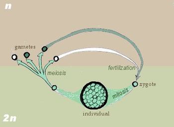
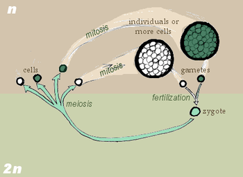
Meiosis occurs in eukaryotic life cycles involving sexual reproduction, consisting of the constant cyclical process of meiosis and fertilization. This takes place alongside normal mitotic cell division. In multicellular organisms, there is an intermediary step between the diploid and haploid transition where the organism grows. At certain stages of the life cycle, germ cells produce gametes. Somatic cells make up the body of the organism and are not involved in gamete production.
Cycling meiosis and fertilization events produces a series of transitions back and forth between alternating haploid and diploid states. The organism phase of the life cycle can occur either during the diploid state (diplontic life cycle), during the haploid state (haplontic life cycle), or both (haplodiplontic life cycle, in which there are two distinct organism phases, one during the haploid state and the other during the diploid state). In this sense there are three types of life cycles that utilize sexual reproduction, differentiated by the location of the organism phase(s).
In the diplontic life cycle (with pre-gametic meiosis), of which humans are a part, the organism is diploid, grown from a diploid cell called the zygote. The organism's diploid germ-line stem cells undergo meiosis to create haploid gametes (the spermatozoa for males and ova for females), which fertilize to form the zygote. The diploid zygote undergoes repeated cellular division by mitosis to grow into the organism.
In the haplontic life cycle (with post-zygotic meiosis), the organism is haploid instead, spawned by the proliferation and differentiation of a single haploid cell called the gamete. Two organisms of opposing sex contribute their haploid gametes to form a diploid zygote. The zygote undergoes meiosis immediately, creating four haploid cells. These cells undergo mitosis to create the organism. Many fungi and many protozoa utilize the haplontic life cycle.
Finally, in the haplodiplontic life cycle (with sporic or intermediate meiosis), the living organism alternates between haploid and diploid states. Consequently, this cycle is also known as the alternation of generations. The diploid organism's germ-line cells undergo meiosis to produce spores. The spores proliferate by mitosis, growing into a haploid organism. The haploid organism's gamete then combines with another haploid organism's gamete, creating the zygote. The zygote undergoes repeated mitosis and differentiation to become a diploid organism again. The haplodiplontic life cycle can be considered a fusion of the diplontic and haplontic life cycles.[23]
In plants and animals
Meiosis occurs in all animals and plants. The end result, the production of gametes with half the number of chromosomes as the parent cell, is the same, but the detailed process is different. In animals, meiosis produces gametes directly. In land plants and some algae, there is an alternation of generations such that meiosis in the diploid sporophyte generation produces haploid spores. These spores multiply by mitosis, developing into the haploid gametophyte generation, which then gives rise to gametes directly (i.e. without further meiosis). In both animals and plants, the final stage is for the gametes to fuse, restoring the original number of chromosomes.[24]
In mammals
In females, meiosis occurs in cells known as oocytes (singular: oocyte). Each primary oocyte divides twice in meiosis, unequally in each case. The first division produces a daughter cell, and a much smaller polar body which may or may not undergo a second division. In meiosis II, division of the daughter cell produces a second polar body, and a single haploid cell, which enlarges to become an ovum. Therefore, in females each primary oocyte that undergoes meiosis results in one mature ovum and one or two polar bodies.
Note that there are pauses during meiosis in females. Maturing oocytes are arrested in prophase I of meiosis I and lie dormant within a protective shell of somatic cells called the follicle. At the beginning of each menstrual cycle, FSH secretion from the anterior pituitary stimulates a few follicles to mature in a process known as folliculogenesis. During this process, the maturing oocytes resume meiosis and continue until metaphase II of meiosis II, where they are again arrested just before ovulation. If these oocytes are fertilized by sperm, they will resume and complete meiosis. During folliculogenesis in humans, usually one follicle becomes dominant while the others undergo atresia. The process of meiosis in females occurs during oogenesis, and differs from the typical meiosis in that it features a long period of meiotic arrest known as the dictyate stage and lacks the assistance of centrosomes.[25][26]
In males, meiosis occurs during spermatogenesis in the seminiferous tubules of the testicles. Meiosis during spermatogenesis is specific to a type of cell called spermatocytes, which will later mature to become spermatozoa. Meiosis of primordial germ cells happens at the time of puberty, much later than in females. Tissues of the male testis suppress meiosis by degrading retinoic acid, a stimulator of meiosis. This is overcome at puberty when cells within seminiferous tubules called Sertoli cells start making their own retinoic acid. Sensitivity to retinoic acid is also adjusted by proteins called nanos and DAZL.
In female mammals, meiosis begins immediately after primordial germ cells migrate to the ovary in the embryo. It is retinoic acid, derived from the primitive kidney (mesonephros) that stimulates meiosis in ovarian oogonia. Tissues of the male testis suppress meiosis by degrading retinoic acid, a stimulator of meiosis. This is overcome at puberty when cells within seminiferous tubules called Sertoli cells start making their own retinoic acid.[27][28]
Variations
Nondisjunction
The normal separation of chromosomes in meiosis I or sister chromatids in meiosis II is termed disjunction. When the segregation is not normal, it is called nondisjunction. This results in the production of gametes which have either too many or too few of a particular chromosome, and is a common mechanism for trisomy or monosomy. Nondisjunction can occur in the meiosis I or meiosis II, phases of cellular reproduction, or during mitosis.
Most monosomic and trisomic human embryos are not viable, but some aneuploidies can be tolerated, such as trisomy for the smallest chromosome, chromosome 21. Phenotypes of these aneuploidies range from severe developmental disorders to asymptomatic. Medical conditions include but are not limited to:
- Down syndrome – trisomy of chromosome 21
- Patau syndrome – trisomy of chromosome 13
- Edwards syndrome – trisomy of chromosome 18
- Klinefelter syndrome – extra X chromosomes in males – i.e. XXY, XXXY, XXXXY, etc.
- Turner syndrome – lacking of one X chromosome in females – i.e. X0
- Triple X syndrome – an extra X chromosome in females
- XYY syndrome – an extra Y chromosome in males.
The probability of nondisjunction in human oocytes increases with increasing maternal age,[29] presumably due to loss of cohesin over time.[30]
Other
Alongside with the variations of meiosis related to the moment when meiosis occur in life cycles, resulting in post-zygotic, pre-gametic and intermediate meiosis (see above), the number of nuclear divisions in meiosis is also variable. The majority of eukaryotes have a two-divisional meiosis (though sometimes achiasmatic), but a very rare form, one-divisional meiosis, occurs in some flagellates (parabasalids and oxymonads) from the gut of the wood-feeding cockroach Cryptocercus.[31]
Comparison to mitosis
In order to understand meiosis, a comparison to mitosis is helpful. The table below shows the differences between meiosis and mitosis.[32]
| Meiosis | Mitosis | |
|---|---|---|
| End result | Normally four cells, each with half the number of chromosomes as the parent | Two cells, having the same number of chromosomes as the parent |
| Function | Production of gametes (sex cells) in sexually reproducing eukaryotes with diplont life cycle | Cellular reproduction, growth, repair, asexual reproduction |
| Where does it happen? | Almost all eukaryotes (animals, plants, fungi, and protists);[33][31] In gonads, before gametes (in diplontic life cycles); After zygotes (in haplontic); Before spores (in haplodiplontic) | All proliferating cells in all eukaryotes |
| Steps | Prophase I, Metaphase I, Anaphase I, Telophase I, Prophase II, Metaphase II, Anaphase II, Telophase II | Prophase, Prometaphase, Metaphase, Anaphase, Telophase |
| Genetically same as parent? | No | Yes |
| Crossing over happens? | Yes, normally occurs between each pair of homologous chromosomes | Very rarely |
| Pairing of homologous chromosomes? | Yes | No |
| Cytokinesis | Occurs in Telophase I and Telophase II | Occurs in Telophase |
| Centromeres split | Does not occur in Anaphase I, but occurs in Anaphase II | Occurs in Anaphase |
See also
References
- 1 2 Freeman, Scott (2011). Biological Science (6th ed.). Hoboken, NY: Pearson. p. 210.
- ↑ Letunic, I; Bork, P (2006). "Interactive Tree of Life". Retrieved 23 July 2011.
- ↑ Bernstein H, Bernstein C, Michod RE (2011). “Meiosis as an evolutionary adaptation for DNA repair." In “DNA Repair", Intech Publ (Inna Kruman, editor), Chapter 19: 357-382 DOI: 10.5772/1751 ISBN 978-953-307-697-3 Available online from: http://www.intechopen.com/books/dna-repair/meiosis-as-an-evolutionary-adaptation-for-dna-repair
- ↑ Bernstein H, Bernstein C (2010). "Evolutionary origin of recombination during meiosis". BioScience. 60 (7): 498–505. doi:10.1525/bio.2010.60.7.5.
- ↑ LODÉ T (2011). "Sex is not a solution for reproduction: the libertine bubble theory". BioEssays. 33 (6): 419–422. doi:10.1002/bies.201000125. PMID 21472739.
- ↑ Hassold, Terry; Hunt, Patricia (1 April 2001). "To err (meiotically) is human: the genesis of human aneuploidy". Nature Reviews Genetics. 2 (4): 280–291. doi:10.1038/35066065. PMID 11283700.
- ↑ J.B. Farmer and J.E.S. Moore, Quarterly Journal of Microscopic Science 48:489 (1905) as quoted in the Oxford English Dictionary, Third Edition, June 2001, s.v.
- ↑ Battaglia E. (1985). Meiosis and mitosis: a terminological criticism. Ann Bot (Rome) 43: 101–140. link.
- ↑ "Mitosis". 2012-10-27. Retrieved 2018-02-09.
- ↑ Monaghan, Floyd; Corcos, Alain (1984-01-01). "On the origins of the Mendelian laws". Journal of Heredity. 75 (1): 67–69. doi:10.1093/oxfordjournals.jhered.a109868. ISSN 0022-1503.
- ↑ Saleem, Muhammad (2001). "Inherited Differences in Crossing Over and Gene Conversion Frequencies Between Wild Strains of Sordaria fimicola From "Evolution Canyon"". Genetics. 159.
- ↑ Zhou, A.; Pawlowski, W.P. (August 2014). "Regulation of meiotic gene expression in plants". Frontiers in Plant Science. 5: Article 413. doi:10.3389/fpls.2014.00413. PMC 4142721. PMID 25202317.

- ↑ Brar GA, Yassour M, Friedman N, Regev A, Ingolia NT, Weissman JS (February 2012). "High-resolution view of the yeast meiotic program revealed by ribosome profiling". Science. 335 (6068): 552–7. doi:10.1126/science.1215110. PMC 3414261. PMID 22194413.
- ↑ Freeman 2005, pp. 244–45
- 1 2 3 Freeman 2005, pp. 249–250
- 1 2 3 4 5 6 Snustad, DP; Simmons, MJ (December 2008). Principles of Genetics (5th ed.). Wiley. ISBN 978-0-470-38825-9.
- ↑ Krebs, JE; Goldstein, ES; Kilpatrick, ST (November 2009). Lewin's Genes X (10th ed.). Jones & Barlett Learning. ISBN 978-0-7637-6632-0.
- ↑ 1950-, Nussbaum, Robert L.,. Thompson & Thompson genetics in medicine. McInnes, Roderick R.,, Willard, Huntington F.,, Hamosh, Ada,, Preceded by: Nussbaum, Robert L., 1950- (Eighth ed.). Philadelphia, PA. p. 19. ISBN 1437706967. OCLC 908336124.
- ↑ Raven, Peter H.; Johnson, George B.; Mason, Kenneth A.; Losos, Jonathan & Singer, Susan. Biology, 8th ed. McGraw-Hill 2007.
- ↑ Petronczki M, Siomos MF, Nasmyth K (February 2003). "Un ménage à quatre: the molecular biology of chromosome segregation in meiosis". Cell. 112 (4): 423–40. doi:10.1016/S0092-8674(03)00083-7. PMID 12600308.
- ↑ http://www.phschool.com/science/biology_place/biocoach/meiosis/metaii.html. Missing or empty
|title=(help) - ↑ Retallack GJ, Krull ES, Thackeray GD, Parkinson D (2013-09-01). "Problematic urn-shaped fossils from a Paleoproterozoic (2.2 Ga) paleosol in South Africa". Precambrian Research. 235: 71–87. Bibcode:2013PreR..235...71R. doi:10.1016/j.precamres.2013.05.015.
- ↑ South, G. R.; Whittick, A. (2009-07-08). An Introduction to Phycology. John Wiley & Sons. ISBN 9781444314205.
- ↑ Bidlack, James E. (2011). Introductory Plant Biology. New York, NY: McGraw HIll. pp. 214–29.
- ↑ Brunet, S.; Verlhac, M. H. (2010). "Positioning to get out of meiosis: The asymmetry of division". Human Reproduction Update. 17 (1): 68–75. doi:10.1093/humupd/dmq044. PMID 20833637.
- ↑ Rosenbusch B (November 2006). "The contradictory information on the distribution of non-disjunction and pre-division in female gametes". Hum. Reprod. 21 (11): 2739–42. doi:10.1093/humrep/del122. PMID 16982661.
- ↑ Lin Y, Gill ME, Koubova J, Page DC (December 2008). "Germ cell-intrinsic and -extrinsic factors govern meiotic initiation in mouse embryos". Science. 322 (5908): 1685–7. doi:10.1126/science.1166340. PMID 19074348.
- ↑ Suzuki A, Saga Y (February 2008). "Nanos2 suppresses meiosis and promotes male germ cell differentiation". Genes Dev. 22 (4): 430–5. doi:10.1101/gad.1612708. PMC 2238665. PMID 18281459.
- ↑ Hassold, T.; Jacobs, P.; Kline, J.; Stein, Z.; Warburton, D. (July 1980). "Effect of maternal age on autosomal trisomies". Annals of Human Genetics. 44 (1): 29–36. doi:10.1111/j.1469-1809.1980.tb00943.x. PMID 7198887.
- ↑ Tsutsumi, M.; Fujiwara, R.; Nishizawa, H.; Ito, M.; Kogo, H.; Inagaki, H.; Ohye, T.; Kato, T.; Fujii, T.; Kurahashi, H. (May 2014). "Age-related decrease of meiotic cohesins in human oocytes". PLOS ONE. 9 (5): Article e96710. doi:10.1371/journal.pone.0096710. PMC 4013030. PMID 24806359.
- 1 2 Raikov, I. B. (1995). "Meiosis in protists: recent advances and persisting problems". Europ J Protistol. 31: 1–7. doi:10.1016/s0932-4739(11)80349-4.
- ↑ "How Cells Divide". PBS. Public Broadcasting Service. Retrieved 6 December 2012.
- ↑ Heywood, P.; Magee, P.T. (1976). "Meiosis in protists. Some structural and physiological aspects of meiosis in algae, fungi, and protozoa". Bacteriological Reviews. 40 (1): 190–240. PMC 413949. PMID 773364.
Cited texts
- Freeman, Scott (2005). Biological Science (3rd ed.). Upper Saddle River, NJ: Pearson Prentice Hall.
External links
| Wikimedia Commons has media related to Meiosis. |
- Meiosis Flash Animation
- Animations from the U. of Arizona Biology Dept.
- Meiosis at Kimball's Biology Pages
- Khan Academy, video lecture
- CCO The Cell-Cycle Ontology
- Stages of Meiosis animation
- *"Abby Dernburg Seminar: Chromosome Dynamics During Meiosis"
| Biological terms | |
|---|---|
| Sexual reproduction | |
| Sexuality | |
| |