Cell division
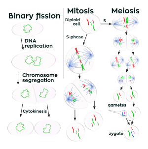
Cell division is the process by which a parent cell divides into two or more daughter cells.[1] Cell division usually occurs as part of a larger cell cycle. In eukaryotes, there are two distinct types of cell division: a vegetative division, whereby each daughter cell is genetically identical to the parent cell (mitosis),[2] and a reproductive cell division, whereby the number of chromosomes in the daughter cells is reduced by half to produce haploid gametes (meiosis). Meiosis results in four haploid daughter cells by undergoing one round of DNA replication followed by two divisions. Homologous chromosomes are separated in the first division, and sister chromatids are separated in the second division. Both of these cell division cycles are used in the process of sexual reproduction at some point in their life cycle. Both are believed to be present in the last eukaryotic common ancestor.
Prokaryotes (bacteria) undergo a vegetative cell division known as binary fission, where their genetic material is segregated equally into two daughter cells. All cell divisions, regardless of organism, are preceded by a single round of DNA replication.
For simple unicellular microorganisms such as the amoeba, one cell division is equivalent to reproduction – an entire new organism is created. On a larger scale, mitotic cell division can create progeny from multicellular organisms, such as plants that grow from cuttings. Mitotic cell division enables sexually reproducing organisms to develop from the one-celled zygote, which itself was produced by meiotic cell division from gametes. After growth, cell division by mitosis allows for continual construction and repair of the organism.[3] The human body experiences about 10 quadrillion cell divisions in a lifetime.[4]
The primary concern of cell division is the maintenance of the original cell's genome. Before division can occur, the genomic information that is stored in chromosomes must be replicated, and the duplicated genome must be separated cleanly between cells.[5] A great deal of cellular infrastructure is involved in keeping genomic information consistent between generations.
Phases of cell division
Interphase
Interphase is the process a cell must go through before mitosis, meiosis, and cytokinesis.[6] Interphase consists of four main stages: G1, S, G0, and G2. G1 is a time of growth for the cell. If the cell does not progress through G1, the cell then enters a stage called G0. In G0, cells are still living but they are put on hold. The cells may later be called back into interphase if needed at a later time. There are checkpoints during interphase that allow the cell to be either progressed or denied further development. In S phase, the chromosomes are replicated in order for the genetic content to be maintained. During G2, the cell undergoes the final stages of growth before it enters the M phase. The M phase, can be either mitosis or meiosis depending on the type of cell. Germ cells undergo meiosis, while somatic cells will undergo mitosis. After the cell proceeds successfully through the M phase, it may then undergo cell division through cytokinesis. The control of each checkpoint is controlled by cyclin and cyclin dependent kinases. The progression of interphase is the result of the increased amount of cyclin. As the amount of cyclin increases, more and more cyclin dependent kinases attach to cyclin signaling the cell further into interphase. The peak of the cyclin attached to the cyclin dependent kinases this system pushes the cell out of interphase and into the M phase, where mitosis, meiosis, and cytokinesis occur.
Prophase
Prophase is the first stage of division. The nuclear envelope is broken down, long strands of chromatin condense to form shorter more visible strands called chromosomes, the nucleolus disappears, and microtubules attach to the chromosomes at the kinetochores present in the centromere.[7] Microtubules associated with the alignment and separation of chromosomes are referred to as the spindle and spindle fibers. Chromosomes will also be visible under a microscope and will be connected at the centromere. During this condensation and alignment period, homologous chromosomes may swap portions of their DNA in a process known as crossing over.
Metaphase
Metaphase is the stage in cell division when the chromosomes line up in the middle of the cell by MTOCs ( microtubule organizing center) by pushing and pulling on centromeres of both chromatids which causes the chromosome to move to the center. The chromosomes are still condensing and are currently at one step away from being the most coiled and condensed they will be.[8] spindle fibre and have already connected to the kinetochores. At this point, the chromosomes are ready to split into opposite poles of the cell towards the spindle to which they are connected. [9]
Anaphase
Anaphase is a very short stage of the cell cycle and occurs after the chromosomes align at the mitotic plate. After the chromosomes line up in the middle of the cell, the spindle fibers will pull them apart. The chromosomes are split apart as the sister chromatids move to opposite sides of the cell.[10]
Telophase
Telophase is the last stage of the cell cycle. Two cells form around the chromatin at the two poles of the cell. Two nuclear membranes begin to reform and the chromatin begin to unwind.[11]
Variants
Cells are broadly classified into two main categories: simple, non-nucleated prokaryotic cells, and complex, nucleated eukaryotic cells. Owing to their structural differences, eukaryotic and prokaryotic cells do not divide in the same way. Also, the pattern of cell division that transforms eukaryotic stem cells into gametes (sperm cells in males or egg cells in females), termed meiosis, is different from that of the division of somatic cells in the body.
Degradation
Multicellular organisms replace worn-out cells through cell division. In some animals, however, cell division eventually halts. In humans this occurs, on average, after 52 divisions, known as the Hayflick limit. The cell is then referred to as senescent. Cells stop dividing because the telomeres, protective bits of DNA on the end of a chromosome required for replication, shorten with each copy, eventually being consumed. Cancer cells, on the other hand, are not thought to degrade in this way, if at all. An enzyme called telomerase, present in large quantities in cancerous cells, rebuilds the telomeres, allowing division to continue indefinitely.
History
.jpg)
A cell division under microscope was first discovered by German botanist Hugo von Mohl in 1835 as he worked over Green algae Cladophora glomerata.[13]
In 1943, cell division was filmed for the first time,[14] by Kurt Michelwith, using a phase-contrast microscope.[15]
See also
- Binary fission
- Cell growth
- Labile cells, cells that constantly divide
- Klerokinesis
References
- ↑ Robert.S Hine, ed. (2008). Oxford Dictionary Biology (6th ed.). New York: Oxford University Press. p. 113. ISBN 978-0-19-920462-5.
- ↑ Griffiths, Anthony J.F.; Wessler, Susan R.; Carroll, Sean B.; Doebley, John (2012). Introduction to Genetic Analysis (10 ed.). New York: W.H. Freeman and Company. p. 35. ISBN 978-1-4292-2943-2.
- ↑ Maton, Anthea (1997). Cells: Building Blocks of Life. New Jersey: Prentice Hall. pp. 70–74. ISBN 0-13-423476-6.
- ↑ Quammen, David (April 2008). "Contagious cancer: The evolution of a killer". Harper's. 316 (1895): 42. Retrieved 24 September 2012.
- ↑ Krylov, Mikhail C. (2010). Cell Division: Theory, Variants, and Degradation. New York: Nova Science Publishers, Inc. p. 137. ISBN 9781608769865.
- ↑ Marieb, Elaine (2000). Essentials of human anatomy and physiology. San Francisco: Benjamin Cummings. ISBN 0-8053-4940-5.
- ↑ Schermelleh, Lothar; Carlton, Peter M.; Haase, Sebastian; Shao, Lin; Winoto, Lukman; Kner, Peter; Burke, Brian; Cardoso, M. Cristin; Agard, David A. (2008-06-06). "Subdiffraction Multicolor Imaging of the Nuclear Periphery with 3D Structured Illumination Microscopy". Science. 320 (5881): 1332–1336. doi:10.1126/science.1156947. ISSN 0036-8075. PMC 2916659. PMID 18535242.
- ↑ "Researchers Shed Light On Shrinking Of Chromosomes". ScienceDaily. June 12, 2007. Retrieved 2017-02-02.
- ↑ Elrod, Susan (2002). Schaum's Outline of Genetics (Fifth ed.). United States of America: McGraw-Hill Companies,Inc. p. 8. ISBN 9780071625036.
- ↑ "The Cell Cycle". www.biology-pages.info. Retrieved 2017-02-02.
- ↑ Hetzer, Martin W. (2017-02-02). "The Nuclear Envelope". Cold Spring Harbor Perspectives in Biology. 2 (3). doi:10.1101/cshperspect.a000539. ISSN 1943-0264. PMC 2829960. PMID 20300205.
- ↑ Phase Holographic Imaging. Cell Division
- ↑ Karl Mägdefrau (1994), "Mohl, Hugo von", Neue Deutsche Biographie (NDB) (in German), 17, Berlin: Duncker & Humblot, pp. 690–691 ; (full text online)
- ↑ Masters, Barry R (2008). "History of the Optical Microscope in Cell Biology and Medicine" (PDF). eLS (formerly Encyclopedia of Life Sciences). John Wiley & Sons, Ltd. p. 3. doi:10.1002/9780470015902.a0003082. ISBN 9780470015902.
- ↑ Video on YouTube
Further reading
- Morgan HI. (2007). "The Cell Cycle: Principles of Control" London: New Science Press.
- J.M.Turner Fetus into Man (1978, 1989). Harvard University Press. ISBN 0-674-30692-9
- Cell division: binary fission and mitosis
External links
| Wikimedia Commons has media related to Cell division. |
- How Cells Divide: Mitosis vs. Meiosis
- The Mitosis and Cell Cycle Control Section from the Landmark Papers in Cell Biology (Gall JG, McIntosh JR, eds.) contains commentaries on and links to seminal research papers on mitosis and cell division. Published online in the Image & Video Library of The American Society for Cell Biology
- The Image & Video Library of The American Society for Cell Biology contains many videos showing the cell division.
- The Cell Division of the Cell Image Library
- Videos of the first cell divisions in Xenopus laevis embryos (side view and top view), acquired by MRI (DOI of paper)
- Images : Calanthe discolor Lindl. - Flavon's Secret Flower Garden
- Tyson's model of cell division and a Description on BioModels Database
- WormWeb.org: Interactive Visualization of the C. elegans Cell Lineage - Visualize the entire set of cell divisions of the nematode C. elegans