Cerebral aqueduct
| Cerebral aqueduct | |
|---|---|
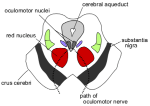 Section through superior colliculus showing path of oculomotor nerve. | |
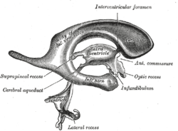 Drawing of a cast of the ventricular cavities, viewed from the side. | |
| Details | |
| Identifiers | |
| Latin |
aqueductus mesencephali (cerebri). aqueductus Sylvii |
| MeSH | D002535 |
| NeuroNames | 509 |
| NeuroLex ID | birnlex_1261 |
| FMA | 78467 |
| Anatomical terms of neuroanatomy | |
The cerebral aqueduct, also known as the aqueductus mesencephali, mesencephalic duct, sylvian aqueduct, or aqueduct of Sylvius, is within the mesencephalon (or midbrain), contains cerebrospinal fluid (CSF), and connects the third ventricle in the diencephalon to the fourth ventricle within the region of the mesencephalon and metencephalon, located dorsal to the pons and ventral to the cerebellum.
Structure
Development
The cerebral aqueduct, as other parts of the ventricular system of the brain, develops from the central canal of the neural tube, and it originates from the portion of the neural tube that is present in the developing mesencephalon, hence the name "mesencephalic duct."[1]
Function
The aqueduct functions to connect the third and fourth ventricles and to ensure the flow of cerebrospinal fluid through these areas.
Clinical significance
Aqueductal stenosis, a narrowing of the cerebral aqueduct, obstructs the flow of CSF and has been associated with non-communicating hydrocephalus. Such narrowing can be congenital, arise via tumor compression, or through cyclical gliosis secondary to an initial partial obstruction.
Additional images
 Transverse section through mid-brain; number 2 indicates the cerebral aqueduct.
 Transverse section of mid-brain at level of inferior colliculi.
 Transverse section of mid-brain at level of superior colliculi.
 MRI section of mid-brain.
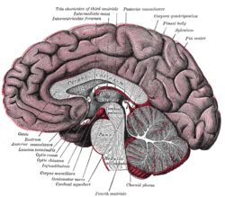 Median sagittal section of brain.
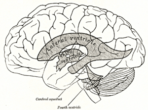 Scheme showing relations of the ventricles to the surface of the brain.
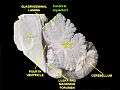 Cerebral aqueduct
 Cerebral aqueduct
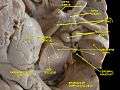 Cerebral peduncle, optic chasm, cerebral aqueduct. Inferior view. Deep dissection.
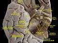 Cerebral peduncle, optic chasm, cerebral aqueduct. Inferior view. Deep dissection.
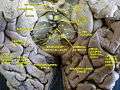 Cerebrum. Inferior view.Deep dissection
See also
References
- ↑ Le, Tao; Bhushan, Vikas; Vasan, Neil (2010). First Aid for the USMLE Step 1: 2010 20th Anniversary Edition. USA: The McGraw-Hill Companies, Inc. p. 126. ISBN 978-0-07-163340-6.
External links
| Wikimedia Commons has media related to Cerebral aqueduct. |
- Atlas image: n2a3p2 at the University of Michigan Health System