Heterotrimeric G protein
| Heterotrimeric G-protein GTPase | |||||||||
|---|---|---|---|---|---|---|---|---|---|
| Identifiers | |||||||||
| EC number | 3.6.5.1 | ||||||||
| CAS number | 9059-32-9 | ||||||||
| Databases | |||||||||
| IntEnz | IntEnz view | ||||||||
| BRENDA | BRENDA entry | ||||||||
| ExPASy | NiceZyme view | ||||||||
| KEGG | KEGG entry | ||||||||
| MetaCyc | metabolic pathway | ||||||||
| PRIAM | profile | ||||||||
| PDB structures | RCSB PDB PDBe PDBsum | ||||||||
| Gene Ontology | AmiGO / QuickGO | ||||||||
| |||||||||
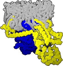
"G protein" usually refers to the membrane-associated heterotrimeric G proteins, sometimes referred to as the "large" G proteins (as opposed to the subclass of smaller, monomeric small GTPases) . These proteins are activated by G protein-coupled receptors and are made up of alpha (α), beta (β) and gamma (γ) subunits,[1] the latter two referred to as the beta-gamma complex.
There are four main families of G proteins: Gi/Go, Gq, Gs, and G12.[2]
Alpha subunits
Reconstitution experiments carried out in the early 1980s showed that purified Gα subunits can directly activate effector enzymes. The GTP form of the α subunit of transducin (Gt) activates the cyclic GMP phosphodiesterase from retinal rod outer segments,[3] and the GTP form of the α subunit of the stimulatory G protein (Gs) activates hormone-sensitive adenylate cyclase.[4][5]
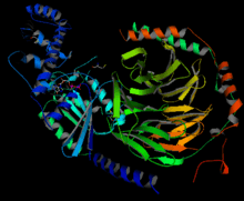
Gα subunits consist of two domains, the GTPase domain, and the alpha-helical domain.
There exist at least 20 different Gα subunits, which are separated into four main families. This nomenclature is based on their sequence homologies:[6]
| G-protein-family | α-subunit | Gene | Signal transduction | Use/Receptors (examples) | Effects (examples) |
|---|---|---|---|---|---|
| Gi-family | |||||
| Gi/o | αi, αo | GNAO1, GNAI1, GNAI2, GNAI3 | Inhibition of adenylate cyclase, opens K+-channels (via β/γ subunits), closes Ca2+-channels | Muscarinic M2 and M4,[7] chemokine receptors, α2-Adrenoreceptors, Serotonin 5-HT1 receptors, Histamine H3 and H4, Dopamine D2-like receptors | Smooth muscle contraction, depress neuronal activity |
| Gt | αt (Transducin) | GNAT1, GNAT2 | Activation of phosphodiesterase 6 | Rhodopsin | Vision |
| Ggust | αgust (Gustducin) | GNAT3 | Activation of phosphodiesterase 6 | Taste receptors | Taste |
| Gz | αz | GNAZ | Inhibition of adenylate cyclase | Platelets | Maintaining the ionic balance of perilymphatic and endolymphatic cochlear fluids. |
| Gs-family | |||||
| Gs | αs | GNAS | Activation of adenylate cyclase | Beta-adrenoreceptors; Serotonin 5-HT4, 5-HT6 and 5-HT7; Dopamine D1-like receptors, Histamine H2 | Increase heart rate, Smooth muscle relaxation, stimulate neuronal activity |
| Golf | αolf | GNAL | Activation of adenylate cyclase | olfactory receptors | Smell |
| Gq-family | |||||
| Gq | αq, α11, α14, α15, α16 | GNAQ, GNA11, GNA14, GNA15 | Activation of phospholipase C | α1-Adrenoreceptors, Muscarinic M1, M3, and M5,[7] Histamine H1, Serotonin 5-HT2 receptors | Smooth muscle contraction, Ca2+ flux |
| G12/13-family | |||||
| G12/13 | α12, α13 | GNA12, GNA13 | Activation of the Rho family of GTPases | Cytoskeletal functions, Smooth muscle contraction | |
G beta-gamma complex
The β and γ subunits are closely bound to one another and are referred to as the G beta-gamma complex. Upon activation of the GPCR, the Gβγ complex is released from the Gα subunit after its GDP-GTP exchange.
Function
The free Gβγ complex can act as a signaling molecule itself, by activating other second messengers or by gating ion channels directly.
For example, the Gβγ complex, when bound to histamine receptors, can activate phospholipase A2. Gβγ complexes bound to muscarinic acetylcholine receptors, on the other hand, directly open G protein-coupled inward rectifying potassium channels (GIRKs)[8]. They can also activate L-type calcium channels, as in H3 receptor pharmacology.
References
- ↑ Hurowitz EH, Melnyk JM, Chen YJ, Kouros-Mehr H, Simon MI, Shizuya H (April 2000). "Genomic characterization of the human heterotrimeric G protein alpha, beta, and gamma subunit genes". DNA Research. 7 (2): 111–20. doi:10.1093/dnares/7.2.111. PMID 10819326.
- ↑ Nature Reviews Drug Discovery GPCR Questionnaire Participants (July 2004). "The state of GPCR research in 2004". Nature Reviews. Drug Discovery (3 ed.). 3 (7): 575, 577–626. doi:10.1038/nrd1458. PMID 15272499.
- ↑ Fung BK, Hurley JB, Stryer L (January 1981). "Flow of information in the light-triggered cyclic nucleotide cascade of vision". Proceedings of the National Academy of Sciences of the United States of America. 78 (1): 152–6. doi:10.1073/pnas.78.1.152. PMC 319009. PMID 6264430.
- ↑ Cerione RA, Sibley DR, Codina J, Benovic JL, Winslow J, Neer EJ, Birnbaumer L, Caron MG, Lefkowitz RJ, et al. (August 1984). "Reconstitution of a hormone-sensitive adenylate cyclase system. The pure beta-adrenergic receptor and guanine nucleotide regulatory protein confer hormone responsiveness on the resolved catalytic unit". The Journal of Biological Chemistry. 259 (16): 9979–82. PMID 6088509.
- ↑ May DC, Ross EM, Gilman AG, Smigel MD (December 1985). "Reconstitution of catecholamine-stimulated adenylate cyclase activity using three purified proteins". The Journal of Biological Chemistry. 260 (29): 15829–33. PMID 2999139.
- ↑ Strathmann MP, Simon MI (July 1991). "G alpha 12 and G alpha 13 subunits define a fourth class of G protein alpha subunits". Proceedings of the National Academy of Sciences of the United States of America. 88 (13): 5582–6. doi:10.1073/pnas.88.13.5582. PMC 51921. PMID 1905812.
- 1 2 Qin K, Dong C, Wu G, Lambert NA (August 2011). "Inactive-state preassembly of G(q)-coupled receptors and G(q) heterotrimers". Nature Chemical Biology. 7 (10): 740–7. doi:10.1038/nchembio.642. PMC 3177959. PMID 21873996.
- ↑ Gulati S, Jin H, Masuho I, Orban T, Cai Y, Pardon E, Martemyanov KA, Kiser PD, Stewart PL, Ford CP, Steyaert J, Palczewski K (2018). "Targeting G protein-coupled receptor signaling at the G protein level with a selective nanobody inhibitor". Nature Communications. 9. doi:10.1038/s41467-018-04432-0. PMID 29777099.
External links
- Heterotrimeric+G-Proteins at the US National Library of Medicine Medical Subject Headings (MeSH)
- EC 3.6.5.1