Ephrin A1
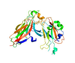


Ephrin A1 is a protein that in humans is encoded by the EFNA1 gene.[5][6][7]
This gene encodes a member of the ephrin (EPH) family. The ephrins and EPH-related receptors comprise the largest subfamily of receptor protein-tyrosine kinases and have been implicated in mediating developmental events, especially in the nervous system and in erythropoiesis. Based on their structures and sequence relationships, ephrins are divided into the ephrin-A (EFNA) class, which are anchored to the membrane by a glycosylphosphatidylinositol linkage, and the ephrin-B (EFNB) class, which are transmembrane proteins. This gene encodes an EFNA class ephrin which binds to the EPHA2, EPHA4, EPHA5, EPHA6, and EPHA7 receptors. Two transcript variants that encode different isoforms were identified through sequence analysis.[7]
Model organisms
| Characteristic | Phenotype |
|---|---|
| Homozygote viability | Normal |
| Fertility | Normal |
| Body weight | Normal |
| Anxiety | Normal |
| Neurological assessment | Normal |
| Grip strength | Normal |
| Hot plate | Normal |
| Dysmorphology | Normal |
| Indirect calorimetry | Normal |
| Glucose tolerance test | Normal |
| Auditory brainstem response | Normal |
| DEXA | Normal |
| Radiography | Abnormal[8] |
| Body temperature | Normal |
| Eye morphology | Normal |
| Clinical chemistry | Normal[9] |
| Plasma immunoglobulins | Normal |
| Haematology | Normal |
| Micronucleus test | Normal |
| Heart weight | Normal |
| Tail epidermis wholemount | Normal |
| Brain histopathology | Normal |
| Salmonella infection | Normal[10] |
| Citrobacter infection | Normal[11] |
| All tests and analysis from[12][13] |
Model organisms have been used in the study of EFNA1 function. A conditional knockout mouse line, called Efna1tm1a(EUCOMM)Wtsi[14][15] was generated as part of the International Knockout Mouse Consortium program—a high-throughput mutagenesis project to generate and distribute animal models of disease to interested scientists.[16][17][18]
Male and female animals underwent a standardized phenotypic screen to determine the effects of deletion.[12][19] Twenty four tests were carried out on homozygous mutant mice and one significant abnormality was observed: a transformation in vertebral number from lumbar vertebrae to sacral vertebrae.[12]
References
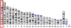
- 1 2 3 GRCh38: Ensembl release 89: ENSG00000169242 - Ensembl, May 2017
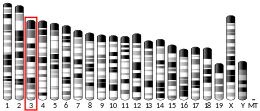
- 1 2 3 GRCm38: Ensembl release 89: ENSMUSG00000027954 - Ensembl, May 2017
- ↑ "Human PubMed Reference:".
- ↑ "Mouse PubMed Reference:".
- ↑ Holzman LB, Marks RM, Dixit VM (Nov 1990). "A novel immediate-early response gene of endothelium is induced by cytokines and encodes a secreted protein". Molecular and Cellular Biology. 10 (11): 5830–8. PMC 361366. PMID 2233719.
- ↑ Cerretti DP, Lyman SD, Kozlosky CJ, Copeland NG, Gilbert DJ, Jenkins NA, Valentine V, Kirstein MN, Shapiro DN, Morris SW (Apr 1996). "The genes encoding the eph-related receptor tyrosine kinase ligands LERK-1 (EPLG1, Epl1), LERK-3 (EPLG3, Epl3), and LERK-4 (EPLG4, Epl4) are clustered on human chromosome 1 and mouse chromosome 3". Genomics. 33 (2): 277–82. doi:10.1006/geno.1996.0192. PMID 8660976.
- 1 2 "Entrez Gene: EFNA1 ephrin-A1".
- ↑ "Radiography data for Efna1". Wellcome Trust Sanger Institute.
- ↑ "Clinical chemistry data for Efna1". Wellcome Trust Sanger Institute.
- ↑ "Salmonella infection data for Efna1". Wellcome Trust Sanger Institute.
- ↑ "Citrobacter infection data for Efna1". Wellcome Trust Sanger Institute.
- 1 2 3 Gerdin AK (2010). "The Sanger Mouse Genetics Programme: High throughput characterisation of knockout mice". Acta Ophthalmologica. 88: 925–7. doi:10.1111/j.1755-3768.2010.4142.x.
- ↑ Mouse Resources Portal, Wellcome Trust Sanger Institute.
- ↑ "International Knockout Mouse Consortium".
- ↑ "Mouse Genome Informatics".
- ↑ Skarnes WC, Rosen B, West AP, Koutsourakis M, Bushell W, Iyer V, Mujica AO, Thomas M, Harrow J, Cox T, Jackson D, Severin J, Biggs P, Fu J, Nefedov M, de Jong PJ, Stewart AF, Bradley A (Jun 2011). "A conditional knockout resource for the genome-wide study of mouse gene function". Nature. 474 (7351): 337–42. doi:10.1038/nature10163. PMC 3572410. PMID 21677750.
- ↑ Dolgin E (Jun 2011). "Mouse library set to be knockout". Nature. 474 (7351): 262–3. doi:10.1038/474262a. PMID 21677718.
- ↑ Collins FS, Rossant J, Wurst W (Jan 2007). "A mouse for all reasons". Cell. 128 (1): 9–13. doi:10.1016/j.cell.2006.12.018. PMID 17218247.
- ↑ van der Weyden L, White JK, Adams DJ, Logan DW (2011). "The mouse genetics toolkit: revealing function and mechanism". Genome Biology. 12 (6): 224. doi:10.1186/gb-2011-12-6-224. PMC 3218837. PMID 21722353.
Further reading
- Pandey A, Lindberg RA, Dixit VM (Sep 1995). "Cell signalling. Receptor orphans find a family". Current Biology. 5 (9): 986–9. doi:10.1016/S0960-9822(95)00195-3. PMID 8542290.
- Flanagan JG, Vanderhaeghen P (1998). "The ephrins and Eph receptors in neural development". Annual Review of Neuroscience. 21: 309–45. doi:10.1146/annurev.neuro.21.1.309. PMID 9530499.
- Zhou R (Mar 1998). "The Eph family receptors and ligands". Pharmacology & Therapeutics. 77 (3): 151–81. doi:10.1016/S0163-7258(97)00112-5. PMID 9576626.
- Holder N, Klein R (May 1999). "Eph receptors and ephrins: effectors of morphogenesis". Development. 126 (10): 2033–44. PMID 10207129.
- Wilkinson DG (2000). "Eph receptors and ephrins: regulators of guidance and assembly". International Review of Cytology. 196: 177–244. doi:10.1016/S0074-7696(00)96005-4. PMID 10730216.
- Xu Q, Mellitzer G, Wilkinson DG (Jul 2000). "Roles of Eph receptors and ephrins in segmental patterning". Philosophical Transactions of the Royal Society of London. Series B, Biological Sciences. 355 (1399): 993–1002. doi:10.1098/rstb.2000.0635. PMC 1692797. PMID 11128993.
- Wilkinson DG (Mar 2001). "Multiple roles of EPH receptors and ephrins in neural development". Nature Reviews. Neuroscience. 2 (3): 155–64. doi:10.1038/35058515. PMID 11256076.
- Mahadevan D, Thanki N, Singh J, McPhie P, Zangrilli D, Wang LM, Guerrero C, LeVine H, Humblet C, Saldanha J (Jul 1995). "Structural studies on the PH domains of Db1, Sos1, IRS-1, and beta ARK1 and their differential binding to G beta gamma subunits". Biochemistry. 34 (28): 9111–7. doi:10.1021/bi00028a021. PMID 7619809.
- Kozlosky CJ, Maraskovsky E, McGrew JT, VandenBos T, Teepe M, Lyman SD, Srinivasan S, Fletcher FA, Gayle RB, Cerretti DP (Jan 1995). "Ligands for the receptor tyrosine kinases hek and elk: isolation of cDNAs encoding a family of proteins". Oncogene. 10 (2): 299–306. PMID 7838529.
- Davis S, Gale NW, Aldrich TH, Maisonpierre PC, Lhotak V, Pawson T, Goldfarb M, Yancopoulos GD (Nov 1994). "Ligands for EPH-related receptor tyrosine kinases that require membrane attachment or clustering for activity". Science. 266 (5186): 816–9. doi:10.1126/science.7973638. PMID 7973638.
- Beckmann MP, Cerretti DP, Baum P, Vanden Bos T, James L, Farrah T, Kozlosky C, Hollingsworth T, Shilling H, Maraskovsky E (Aug 1994). "Molecular characterization of a family of ligands for eph-related tyrosine kinase receptors". The EMBO Journal. 13 (16): 3757–62. PMC 395287. PMID 8070404.
- Gale NW, Holland SJ, Valenzuela DM, Flenniken A, Pan L, Ryan TE, Henkemeyer M, Strebhardt K, Hirai H, Wilkinson DG, Pawson T, Davis S, Yancopoulos GD (Jul 1996). "Eph receptors and ligands comprise two major specificity subclasses and are reciprocally compartmentalized during embryogenesis". Neuron. 17 (1): 9–19. doi:10.1016/S0896-6273(00)80276-7. PMID 8755474.
- Ephnomenclaturecommittee (Aug 1997). "Unified nomenclature for Eph family receptors and their ligands, the ephrins. Eph Nomenclature Committee". Cell. 90 (3): 403–4. doi:10.1016/S0092-8674(00)80500-0. PMID 9267020.
- Nagel W, Schilcher P, Zeitlmann L, Kolanus W (Aug 1998). "The PH domain and the polybasic c domain of cytohesin-1 cooperate specifically in plasma membrane association and cellular function". Molecular Biology of the Cell. 9 (8): 1981–94. doi:10.1091/mbc.9.8.1981. PMC 25450. PMID 9693361.