Depth gauge
A depth gauge is a pressure gauge that displays the equivalent depth in water. It is a piece of diving equipment often used by SCUBA divers.
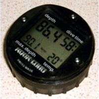
Most modern diving depth gauges have an electronic mechanism and digital display. Older types used a mechanical mechanism and analogue display.
A diver uses a depth gauge with decompression tables and a watch to avoid decompression sickness. A common alternative to the depth gauge, watch and decompression tables is a dive computer. A depth gauge and an oxygen analyser/oxygen sensor can be used to measure the partial pressure of oxygen of the breathing gas, which is necessary to avoid oxygen toxicity.
Digital depth gauges commonly also include a timer showing the interval of time that the diver has been submerged. Some show the diver's rate of ascent and descent, which can be is useful for avoiding barotrauma.
As the gauge only measures water pressure, there is an inherent inaccuracy in the depth displayed by gauges that are used in both fresh water and seawater due to the difference in the densities of fresh water and seawater.
Mode of operation
With water depth, the ambient pressure increases (1 bar every 10 m). Therefore, the exact depth can be determined by meassuring the pressure and comparing it to the pressure at the surface.
Types
Boyle-Mariott's Depth Gauge
The Boyle-Mariottesche depth gauge consists of a circular curved glass tube open at one side. It has no moving parts. While diving, the water goes into the tube and compresses an air bubble inside depending on the depth. The edge of the bubble shows on a scale the depth. For a depth up to 10 m, this depth gauge is quite accurate, because in this range, the pressure doubles from 1 bar to 2 bar, and so it uses half of the scale. At greater depths, it becomes inaccurate. The maximum depth cannot be recorded with this depth gauge.
Bourdon tube depth gauge
The Bourdon tube depth gauge consists of a curved tube made of elastic metal, the Bourdon tube. On the tube, the water presses depending on the design from the inside or the outside. When the pressure increases, the tube stretches; when it decreases the tube recovers to the original curvature. This movement is transferred to a pointer, which can mark the maximum depth reached with a trailing pointer. Its accuracy is good.
Membrane Depth Gauge
At the membrane depth gauge, the water presses onto a flexible metal can, which is compressed with increasing pressure. It also has a membrane, whose movement is also transferred to a pointer and possibly a trailing pointer. It is very accurate.
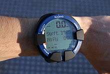
Dive Computer
Dive computers have an integrated depth gauge, whose measured values are digitized. They can calculate the current decompression time needed. The dive depth is displayed along with other values on the display and recorded by the computer for continuous simulation of the decompression model. Most dive computers contain a piezoelectric pressure sensor. Rarely, capacitive or inductive pressure sensors are used.
Light based depth gauges in Biology
A depth gauge can also be based on light: The brightness decreases with depth, but depends on the weather (e.g. whether it is sunny or cloudy) and the time of the day. Also the color depends on the water depth.[1][2]
In water, light attenuates for each wavelength, differently. The UV, violet (> 420 nm), and red (< 500 nm) wavelengths disappear before blue light (470 nm), which penetrates clear water the deepest.[3][4] The wavelength composition is constant for each depth and is almost independent of time of the day and the weather. To gauge depth, an animal would need two photopigments sensitive to different wavelengths to compare different ranges of the spectrum.[1][2] Such pigments may be expressed in different structures.
Such different structures are found in the polychaete Torrea candida. Its eyes have a main and two accessory retinae. The accessory retinae sense UV-light (λmax = 400 nm) and the main retina senses blue-green light (λmax = 560 nm). If the light sensed from all retinae is compared, the depth can be estimated, and so for Torrea candida such a ratio-chromatic depth gauge has been proposed.[5]
A ratio chromatic depth gauge has been found in larvae of the polychaete Platynereis dumerilii.[6] The larvae have two structures: The rhabdomeric photoreceptor cells of the eyes[7] and in the deep brain the ciliary photoreceptor cells. The ciliary photoreceptor cells express a ciliary opsin,[8] which is a photopigment maximally sensitive to UV-light (λmax = 383 nm).[9] Thus, the ciliary photoreceptor cells react on UV-light and make the larvae swimming down gravitactically. The gravitaxis here is countered by phototaxis, which makes the larvae swimming up to the light coming from the surface.[4] Phototaxis is mediated by the rhabdomeric eyes.[10][11][6] The eyes express at least three opsins (at least in the older larvae),[12] and one of them is maximally sensitive to cyan light (λmax = 483 nm) so that the eyes cover a broad wavelength range with phototaxis.[4] When phototaxis and gravitaxis have leveled out, the larvae have found their preferred depth.[6]
See also
- Bathymetry
- Altimeter: A device used in surveying, aviation, and mountain sports to measure terrain elevation.
External links
Articles on depth gauges hosted by the Rubicon Foundation
References
- 1 2 Nilsson, Dan-Eric (31 August 2009). "The evolution of eyes and visually guided behavior". Philosophical Transactions of the Royal Society B: Biological Sciences. 364 (1531): 2833–2847. doi:10.1098/rstb.2009.0083. PMID 19720648.
- 1 2 Nilsson, Dan-Eric (12 April 2013). "Eye evolution and its functional basis". Visual Neuroscience. 30 (1–2): 5–20. doi:10.1017/S0952523813000035. PMID 23578808.
- ↑ Lythgoe, John N. (1988). "Light and Vision in the Aquatic Environment". Sensory Biology of Aquatic Animals. Springer New York: 57–82. doi:10.1007/978-1-4612-3714-3_3.
- 1 2 3 Gühmann, Martin; Jia, Huiyong; Randel, Nadine; Verasztó, Csaba; Bezares-Calderón, Luis A.; Michiels, Nico K.; Yokoyama, Shozo; Jékely, Gáspár (August 2015). "Spectral Tuning of Phototaxis by a Go-Opsin in the Rhabdomeric Eyes of Platynereis". Current Biology. 25 (17): 2265–2271. doi:10.1016/j.cub.2015.07.017.
- ↑ Wald, George; Rayport, Stephen (24 June 1977). "Vision in Annelid Worms". Science. 196 (4297): 1434–1439. doi:10.1126/science.196.4297.1434. PMID 17776921.
- 1 2 3 Verasztó, Csaba; Gühmann, Martin; Jia, Huiyong; Rajan, Vinoth Babu Veedin; Bezares-Calderón, Luis A.; Piñeiro-Lopez, Cristina; Randel, Nadine; Shahidi, Réza; Michiels, Nico K.; Yokoyama, Shozo; Tessmar-Raible, Kristin; Jékely, Gáspár (29 May 2018). "Ciliary and rhabdomeric photoreceptor-cell circuits form a spectral depth gauge in marine zooplankton". eLife. 7. doi:10.7554/eLife.36440. PMID 29809157.
- ↑ Rhode, Birgit (April 1992). "Development and differentiation of the eye in Platynereis dumerilii (Annelida, Polychaeta)". Journal of Morphology. 212 (1): 71–85. doi:10.1002/jmor.1052120108.
- ↑ Arendt, D.; Tessmar-Raible, K.; Snyman, H.; Dorresteijn, A.W.; Wittbrodt, J. (29 October 2004). "Ciliary Photoreceptors with a Vertebrate-Type Opsin in an Invertebrate Brain". Science. 306 (5697): 869–871. Bibcode:2004Sci...306..869A. doi:10.1126/science.1099955. PMID 15514158.
- ↑ Tsukamoto, Hisao; Chen, I-Shan; Kubo, Yoshihiro; Furutani, Yuji (4 August 2017). "A ciliary opsin in the brain of a marine annelid zooplankton is ultraviolet-sensitive, and the sensitivity is tuned by a single amino acid residue". Journal of Biological Chemistry. 292 (31): 12971–12980. doi:10.1074/jbc.M117.793539. ISSN 0021-9258. PMID 28623234.
- ↑ Randel, Nadine; Asadulina, Albina; Bezares-Calderón, Luis A; Verasztó, Csaba; Williams, Elizabeth A; Conzelmann, Markus; Shahidi, Réza; Jékely, Gáspár (27 May 2014). "Neuronal connectome of a sensory-motor circuit for visual navigation". eLife. 3. doi:10.7554/eLife.02730.
- ↑ Jékely, Gáspár; Colombelli, Julien; Hausen, Harald; Guy, Keren; Stelzer, Ernst; Nédélec, François; Arendt, Detlev (20 November 2008). "Mechanism of phototaxis in marine zooplankton". Nature. 456 (7220): 395–399. doi:10.1038/nature07590.
- ↑ Randel, N.; Bezares-Calderon, L. A.; Gühmann, M.; Shahidi, R.; Jekely, G. (2013-05-10). "Expression Dynamics and Protein Localization of Rhabdomeric Opsins in Platynereis Larvae". Integrative and Comparative Biology. 53 (1): 7–16. doi:10.1093/icb/ict046. PMC 3687135. PMID 23667045.