Amelogenesis imperfecta
| Amelogenesis imperfecta | |
|---|---|
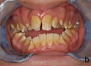 | |
| Amelogenesis imperfecta, hypoplastic type. Note the association of pitted enamel and open bite. | |
| Specialty |
Dentistry |
Amelogenesis imperfecta (AI) is a congenital disorder that presents with a rare abnormal formation of the enamel[1] or external layer of the crown of teeth, unrelated to any systemic or generalized conditions.[2] Enamel is composed mostly of mineral, that is formed and regulated by the proteins in it. Amelogenesis imperfecta is due to the malfunction of the proteins in the enamel (ameloblastin, enamelin, tuftelin and amelogenin) as a result of abnormal enamel formation via amelogenesis.
People afflicted with amelogenesis imperfecta have teeth with abnormal color: yellow, brown or grey; this disorder can afflict any number of teeth of both dentitions. The teeth have a higher risk for dental cavities and are hypersensitive to temperature changes as well as rapid attrition, excessive calculus deposition, and gingival hyperplasia.[3]
Genetics
Several gene expression is needed for enamel formation where the relevant matrix proteins & proteinases are transcribed for regular crystal growth & enamel mineralization.
Mutations in the AMELX,[4] ENAM,[5] MMP20,[6] KLK-4,[7] FAM83H,[8] WDR72,[9] C4orf26,[10] SLC24A4[11][12] LAMB3[13] and ITGB6[14] genes have been found to cause amelogenesis imperfecta (non-syndromic form). AMELX and ENAM encode extracellular matrix proteins of the developing tooth enamel and KLK-4 and MMP20 encode proteases that help degrade organic matter from the enamel matrix during the maturation stage of amelogenesis. SLC24A4 encodes a calcium transporter that mediates calcium transport to developing enamel during tooth development. Less is known about the function of other genes implicated in amelogenesis imperfecta.
Researchers expect that mutations in further genes are likely to be identified as causes of amelogenesis imperfecta.Types include:
| Type | OMIM | Gene | Locus |
|---|---|---|---|
| AI1B | 104500 | ENAM | 4q21 |
| AI1C | 204650 | ENAM | 4q21 |
| AI2A1 | 204700 | KLK4 | 19q13.4 |
| AI2A2 | 612529 | MMP20 | 11q22.3-q23 |
| AI2A3 | 613211 | WDR72 | 15q21.3 |
| AI2A4 | 614832 | C4orf26 | 4q21.1 |
| AI2A5 | 609840 | SLC24A4 | 14q32.12 |
| AI3 | 130900 | FAM83H | 8q24.3 |
| AIH1 | 301200 | AMELX | Xp22.3-p22.1 |
| AIGFS | 614253 | FAM20A | 17q24.2 |
Amelogenesis imperfecta can have different inheritance patterns depending on the gene that is altered. Mutations in the ENAM gene are the most frequent known cause and are most commonly inherited in an autosomal dominant pattern. This type of inheritance means one copy of the altered gene in each cell is sufficient to cause the disorder.
Amelogenesis imperfecta is also inherited in an autosomal recessive pattern; this form of the disorder can result from mutations in the ENAM, MMP20, KLK4, FAM20A, C4orf26 or SLC24A4 genes. Autosomal recessive inheritance means two copies of the gene in each cell are altered.
About 5% of amelogenesis imperfecta cases are caused by mutations in the AMELX gene and are inherited in an X-linked pattern. A condition is considered X-linked if the mutated gene that causes the disorder is located on the X chromosome, one of the two sex chromosomes. In most cases, males with an X-linked form of this condition experience more severe dental abnormalities than affected females.Recent genetic studies suggest that the cause of a significant proportion of amelogenesis imperfecta cases remains to be discovered.
Diagnosis
AI can be classified according to their clinical appearances:[15]
- Type 1 - Hypoplastic
Enamel of abnormal thickness due to malfunction in enamel matrix formation. Enamel is very thin but hard & translucent, and may have random pits & grooves. Condition is of autosomal dominant, autosomal recessive, or x-linked pattern. Enamel differs in appearance from dentine radiographically as normal functional enamel.[16]
- Type 2 - Hypomaturation
Enamel has sound thickness, with a pitted appearance. It is less hard compared to normal enamel, and are prone to rapid wear, although not as intense as Type 3 AI. Condition is of autosomal dominant, autosomal recessive, or x-linked pattern. Enamel appears to be comparable to dentine in its radiodensity on radiograpshs.
- Type 3 - Hypocalcified
Enamel defect due to malfunction of enamel calcification, therefore enamel is of normal thickness but is extremely brittle, with an opaque/chalky presentation. Teeth are prone to staining and rapid wear, exposing dentine. Condition is of autosomal dominant and autosomal recessive pattern. Enamel appears less radioopaque compared to dentine on radiographs.
- Type 4: Hypomature hypoplastic enamel with taurodontism
Enamel has a variation in appearance, with mixed features from Type 1 and Type 2 AI. All Type 4 AI has taurodontism in common. Condition is of autosomal dominant pattern.
Other common features may include an anterior open bite,[17] taurodontism, sensitivity of teeth.
Differential diagnosis would include dental fluorosis, molar-incisor hypomineralization, chronological disorders of tooth development.[18]
Treatment

Preventive and restorative dental care is very important as well as considerations for esthetic issues since the crown are yellow from exposure of dentin due to enamel loss.[3] The main objectives of treatment is pain relief, preserving patient's remaining dentition, and to treat and preserve the patient's occlusal vertical height.[16]
Many factors are to be considered to decide on treatment options such as the classification and severity of AI, the patient's social history, clinical findings etc. There are many classifications of AI but the general management of this condition is similar.
Full-coverage crowns are sometimes being used to compensate for the abraded enamel in adults, tackling the sensitivity the patient experiences. Usually stainless steel crowns are used in children which may be replaced by porcelain once they reach adulthood.[19] These aid with maintaining occlusal vertical dimension.
Aesthetics may be addressed via placement of composite or porcelain veneers, depending on patient factors e.g. age. If the patient has primary or mixed dentition, lab-made composite veneers may be provided temporarily, to be replaced by permanent porcelain veneers once the patient has stabilized permanent dentition. The patient's oral hygiene and diet should be controlled as well as they play a factor in the success of retaining future restorations.
In the worst-case scenario, the teeth may have to be extracted and implants or dentures are required. Loss of nerves in the affected teeth may occur.
Epidemiology
The exact incidence of amelogenesis imperfecta is uncertain. Estimates vary widely, from 1 in 700 people in northern Sweden to 1 in 14,000 people in the United States.[20]
This condition is neither caused by nor the equivalent of dental fluorosis. A manifestation of amelogenesis imperfecta known as "snow capping" is confined to the outer prismless enamel layer. It may superficially resemble dental fluorosis, and indeed "snow capping" may be used as a descriptive term in some incidents of dental fluorosis.[21][22]
References
- ↑ Pieter J. Slootweg (2007). Dental pathology: a practical introduction. Springer Science & Business Media. pp. 19–. ISBN 978-3-540-71690-7. Retrieved 28 December 2010.
- ↑ M, Kida (2002). "Autosomal-dominant hypoplastic form of amelogenesis imperfecta caused by an enamelin gene mutation at the exon-intron boundary". Journal of Dental Research. 81 (11): 738–42. doi:10.1177/154405910208101103.
- 1 2 American Academy of Pediatric Dentistry, Guideline on Dental Management of Heritable Dental Developmental Anomalies, 2013, http://www.aapd.org/media/Policies_Guidelines/G_OHCHeritable.pdf
- ↑ Lagerström M, Dahl N, Nakahori Y, Nakagome Y, Bäckman B, Landegren U, Pettersson U (1991). "A deletion in the amelogenin gene (AMG) causes X-linked amelogenesis imperfecta (AIH1)". Genomics. 10 (4): 971–975. doi:10.1016/0888-7543(91)90187-j. PMID 1916828.
- ↑ Rajpar MH.; Harley K; Laing C; Davies RM; Dixon MJ (2001). "Mutation of the gene encoding the enamel-specific protein, enamelin, causes autosomal-dominant amelogenesis imperfecta". Hum. Mol. Genet. 10 (16): 1673–1677. doi:10.1093/hmg/10.16.1673. PMID 11487571.
- ↑ Kim JW, Simmer JP, Hart TC, Hart PS, Ramaswami MD, Bartlett JD, Hu JC (2005). "MMP-20 mutation in autosomal recessive pigmented hypomaturation amelogenesis imperfecta". J. Med. Genet. 42: 271–275. doi:10.1136/jmg.2004.024505. PMC 1736010. PMID 15744043.
- ↑ Hart PS, Hart TC, Michalec MD, Ryu OH, Simmons D, Hong S (2004). "Mutation in kallikrein 4 causes autosomal recessive hypomaturation amelogenesis imperfecta". J. Med. Genet. 41 (7): 545–549. doi:10.1136/jmg.2003.017657. PMC 1735847. PMID 15235027.
- ↑ Kim JW, Lee SK, Lee ZH, Park JC, Lee KE, Lee MH, Park JT, Seo BM, Hu JC, Simmer JP (2008). "FAM83H mutations in families with autosomal-dominant hypocalcified amelogenesis imperfecta". Am. J. Hum. Genet. 82: 489–494. doi:10.1016/j.ajhg.2007.09.020. PMC 2427219. PMID 18252228.
- ↑ El-Sayed W, Parry DA, Shore RC, Ahmed M, Jafri H, Rashid Y, Al-Bahlani S, Al Harasi S, Kirkham J, Inglehearn CF, Mighell AJ (2009). "Mutations in the beta propeller WDR72 cause autosomal-recessive hypomaturation amelogenesis imperfecta". Am. J. Hum. Genet. 85: 699–705. doi:10.1016/j.ajhg.2009.09.014. PMC 2775821. PMID 19853237.
- ↑ Parry DA, Brookes SJ, Logan CV, Poulter JA, El-Sayed W, Al-Bahlani S, Al Harasi S, Sayed J, Raïf M, Shore RC (2012). "Mutations in C4orf26, Encoding a Peptide with In Vitro Hydroxyapatite Crystal Nucleation and Growth Activity, Cause Amelogenesis Imperfecta". Am. J. Hum. Genet. 91: 565–571. doi:10.1016/j.ajhg.2012.07.020. PMC 3511980. PMID 22901946.
- ↑ Parry DA, Poulter JA, Logan CV, Brookes SJ, Jafri H, Ferguson CH, Anwari BM, Rashid Y, Zhao H, Johnson CA, Inglehearn CF, Mighell AJ (2013). "Identification of mutations in SLC24A4, encoding a potassium-dependent sodium/calcium exchanger, as a cause of amelogenesis imperfecta". Am. J. Hum. Genet. 92 (2): 307–312. doi:10.1016/j.ajhg.2013.01.003. PMC 3567274. PMID 23375655.
- ↑ Herzog, Curtis R.; Reid, Bryan M.; Seymen, Figen; Koruyucu, Mine; Tuna, Elif Bahar; Simmer, James P.; Hu, Jan C.-C. (2015-02-01). "Hypomaturation amelogenesis imperfecta caused by a novel SLC24A4 mutation". Oral Surgery, Oral Medicine, Oral Pathology and Oral Radiology. 119 (2): e77–81. doi:10.1016/j.oooo.2014.09.003. ISSN 2212-4411. PMC 4291293. PMID 25442250.
- ↑ Poulter JA, El-Sayed W, Shore RC, Kirkham J, Inglehearn CF, Mighell AJ (2013). "Whole-exome sequencing, without prior linkage, identifies a mutation in LAMB3 as a cause of dominant hypoplastic amelogenesis imperfecta". Eur. J. Hum. Genet. 22 (1): 132–5. doi:10.1038/ejhg.2013.76. PMID 23632796.
- ↑ Wang SK, Choi M, Richardson AS, Reid BM, Lin BP, Wang SJ, Kim JW, Simmer JP, Hu JC (2014). "ITGB6 loss-of-function mutations cause autosomal recessive amelogenesis imperfecta". Hum. Mol. Genet. 23 (8): 2157–63. doi:10.1093/hmg/ddt611. PMC 3959820. PMID 24305999.
- ↑ Fonseca, RB (2006). "Enamel hypoplasia or amelogenesis imperfecta – a restorative approach". Brazilian Journal of Oral Sciences. 5 (16): 941–3.
- 1 2 Visram, Salima (2006). "Amelogenesis Imperfecta - Clinical Presentation & Management: A Case Report". Dental Update. 33: 612–616. doi:10.12968/denu.2006.33.10.612.
- ↑ Bouvier D, Duprez JP, Bois D (1996). "Rehabilitation of young patients with amelogenesis imperfect: a report of two cases". J Dent Children. 63 (6): 443–447.
- ↑ Peter JM Crawford, Michael Aldred, Agnes Bloch-Zupan (4 April 2007). "Amelogenesis Imperfecta". Orphanet J Rare Dis. 2: 17. doi:10.1186/1750-1172-2-17.
- ↑ Illustrated Dental Embryology, Histology, and Anatomy, Bath-Balogh and Fehrenbach, Elsevier, 2011, page 64
- ↑ Hoppenreijs, TJ (1998). "Open bite deformity in amelogenesis imperfecta. Part 1: An analysis of contributory factors and implications for treatment". Journal of Cranio-Maxillo-Facial Surgery. 26 (4): 260–6. doi:10.1016/s1010-5182(98)80023-1.
- ↑ Chaudhary M, Dixit S, Singh A, Kunte S (2009). "Amelogenesis imperfecta: Report of a case and review of literature". Journal of Oral and Maxillofacial Pathology : JOMFP. 13 (2): 70–77. doi:10.4103/0973-029X.57673. PMC 3162864. PMID 21887005.
- ↑ Hu JCC, Chan HC, Simmer SG, Seymen F, Richardson AS; et al. (2012). "Amelogenesis Imperfecta in Two Families with Defined AMELX Deletions in ARHGAP6". PLOS ONE. 7 (12): e52052. doi:10.1371/journal.pone.0052052.
Further reading
- Winter GB, Brook AH (1975). "Enamel hypoplasia and anomalies of the enamel". Dent Clin North Am. 19 (1): 3–24. PMID 162891.
- Simmer JP, Hu JC (2001). "Dental enamel formation and its impact on clinical dentistry". J Dent Educ. 65 (9): 896–905. PMID 11569606.
- Aldred MJ, Savarirayan R, Crawford PJ (2003). "Amelogenesis imperfecta: a classification and catalogue for the 21st century". Oral Dis. 9 (1): 19–23. doi:10.1034/j.1601-0825.2003.00843.x. PMID 12617253.
- Nusier M, Yassin O, Hart TC, Samimi A, Wright JT (2004). "Phenotypic diversity and revision of the nomenclature for autosomal recessive amelogenesis imperfecta". Oral Surg Oral Med Oral Pathol Oral Radiol Endod. 97 (2): 220–30. doi:10.1016/j.tripleo.2003.08.007. PMID 14970781.
- Stephanopoulos G, Garefalaki ME, Lyroudia K (2005). "Genes and related proteins involved in amelogenesis imperfecta". J Dent Res. 84 (12): 1117–26. doi:10.1177/154405910508401206. PMID 16304440.
External links
| Classification | |
|---|---|
| External resources |