Tooth impaction
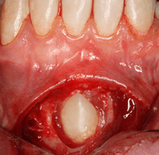

An impacted tooth is one that fails to erupt into the dental arch within the expected developmental window. Because impacted teeth do not erupt, they are retained throughout the individual's lifetime unless extracted or exposed surgically. Teeth may become impacted because of adjacent teeth, dense overlying bone, excessive soft tissue or a genetic abnormality. Most often, the cause of impaction is inadequate arch length and space in which to erupt. That is the total length of the alveolar arch is smaller than the tooth arch (the combined mesiodistal width of each tooth). The wisdom teeth (third molars) are frequently impacted because they are the last teeth to erupt in the oral cavity. Mandibular third molars are more commonly impacted than their maxillary counterparts.
As a general rule, all impacted teeth must be removed[1] except, in certain cases, canine teeth: canines may just remain buried and give no further problems, thus not requiring surgical intervention.[2] However, removal of an asymptomatic impacted tooth isn't a medical consensus: watchful monitoring may be a more prudent strategy[3] and open path for a future placement of implant on such impacted tooth.[4]
Classification
Classifications enable the oral surgeon to determine the difficulty in removal of the impacted tooth.[5] The primary factor determining the difficulty is accessibility, which is determined by adjacent teeth or other structures that impair access or delivery pathway. The majority of classification schemes are based on analysis on a radiograph. The most frequently considered factors are discussed below.
Angulation of tooth
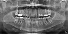
Most commonly used classification system with respect to treatment planning. Depending on the angulation the tooth might be classified as:
- Mesioangular
- Horizontal
- Vertical
- Distoangular
- Palatal
- Buccal
- Lingual
Relationship of tooth to anterior border of ramus
This type of classification is based on the amount of impacted tooth that is covered with the mandibular ramus. It is known as the Pell and Gregory classification, classes 1, 2, and 3.[6]
Relationship of tooth to occlusal plane
The depth of the impacted tooth compared with the adjacent second molar gives the basis for this type of classification. This was also given by Pell and Gregory and is called as Pell and Gregory A, B and C classification.
Complications
Erupted teeth that are adjacent to impacted teeth are predisposed to periodontal disease. Since the most difficult tooth surface to be cleaned is the distal surface of the last tooth, in the presence of an impacted tooth there is always gingival inflammation around the second molar that is invariably present. Even this minor amount of inflammation can provide bacteria access to a larger portion of the root surface that results in early formation of periodontitis compromising the tooth. Even in situations in which no obvious communication exists between the mouth and the impacted third molar there may be enough communication to initiate dental caries (tooth decay).
Pericoronitis
Pericoronitis is an infection of the soft tissue that covers the crown of an impacted tooth and is usually caused by the normal oral microbiota. For most people there exists a balance between the host defenses and the oral micriobiota but if the host defenses are compromised like during minor illness such as influenza or an upper respiratory tract infection, pericoronitis results. Another common cause is entrapment of food beneath the gum flap (also called an operculum). Pericoronitis can present as a mild infection or severe infection. In its mildest form it is just a localized tissue swelling and soreness whereas in severe forms the swelling is slightly larger even sometimes creating trismus (difficulty opening the mouth).
Occasionally, an impacted tooth causes sufficient pressure on the roots of adjacent teeth causing it to resorb.
An impacted tooth occupies space that is usually filled with bone. This weakens that area of bone and renders the jaw more susceptible to fracture.
When impacted teeth are retained completely within the alveolar process, the associated follicular sac is also retained along with it. Though in most persons the dental follicle maintains its original size sometimes it may undergo cystic degeneration and become a dentigerous cyst or a keratocyst.
Symptoms
Most commonly the individual complains of food getting lodged beneath the gums and a soreness that is usually confused with throat infections. In slightly milder forms a swelling is visible and mouth opening becomes difficult in severe cases. Pain is invariably present.
Management
All impacted teeth, unless otherwise contraindicated, are considered for surgical removal. Thus, dental extractions will often take place. The type of extraction (simple or surgical) often depends on the location of the teeth.
In some cases, for aesthetic purposes, a surgeon may wish to expose the canine. This may be achieved through open or closed exposure. Studies show no advantage of one method over another.[7]
References
- ↑ Miloro, M.; Ghali, G.E.; Larsen, P.; Waite, P. (2004). Peterson's Principles of Oral and Maxillofacial Surgery. 1. B C Decker. ISBN 9781550092349. Retrieved 2015-08-22.
- ↑ "Impacted teeth including surgery for canine teeth | Cambridge University Hospitals". cuh.org.uk. Retrieved 2015-08-22.
- ↑ Mettes, Theodorus Dirk G.; et al. (2016-08-31). Cochrane Oral Health Group, ed. "Surgical removal versus retention for the management of asymptomatic disease-free impacted wisdom teeth". Scientific Institute for Quality of Healthcare, Radboud University Nijmegen Medical Center, Nijmegen, Netherlands. doi:10.1002/14651858.CD003879.pub4. PMID 27578151.
- ↑ Mithridade, Davarpanah; et al. (2015-01-30). "Unconventional Implant Placement IV. Implant Placement through Impacted Teeth to Avoid Invasive Surgery. Long-term Results of 3 Cases". The Open Dentistry Journal. 12: 15–20. doi:10.2174/1874210601509010015. ISSN 1874-2106. PMC 4319210.
- ↑ Journal of the Medical Association of Thailand. Index Copernicus. September 2006. ISSN 0125-2208. Retrieved 2015-08-22.
- ↑ Fragiskos, F.D. (2007). Oral Surgery. Springer. p. 126. ISBN 9783540499756. Retrieved 2015-08-22.
- ↑ Parkin N, Benson PE, Thind B, Shah A, Khalil I, Ghafoor S. (August 2017). "Open versus closed surgical exposure of canine teeth that are displaced in the roof of the mouth". Cochrane Database of Systematic Reviews (8).