A-DNA
A-DNA is one of the possible double helical structures which DNA can adopt. A-DNA is thought to be one of three biologically active double helical structures along with B-DNA and Z-DNA. It is a right-handed double helix fairly similar to the more common B-DNA form, but with a shorter, more compact helical structure whose base pairs are not perpendicular to the helix-axis as in B-DNA. It was discovered by Rosalind Franklin, who also named the A and B forms. She showed that DNA is driven into the A form when under dehydrating conditions. Such conditions are commonly used to form crystals, and many DNA crystal structures are in the A form.[1] The same helical conformation occurs in double-stranded RNAs, and in DNA-RNA hybrid double helices.
Structure
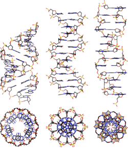
A-DNA is fairly similar to B-DNA given that it is a right-handed double helix with major and minor grooves. However, as shown in the comparison table below, there is a slight increase in the number of base pairs (bp) per turn (resulting in a smaller twist angle), and smaller rise per base pair (making A-DNA 20-25% shorter than B-DNA). The major groove of A-DNA is deep and narrow, while the minor groove is wide and shallow. A-DNA is broader and apparently more compressed along its axis than B-DNA.[2]
Comparison geometries of the most common DNA forms
| Geometry attribute: | A-form | B-form | Z-form |
|---|---|---|---|
| Helix sense | right-handed | right-handed | left-handed |
| Repeating unit | 1 bp | 1 bp | 2 bp |
| Rotation/bp | 32.7° | 34.3° | 60°/2 |
| Mean bp/turn | 11 | 10.5 | 12 |
| Inclination of bp to axis | +19° | −1.2° | −9° |
| Rise/bp along axis | 2.6 Å (0.26 nm) | 3.4 Å (0.34 nm) | 3.7 Å (0.37 nm) |
| Rise/turn of helix | 28.6 Å (2.86 nm) | 35.7 Å (3.57 nm) | 45.6 Å (4.56 nm) |
| Mean propeller twist | +18° | +16° | 0° |
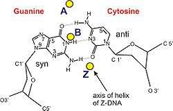
| Glycosyl angle | anti | anti | pyrimidine: anti, purine: syn |
| Nucleotide phosphate to phosphate distance | 5.9 Å | 7.0 Å | C: 5.7 Å, G: 6.1 Å |
| Sugar pucker | C3'-endo | C2'-endo | C: C2'-endo, G: C3'-endo |
| Diameter | 23 Å (2.3 nm) | 20 Å (2.0 nm) | 18 Å (1.8 nm) |
Biological function
Dehydration of DNA drives it into the A form, and this apparently protects DNA under conditions such as the extreme desiccation of bacteria.[3] Protein binding can also strip solvent off of DNA and convert it to the A form, as revealed by the structure of a rod-shaped virus.[4]
It has been proposed that the motors that package double-stranded DNA in bacteriophages exploit the fact that A-DNA is shorter than B-DNA, and that conformational changes in the DNA itself are the source of the large forces generated by these motors.[5] Experimental evidence for A-DNA as an intermediate in viral biomotor packing comes from double dye Förster resonance energy transfer measurements showing that B-DNA is shortened by 24% in a stalled ("crunched") A-form intermediate.[6][7] In this model, ATP hydrolysis is used to drive protein conformational changes that alternatively dehydrate and rehydrate the DNA, and the DNA shortening/lengthening cycle is coupled to a protein-DNA grip/release cycle to generate the forward motion that moves DNA into the capsid.
See also
References
- ↑ Rosalind, Franklin (1953). "The Structure of Sodium Thymonucleate Fibres. I. The Influence of Water Content" (PDF). Acta Crystallographica. 6: 673–677. doi:10.1107/s0365110x53001939.
- ↑ Dickerson, Richard E. (1992). "DNA Structure From A to Z" (PDF). Methods in Enzymology. 211: 67–111. doi:10.1016/0076-6879(92)11007-6 – via Elsevier Science Direct.
- ↑ Whelan DR, et al. (2014). "Detection of an en masse and reversible B- to A-DNA conformational transition in prokaryotes in response to desiccation". J R Soc Interface. 11: 20140454. doi:10.1098/rsif.2014.0454. PMC 4208382. PMID 24898023.
- ↑ Di Maio F, Egelman EH, et al. (2015). "A virus that infects a hyperthermophile encapsidates A-form DNA". Science. 348: 914–917. doi:10.1126/science.aaa4181. PMC 5512286. PMID 25999507.
- ↑ Harvey, SC (2015). "The scrunchworm hypothesis: Transitions between A-DNA and B-DNA provide the driving force for genome packaging in double-stranded DNA bacteriophages". Journal of Structural Biology. 189: 1–8. doi:10.1016/j.jsb.2014.11.012. PMC 4357361. PMID 25486612.
- ↑ Oram, M (2008). "Modulation of the packaging reaction of bacteriophage t4 terminase by DNA structure". J Mol Biol. 381: 61–72. doi:10.1016/j.jmb.2008.05.074. PMC 2528301.
- ↑ Ray, K (2010). "DNA crunching by a viral packaging motor: Compression of a procapsid-portal stalled Y-DNA substrate". Virology. 398: 224–232. doi:10.1016/j.virol.2009.11.047.