Oogenesis
Oogenesis, ovogenesis, or oögenesis /ˌoʊ.əˈdʒɛnɪsɪs/[1] is the differentiation of the ovum (egg cell) into a cell competent to further develop when fertilized.[2] It is developed from the primary oocyte by maturation. Oogenesis is initiated in the embryonic stage.
| Oogenesis | |
|---|---|
| Identifiers | |
| MeSH | D009866 |
| Anatomical terminology | |
Oogenesis in non-human mammals
In mammals, the first part of oogenesis starts in the germinal epithelium, which gives rise to the development of ovarian follicles, the functional unit of the ovary.
Oogenesis consists of several sub-processes: oocytogenesis, ootidogenesis, and finally maturation to form an ovum (oogenesis proper). Folliculogenesis is a separate sub-process that accompanies and supports all three oogenetic sub-processes.
| Cell type | ploidy/chromosomes | chromatids | Process | Time of completion |
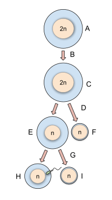
| Oogonium | diploid/46(2N) | 2C | Oocytogenesis (mitosis) | Third trimester |
| primary oocyte | diploid/46(2N) | 4C | Ootidogenesis (meiosis I) (Folliculogenesis) | Dictyate in prophase I for up to 50 years |
| secondary oocyte | haploid/23(1N) | 2C | Ootidogenesis (meiosis II) | Halted in metaphase II until fertilization |
| Ootid | haploid/23(1N) | 1C | Ootidogenesis (meiosis II) | Minutes after fertilization |
| Ovum | haploid/23(1N) | 1C |
Oogonium —(Oocytogenesis)—> Primary Oocyte —(Meiosis I)—> First Polar body (Discarded afterward) + Secondary oocyte —(Meiosis II)—> Second Polar Body (Discarded afterward) + Ovum
Oocyte meiosis, important to all animal life cycles yet unlike all other instances of animal cell division, occurs completely without the aid of spindle-coordinating centrosomes.[3][4]
The creation of oogonia
The creation of oogonia traditionally doesn't belong to oogenesis proper, but, instead, to the common process of gametogenesis, which, in the female human, begins with the processes of folliculogenesis, oocytogenesis, and ootidogenesis. Oogonia enter meiosis during embryonic development, becoming oocytes. Meiosis begins with DNA replication and meiotic crossing over. It then stops in early prophase.
Maintenance of meiotic arrest
Mammalian oocytes are maintained in meiotic prophase arrest for a very long time -- months in mice, years in humans. Initially the arrest is due to lack of sufficient cell cycle proteins to allow meiotic progression. However, as the oocyte grows, these proteins are synthesized, and meiotic arrest becomes dependent on cyclic AMP [5]. The cyclic AMP is generated by the oocyte by adenylyl cyclase in the oocyte membrane. The adenylyl cyclase is kept active by a constitutively active G-protein-coupled receptor known as GPR3 and a G-protein, Gs, also present in the oocyte membrane [6].
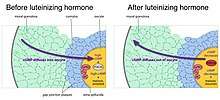
Maintenance of meiotic arrest also depends on the presence of a multilayered complex of cells, known as a follicle, that surrounds the oocyte. Removal of the oocyte from the follicle causes meiosis to progress in the oocyte.[7] The cells that comprise the follicle, known as granulosa cells, are connected to each other by proteins known as gap junctions, that allow small molecules to pass between the cells. The granulosa cells produce a small molecule, cyclic GMP, that diffuses into the oocyte through the gap junctions. In the oocyte, cyclic GMP prevents the breakdown of cyclic AMP by the phosphodiesterase PDE3, and thus maintains meiotic arrest.[8] The cyclic GMP is produced by the guanylyl cyclase NPR2.[9]
Reinitiation of meiosis and stimulation of ovulation by luteinizing hormone
As follicles grow, they acquire receptors for luteinizing hormone, a pituitary hormone that reinitiates meiosis in the oocyte and causes ovulation of a fertilizable egg. Luteinizing hormone acts on receptors in the outer layers of granulosa cells of the follicle, causing a decrease in cyclic GMP in the granulosa cells [10]. Because the granulosa cells and oocyte are connected by gap junctions, cyclic GMP also decreases in the oocyte, causing meiosis to resume [11]. Meiosis then proceeds to second metaphase, where it pauses again until fertilization. Luteinizing hormone also stimulates gene expression leading to ovulation [12].
Human oogenesis
Oogenesis
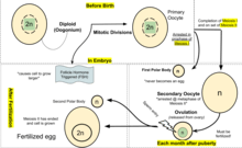
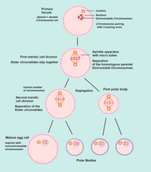
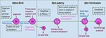
Oogenesis starts with the process of developing primary oocytes, which occurs via the transformation of oogonia into primary oocytes, a process called oocytogenesis.[13] Oocytogenesis is complete either before or shortly after birth.
Number of primary oocytes
It is commonly believed that, when oocytogenesis is complete, no additional primary oocytes are created, in contrast to the male process of spermatogenesis, where gametocytes are continuously created. In other words, primary oocytes reach their maximum development at ~20[14] weeks of gestational age, when approximately seven million primary oocytes have been created; however, at birth, this number has already been reduced to approximately 1-2 million.
Two publications have challenged the belief that a finite number of oocytes are set around the time of birth.[15][16] The renewal of ovarian follicles from germline stem cells (originating from bone marrow and peripheral blood) has been reported in the postnatal mouse ovary. In contrast, DNA clock measurements do not indicate ongoing oogenesis during human females' lifetimes.[17] Thus, further experiments are required to determine the true dynamics of small follicle formation.
Ootidogenesis
The succeeding phase of ootidogenesis occurs when the primary oocyte develops into an ootid. This is achieved by the process of meiosis. In fact, a primary oocyte is, by its biological definition, a cell whose primary function is to divide by the process of meiosis.[18]
However, although this process begins at prenatal age, it stops at prophase I. In late fetal life, all oocytes, still primary oocytes, have halted at this stage of development, called the dictyate. After menarche, these cells then continue to develop, although only a few do so every menstrual cycle.
Meiosis I
Meiosis I of ootidogenesis begins during embryonic development, but halts in the diplotene stage of prophase I until puberty. The mouse oocyte in the dictyate (prolonged diplotene) stage actively repairs DNA damage, whereas DNA repair is not detectable in the pre-dictyate (leptotene, zygotene and pachytene) stages of meiosis.[19] For those primary oocytes that continue to develop in each menstrual cycle, however, synapsis occurs and tetrads form, enabling chromosomal crossover to occur. As a result of meiosis I, the primary oocyte has now developed into the secondary oocyte and the first polar body.
Meiosis II
Immediately after meiosis I, the haploid secondary oocyte initiates meiosis II. However, this process is also halted at the metaphase II stage until fertilization, if such should ever occur. If the egg is not fertilized, it is disintegrated and released (menstruation) and the secondary oocyte does not complete meiosis II (and doesn't become an ovum). When meiosis II has completed, an ootid and another polar body have now been created.
Folliculogenesis
Synchronously with ootidogenesis, the ovarian follicle surrounding the ootid has developed from a primordial follicle to a preovulatory one.
Maturation into ovum
Both polar bodies disintegrate at the end of Meiosis II, leaving only the ootid, which then eventually undergoes maturation into a mature ovum.
The function of forming polar bodies is to discard the extra haploid sets of chromosomes that have resulted as a consequence of meiosis.
In vitro maturation
In vitro maturation (IVM) is the technique of letting ovarian follicles mature in vitro. It can potentially be performed before an IVF. In such cases, ovarian hyperstimulation isn't essential. Rather, oocytes can mature outside the body prior to IVF. Hence, no (or at least a lower dose of) gonadotropins have to be injected in the body.[20] Immature eggs have been grown until maturation in vitro at a 10% survival rate, but the technique is not yet clinically available.[21] With this technique, cryopreserved ovarian tissue could possibly be used to make oocytes that can directly undergo in vitro fertilization.[21]
Oogenesis in non-mammals
_(diagram).png)
Some algae and the oomycetes produce eggs in oogonia. In the brown alga Fucus, all four egg cells survive oogenesis, which is an exception to the rule that generally only one product of female meiosis survives to maturity.
In plants, oogenesis occurs inside the female gametophyte via mitosis. In many plants such as bryophytes, ferns, and gymnosperms, egg cells are formed in archegonia. In flowering plants, the female gametophyte has been reduced to an eight-celled embryo sac within the ovule inside the ovary of the flower. Oogenesis occurs within the embryo sac and leads to the formation of a single egg cell per ovule.
In ascaris, the oocyte does not even begin meiosis until the sperm touches it, in contrast to mammals, where meiosis is completed in the estrus cycle.
In female Drosophila flies, genetic recombination occurs during meiosis. This recombination is associated with formation of DNA double-strand breaks and the repair of these breaks. [22] The repair process leads to crossover recombinants as well as at least three times as many noncrossover recombinants (e.g. arising by gene conversion without crossover).[22]
See also
References
Cho WK, Stern S, Biggers JD. 1974. Inhibitory effect of dibutyryl cAMP on mouse oocyte maturation in vitro. J Exp Zool.187:383-386
- Merriam-Webster Online Dictionary Definition: Oogenesis
- Gilbert, Scott F. (2000-01-01). "Oogenesis". Cite journal requires
|journal=(help) - Szollosi D, Calarco P, Donahue RP (1972). "Absence of centrioles in the first and second meiotic spindles of mouse oocytes". J Cell Sci. 11 (2): 521–541. PMID 5076360.
- Manandhar G, Schatten H, Sutovsky P (January 2005). "Centrosome reduction during gametogenesis and its significance". Biol. Reprod. 72 (1): 2–13. doi:10.1095/biolreprod.104.031245. PMID 15385423.
- Jaffe LA, Egbert JR. 2017. Regulation of Mammalian Oocyte Meiosis by Intercellular Communication Within the Ovarian Follicle. Ann. Rev. Physiol. 79:237-260.
- Mehlmann LM, Saeki Y, Tanaka S, Brennan TJ, Evsikov AV, Pendola FL, Knowles BB, Eppig JJ, Jaffe LA. 2004. The Gs-linked receptor GPR3 maintains meiotic arrest in mammalian oocytes. Science 306:1947-1950
- Edwards RG. 1965. Maturation in vitro of mouse, sheep, cow, pig, rhesus monkey and human ovarian oocytes. Nature 20:349-351.
- Norris RP, Ratzan WJ, Freudzon M, Mehlmann LM, Krall J, Movsesian MA, Wang H, Ke H, Nikolaev VO, Jaffe LA. 2009. Cyclic GMP from the surrounding somatic cells regulates cyclic AMP and meiosis in the mouse oocyte. Development 136:1869-1878.
- Zhang M, Su YQ, Sugiura K, Xia G, Eppig JJ. 2010. Granulosa cell ligand NPPC and its receptor NPR2 maintain meiotic arrest in mouse oocytes. Science 330:366-369
- Jaffe LA, Egbert JR. 2017. Regulation of Mammalian Oocyte Meiosis by Intercellular Communication Within the Ovarian Follicle. Ann. Rev. Physiol. 79:237-260.
- Shuhaibar LC, Egbert JR, Norris RP, Lampe PD, Nikolaev VO, Thunemann M, Wen L, Feil R, Jaffe LA Intercellular signaling via cyclic GMP diffusion through gap junctions restarts meiosis in mouse ovarian follicles. Proc. Natl. Acad. Sci. USA 112: 5527-5532.
- Richards JS, Ascoli M. 2018. Endocrine, paracrine, and autocrine signaling pathways that regulate ovulation. Trends Endocrinol. Metab. 29:313-325.
- NCBI - The saga of the germ line
- Lobo RA (September 2003). "Early ovarian ageing: a hypothesis. What is early ovarian ageing?". Hum. Reprod. 18 (9): 1762–4. doi:10.1093/humrep/deg377. PMID 12923124.
- Johnson J, Bagley J, Skaznik-Wikiel M, et al. (July 2005). "Oocyte generation in adult mammalian ovaries by putative germ cells in bone marrow and peripheral blood". Cell. 122 (2): 303–15. doi:10.1016/j.cell.2005.06.031. PMID 16051153.
- Johnson J, Canning J, Kaneko T, Pru J, Tilly J (2004). "Germline stem cells and follicular renewal in the postnatal mammalian ovary". Nature. 428 (6979): 145–50. doi:10.1038/nature02316. PMID 15014492.
- Forster P, Hohoff C, Dunkelmann B, Schürenkamp M, Pfeiffer H, Neuhuber F, Brinkmann B (2015). "Elevated germline mutation rate in teenage fathers". Proc R Soc B. 282 (1803): 20142898. doi:10.1098/rspb.2014.2898. PMC 4345458. PMID 25694621.
- Biochem
- Guli CL, Smyth DR (1988). "UV-induced DNA repair is not detectable in pre-dictyate oocytes of the mouse". Mutat Res. 208 (2): 115–119. doi:10.1016/s0165-7992(98)90010-0. PMID 3380109.
- "Vejledning om kunstig befrugtning 2006 (Danish)" (PDF). Archived from the original (PDF) on 2012-03-09. Retrieved 2011-01-29.
-
- McLaughlin, M; Albertini, D F; Wallace, W H B; Anderson, R A; Telfer, E E (2018). "Metaphase II oocytes from human unilaminar follicles grown in a multi-step culture system". MHR: Basic Science of Reproductive Medicine. 24 (3): 135–142. doi:10.1093/molehr/gay002. ISSN 1360-9947. PMID 29390119.
- Further comments in BBC news article: James Gallagher (2018-02-09). "First human eggs grown in laboratory". BBC News.
- Mehrotra S, McKim KS. Temporal analysis of meiotic DNA double-strand break formation and repair in Drosophila females. PLoS Genet. 2006 Nov 24;2(11):e200. PMID:17166055
- Bibliography
- Manandhar G, Schatten H and Sutovsky P (2005). Centrosome reduction during gametogenesis and its significance. Biol Reprod, 72(1)2-13.
External links
| Biological terms | |
|---|---|
| Sexual reproduction |
|
| Sexuality | |
| |