Renal pelvis
| Renal pelvis | |
|---|---|
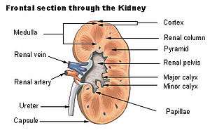 Cross-section of the kidney, with major structures labelled. The renal pelvis, located in the middle of the image, collects urine from the urinary calices, and labelled on the right. | |
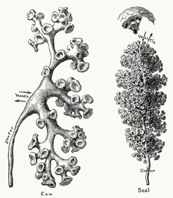 An image showing just the pelvis and calices of the kidneys, with the rest of the kidney removed, from a dissected cow and seal specimen. These vary greatly in size and number depending on species. | |
| Details | |
| Precursor | Ureteric bud |
| System | Urinary system |
| Identifiers | |
| Latin | pelvis renalis |
| MeSH | D007682 |
| TA | A08.1.05.001 |
| FMA | 15575 |
| Anatomical terminology | |
The renal pelvis or pelvis of the kidney is the funnel-like dilated part of the ureter in the kidney. In humans, the renal pelvis is the point where the two or three major calyces join together. It has a mucous membrane is covered with transitional epithelium, and an underlying lamina propria of loose to dense connective tissue.
The renal pelvis functions as a funnel for urine flowing to the ureter.
The renal pelvis is the location of several kinds of kidney cancer and is affected by infection in pyelonephritis.
Clinical significance
The renal pelvis is the location of several kinds of kidney cancer and is affected by infection in pyelonephritis. A large "staghorn" kidney stone may block all or part of the renal pelvis.
The size of the renal pelvis plays a major role in the grading of hydronephrosis. Normally, the anteroposterior diameter of the renal pelvis is less than 4 mm in fetuses up to 32 weeks of gestational age and 7 mm afterwards.[1] In adults, 13% of the normal population have a transverse pelvic diameter of over 10 mm.[2]
Etymology and pronunciation
Like the bony pelvis, the renal pelvis (/ˈriːnəl
Additional images
 Depiction of the developing renal pelvis.
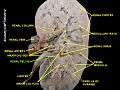 Renal pelvis in a cadaveric specimen.
See also
- Renal sinus
- Pyelectasis, the dilation of the renal pelvis
References
- ↑ Page 189 in: V. D'Addario (2014). Donald School Basic Textbook of Ultrasound in Obstetrics & Gynecology. JP Medical Ltd. ISBN 9789351523376.
- ↑ Emamian SA, Nielsen MB, Pedersen JF, Ytte L (1993). "Sonographic evaluation of renal appearance in 665 adult volunteers. Correlation with age and obesity". Acta Radiol. 34 (5): 482–5. doi:10.3109/02841859309175388. PMID 8369185.
- ↑ Merriam-Webster, Merriam-Webster's Unabridged Dictionary, Merriam-Webster.
External links
- Anatomy figure: 40:03-07 at Human Anatomy Online, SUNY Downstate Medical Center—"Section of the kidney, anterior view."
- Anatomy image:8962 at the SUNY Downstate Medical Center
- Anatomy photo: Urinary/mammal/pelvis0/pelvis1 - Comparative Organology at University of California, Davis—"Mammal, renal pelvis (Gross, Medium)"
- Anatomy photo: Urinary/mammal/pelvis1/pelvis1 - Comparative Organology at University of California, Davis—"Mammal, renal pelvis (LM, Medium)"
Anatomy of the urinary system | |||||||||||||
|---|---|---|---|---|---|---|---|---|---|---|---|---|---|
| Kidneys |
| ||||||||||||
| Ureters | |||||||||||||
| Bladder | |||||||||||||
| Urethra |
| ||||||||||||
| |||||||||||||