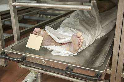
Cadaver
A cadaver, also referred to as a corpse (singular) in medical, literary, and legal usage, or when intended for dissection, is a deceased body.[1]
Human decay
Observation of the various stages of decomposition can help determine how long a body has been dead.
Stages of decomposition
- The first stage is autolysis, more commonly known as self-digestion, during which the body's cells are destroyed through the action of their own digestive enzymes. However, these enzymes are released into the cells because of active processes ceasing in the cells, not as an active process. In other words, though autolysis resembles the active process of digestion of nutrients by live cells, the dead cells are not actively digesting themselves as is often claimed in popular literature and as the synonym of autolysis self-digestion seems to imply. As a result of autolysis, liquid is created that gets between the layers of skin and makes the skin peel off. During this stage, flies (when present) start to lay eggs in the openings of the body: eyes, nostrils, mouth, ears, open wounds, and other orifices. Hatched larvae (maggots) of blowflies, subsequently get under the skin and start to eat the body.
- The second stage of decomposition is bloating; bacteria in the gut begin to break down the tissues of the body, releasing gas that accumulates in the intestines, which becomes trapped because of the early collapse of the small intestine. This bloating occurs largely in the abdomen, and sometimes in the mouth and genitals. The tongue may swell. This usually happens in about the second week of decomposition. Gas accumulation and bloating will continue until the body is decomposed sufficiently for the gas to escape.
- The third stage is putrefaction. It is the last and longest stage. Putrefaction is where the larger structures of the body break down, and tissues liquefy. The digestive organs, the brain, and lungs are the first to disintegrate. Under normal conditions, the organs are unidentifiable after three weeks. The muscles can be eaten by bacteria or devoured by animals. Eventually, sometimes after several years, all that remains is the skeleton. In acid-rich soils, the skeleton will eventually dissolve into its base chemicals.
The rate of decomposition depends on many factors including temperature and the environment. The warmer and more humid the environment, the faster the body is broken down.[2]
History

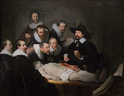
Greek physicians Herophilus (335–280 BC) and Erasistratus (c.304–c.250 BC) were among the first on record to have dissected bodies. Andreas Vesalius (1514–1564), author of De humani corporis fabrica, who was able to dispel many misconceptions by dissecting human cadavers, is regarded as the father of modern human anatomy.[3][4] Indian ancient texts Sushruta Samhita (2nd century BCE) and Charaka Samhita have mentioned the dissection procedure.[5][6]
The tradition of dissecting criminals was carried up into the eighteenth and nineteenth century when anatomy schools became popular in England and Scotland. Criminals who were executed for their crimes were used as the first cadavers. From the 16th century until 1832, and the passage of the Anatomy Act, in Britain the only cadavers legally available for dismemberment came from executed murderers.[7] The demand for cadavers increased when the number of criminals being executed decreased. Since corpses were in such high demand, it became commonplace to steal bodies from graves in order to keep the market supplied.
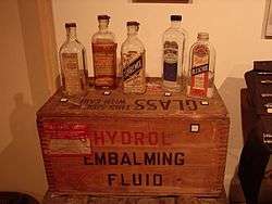
The methods of preserving cadavers have changed over the last 200 years. At that time, cadavers had to be used immediately because there were no adequate methods to keep the body from quickly decaying. Preservation was needed in order to carry out classes and lessons about the human body. Glutaraldehyde was the first main chemical used for embalming and preserving the body although it leaves a yellow stain in the tissues, which can interfere with observation and research.
Formaldehyde is the chemical that is used as the main embalming chemical now. It is a colorless solution that maintains the tissue in its lifelike texture and can keep the body well preserved for an extended period.
Cadavers in science
Cadavers have contributed to body science and medical students often use cadavers to study anatomy. Cadavers are often used to verify surgical techniques before moving on to living patients.[8] While many schools have moved to using technology and surgical models to teach students, cadavers are still needed for hands on learning. However, the expense of maintaining cadaveric dissection facilities has limited the time and resources available for gross anatomy teaching in many medical schools, with some adopting alternative prosection-based or simulated teaching.[9] This, coupled with decreasing time dedicated to gross anatomical courses within the growing greater medical school curriculum, has caused controversy surrounding the sufficiency of anatomical teaching with nearly half of newly qualified doctors believing they received insufficient anatomy teaching.[10]
Appendectomies, the removal of the appendix, are performed 28,000 times a year in the United States and are still practiced on human cadavers and not with technology simulations.[11] Gross anatomy, a common course in medical school studying the visual structures of the body, gives students the opportunity to have a hands-on learning environment. The need for cadavers has also grown outside of academic programs for research. Organizations like Science Care and the Anatomy Gifts Registry help send bodies where they are needed most.[11]
Preservation techniques and cadaver care
Once a person has deceased, the body is injected with embalming fluid through the arterial system. The embalming fluid contains a multitude of different chemicals: Cells conditioners, Cavity Fluid, Dyes, Preservative chemical, Water, Humectants, etc. The embalming fluid is a mixture of about nine plus chemicals that make it up. The well known chemicals are known as methanol and formaldehyde, also another known as glutaraldehyde. These chemicals, like methanol, are extremely toxic to people, and reasonable caution must be taken. Also, diseases can still be transmissible by a cadaver. Although rare, diseases like HIV, TB, and Hep A/B, can still be a threat. When handling cadavers latex gloves must be on at all times. A mask is suggested to be worn, as the chemicals such as formaldehyde, evaporate quickly into the air as well as, fungal spores and aerosols.[12] Another precaution is to have the cadaver in a well ventilated area and to make sure fresh air is available. Eye protection is needed, for the possibility of any liquids/splashes/fumes entering the eye.[12]
Although, taking precautions and being in a ventilated area is needed, it also subjects the cadaver to drying out quicker. Standard precautions to slow this process down are important for both, students and professors. Precautions, include plastic wrap and a body bag to help keep the moisture in longer, when the body is not being used for scientific learning purposes.[12] Other steps require an antifungal and moistening fluid. When or if, mold starts to grow on the cadaver, it is suggested to use an antifungal and get rid of all plastic/wrapping/sheets covering the cadaver. Phenol has been used to wipe away the fungus. (WARNING: only trained personnel should handle phenol) (WARNING: never touch phenol with or without gloves, use another object to handle phenol cloth) Wear gloves and use FORCEPS to handle cloth, when wiping away fungus.[13] All areas that appear to still be affected by the mold after being wiped are encouraged to be cut away and disposed of in the proper way. Mold is rare, but should be taken seriously as well as the chemicals used to get rid of the mold.[13]
To prevent any sort of growth (e.g. fungus), the use of a moistening solution is recommended. Nebanol is a Moistening Agent/Antifungal used to keep the cadaver bodies moist and pliable, longer. Moistening Solution/Nebanol can be purchased from Nebraska Scientific.[13] Nebanol is a non-hazardous solution, as per OSHA approval. Using a Moistening Solution regularly and often, prevents mold from ever-growing and is safer than the alternative of chemicals like phenol.[13]
Body snatching
While the term "grave robber" was technically used for individuals who stole jewelry from the deceased, some respected anatomy instructors exhumed bodies themselves. Famous anatomist Thomas Sewall, who later became the personal physician for three U.S. presidents, was convicted in 1818 of digging up a corpse for dissection.
There are cases in which some anatomists would even dissect members of their own family. William Harvey, the man famous for discovering the circulatory system, was so dedicated to his study that he even went as far to dissect his father and sister. From 1827 to 1828 in Scotland, a number of murders were carried out, so that the bodies could be sold to medical schools for research purposes. These became known as the West Port murders. The Anatomy Act of 1832 was formed and passed because of the murders. H. H. Holmes, a noted serial killer in Chicago, Illinois, USA, sold the skeletons of some of his victims to medical schools.
By 1828 some anatomists were paying others to perform the exhumation. At that time, some London anatomy schools employed ten full-time body snatchers and about 200 part-time workers during the dissection season. This period ran from October to May, when the winter cold slowed down the decomposition of the bodies. At the time, a crew of six or seven could dig up about 310 bodies.
Disposing of the dissected body was difficult, and over the years, rumors have appeared about how anatomists might have managed to do so. One possibility was secretly burying the remains behind their school, whilst another rumored possibility was that they gave the bodies to zoo keepers, as feed for carnivorous animals or burial beneath elephant grazing pens, or fed the bodies to vultures kept specifically for this purpose.
Stories appeared of people murdering for the money they could make off cadaver sales. Two of the most famous are that of Burke and Hare, and that of Bishop, May, and Williams.
- Burke and Hare — Burke and Hare ran a boarding house. When one of their tenants died, they brought him to Robert Knox's anatomy classroom in Edinburgh where they were paid seven pounds for the body. Realizing the possible profit, they murdered 16 people by asphyxiation over the next year and sold their bodies to Knox. They were eventually caught when a tenant returned to her bed only to encounter a corpse. Hare testified against Burke in exchange for amnesty and Burke was found guilty, hanged, and publicly dissected.
- London Burkers, Bishop, May and Williams — These body snatchers also killed three boys, ages ten, 11 and 14 years old. The anatomist that they sold the cadavers to was suspicious. To delay their departure the anatomist said he needed to break a 50-pound note. He sent for the police who arrested the men. In Bishop's confession he stated, "I have followed the course of obtaining a livelihood as a body snatcher for 12 years, and have obtained and sold, I think from 500 to 1,000 bodies.
Body snatching, an act of the past, is said to be the initial controversy amongst medical ethics. Medical practice is viewed by the public as a source of treatment and healing, making the learning process overshadowed. This caused past physicians to resort to unlawful ways to fulfill their passion for knowledge.[14]
Making cars safer
Since the 1930s cadavers have been essential in making vehicles safer. Cadavers have helped set guidelines on the safety features of vehicles ranging from laminated windshields to seat belt airbags. After the crash tests, the cadavers are taken in to get x-rayed and autopsied to examine the damage. Cadavers have helped Ford promote inflatable rear seat belts in the 2011 Explorer.[15] Cadavers can indicate how seat belts will create soft tissue damage, which is something a crash test dummy cannot do. An example that occurred in an experiment in Europe was that scientists were testing a new seat belt and thought it was unflawed. Then, after testing it on a dummy, they brought in a cadaver. They later found out that the seat belt protected the sternum, but not the pelvis. They were unable to find this out in the dummy because it did not have working parts located in the pelvic area, like the human cadaver.[16]
Embalming
When a corpse is buried, the body will decompose by the actions of anaerobic bacteria. In many countries, corpses buried in coffins are embalmed. An embalmer may prepare the corpse for a lifelike appearance. Embalming fluid is then pumped into the body via an artery (commonly carotid, or femoral). This rehydrates the tissues and severely reduces the pace of decomposition.
Embalming is used to preserve the corpse temporarily, but may last for years. In some countries, such as the United States and Japan, make-up is applied to the corpse to prepare the body for public presentation. About 70 percent of Americans now die at hospitals or other facilities, rather than at home, and the bodies that do go through a formal viewing are preserved with embalming fluid and covered with makeup, then sealed in caskets to decompose deep underground.[17] The first step to embalming is surgical. Bodily fluids are removed and replaced with formaldehyde-based chemicals. The second step is cosmetic. During this step the body is prepared for viewing. This process consists of styling the hair, applying make-up, and setting the facial features. The corpse is then ready to be placed into a coffin. The embalmers then lower the corpse into the coffin, and then lower the coffin into the grave.
See also
- Anatomy Act 1832
- Autopsy
- Body farm
- Morgue
- Cadaverine, a foul-smelling chemical released during decomposition
- Conservation and restoration of human remains
- Dissection
- Eloise Cemetery
- Kadaververwertungsanstalt
- Andreas Vesalius
References
- ↑ New Oxford Dictionary of English, 1999. cadaver Medicine: or poetic/literary: a cait.
- ↑ "Decomposition – The Forensics Library". aboutforensics.co.uk. Retrieved 2017-02-06.
- ↑ O'Malley, C. D. (1964). "Andreas Vesalius 1514–1564". Medical History. 8 (4): 299–308. doi:10.1017/s002572730002977x. ISSN 0025-7273. PMC 1033406. PMID 14230135.
- ↑ "Andreas Vesalius – Biography, Facts and Pictures". www.famousscientists.org.
- ↑ "Here's why science declined in India".
- ↑ "dissection of corpses".
- ↑ Roach, Mary (2004). Stiff: The Curious Lives of Human Cadavers. New York, NY: W. W. Norton & Company. pp. 40–41. ISBN 0-393-05093-9.
- ↑ Eizenberg, Norman (December 30, 2017). "Anatomy and its impact on medicine: Will it continue?". The Australasian Medical Journal. 8: 373–7. doi:10.4066/AMJ.2015.2550. PMC 4701898. PMID 26759611.
- ↑ Older, J (2004). "Anatomy: a must for teaching the next generation". Surgeon J R Coll Surg Edinb Irel. 2: 79–90. doi:10.1002/ca.20662.
- ↑ Fitzgerald, J.; White, M; Tang, S; CMaxwell-Armstrong, C; James, D (2008). "Are we teaching sufficient anatomy at medical school? The opinions of newly qualified doctors". Clinical Anatomy. 21: 718–724. doi:10.1002/ca.20662.
- 1 2 McCall, Matt (July 29, 2016). "The Secret Lives of Cadavers". National Geographic.
- 1 2 3 Demiryürek, Deniz; Bayramoǧlu, Alp; Ustaçelebi, Şemsettin (26 December 2002). "Infective agents in fixed human cadavers: A brief review and suggested guidelines". The Anatomical Record. John Wiley & Sons, Inc. doi:10.1002/ar.10143.
- 1 2 3 4 Druecker, Jay (25 January 2000). "Cadaver Care HAPP-L". Chadron State College and Imperial Valley College. Retrieved 2 October 2018.
- ↑ Frank, Julia Bess (1976). "Body snatching: a grave medical problem". The Yale Journal of Biology and Medicine. 49: 409–410.
- ↑ Hyde, Justin (August 21, 2010). "How Cadavers Made Your Car Safer". WIRED. Archived from the original on April 20, 2015. Retrieved April 13, 2017.
- ↑ Hadden, Gerry (February 10, 2014). "Europe takes a cue from US and decides to use cadavers to make cars safer". PRI's The World. Archived from the original on April 13, 2017. Retrieved April 13, 2017.
- ↑ Stromberg, Joseph (October 28, 2014). "The science of human decay: Inside the world's largest body farm". Vox. Retrieved February 6, 2017.
Further reading
- Jones, D. Gareth (2000). Speaking for the Dead: Cadavers in Biology and Medicine. Aldershot: Ashgate. ISBN 0-7546-2073-5.
- Roach, Mary (2003). Stiff: The Curious Lives of Human Cadavers. New York, NY: W. W. Norton and Company Inc.
- Shultz, Suzanne (1992). Body Snatching: the Robbing of Graves for the Education of Physicians. Jefferson, North Carolina: McFarland & Company Inc.
- Wright-St. Clair, R. E. (February 1961). "Murder For Anatomy". New Zealand Medical Journal. 60: 64–69.
External links
| Wikimedia Commons has media related to Human corpses. |
| Look up cadaver, corpse, or lich in Wiktionary, the free dictionary. |
- Documents: Cadavers Netted Hundreds of Thousands
- Selling Bodies, Making Profits
- Medicos Foil Bid to Sell Cadavers
- Origins of Exhibited Cadavers Questioned