Median umbilical ligament
| Median umbilical ligament | |
|---|---|
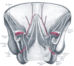 Posterior view of the anterior abdominal wall in its lower half. The peritoneum is in place, and the various cords are shining through. Median umbilical ligament isn't labeled, but it is located just underneath the median umbilical fold, seen in the center of the diagram | |
| Details | |
| From | Apex of urinary bladder |
| To | umbilicus |
| Identifiers | |
| Latin |
Ligamentum umbilicale medianum, ligamentum suspensorium vesicae urinariae |
| TA | A08.3.01.007 |
| FMA | 16568 |
| Anatomical terminology | |
The median umbilical ligament is a structure in human anatomy. It is a shrivelled piece of tissue that represents the remnant of the embryonic urachus.
It extends from the apex of the bladder to the umbilicus, on the deep surface of the anterior abdominal wall. It is unpaired.
It is covered by the median umbilical fold.
Lateral to this structure are the medial umbilical ligament (which is a different structure, not to be confused) and the lateral umbilical ligament.
Significance
It may be used as a landmark for surgeons who are performing laparoscopy, such as laparoscopic inguinal hernia repair. Other than this, it has no function in a born human and may be cut or removed with impunity.
References
External links
- Median umbilical ligament
- Anatomy figure: 36:01-01 at Human Anatomy Online, SUNY Downstate Medical Center - "The inguinal canal and derivation of the layers of the spermatic cord."
- Anatomy photo:44:04-0105 at the SUNY Downstate Medical Center - "The Male Pelvis: The Urinary Bladder"
- Anatomy image:7576 at the SUNY Downstate Medical Center
- Median umbilical fold
- Anatomy figure: 36:03-12 at Human Anatomy Online, SUNY Downstate Medical Center - "Internal surface of the anterior abdominal wall."
Anatomy of the urinary system | |||||||||||||
|---|---|---|---|---|---|---|---|---|---|---|---|---|---|
| Kidneys |
| ||||||||||||
| Ureters | |||||||||||||
| Bladder | |||||||||||||
| Urethra |
| ||||||||||||
| |||||||||||||
This article is issued from
Wikipedia.
The text is licensed under Creative Commons - Attribution - Sharealike.
Additional terms may apply for the media files.