Pseudopodia
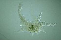
A pseudopod or pseudopodium (plural: pseudopods or pseudopodia) (from the Greek word ψευδοποδός, ψευδός "false" + ποδός "foot") is a temporary cytoplasm-filled projection of an eukaryotic cell membrane or a unicellular protist. Pseudopods may be used for motility, or for ingesting nutrients or other particulate matter. Cells that make pseudopods are generally referred to as amoeboids. Pseudopods extend and contract by the reversible assembly of actin subunits into many microfilaments. Filaments near the cell's end interact with myosin which causes contraction. The pseudopod extends itself until the actin reassembles itself into a network.
It is supposed that actin polymerization is at the origin of the force propelling the cell forward. Generally several pseudopodia arise from the surface of the body (polypodial, e.g. Amoeba proteus), or a single pseudopod may form on the surface of the body (monopodial, e.g. Entamoeba histolytica).
Pseudopodia are formed by microtubule and filament structures. The cell surface projects a membrane process called the lamellipodium, which is supported inside by filaments that form at the leading edge, turning into networks as they merge. Cytoplasm flows into the lamellipodium, forming the pseudopodia.
The functions of pseudopodia include locomotion and the capturing of prey. Pseudopodia are critical in sensing prey that can then be engulfed; the engulfing pseudopodia are called phagocytosis pseudopodia. A common example of this sort of amoeboid cell is the human white blood cell. Human mesenchymal stem cells are a good example of a cell type which uses pseudopodia for locomotive reasons: these migratory cells are responsible for in-utero remodeling for example in the formation of the trilaminar germ disc during gastrulation.[1]
Pseudopodia do not all look like amorphous blobs; instead, they can be classified by their distinct appearances.[2] Lobopodia are bulbous and amoebic. Filopodia are slender, thread-like, and are supported largely by microfilaments. Reticulopodia are very complex and bear individual pseudopodia that form irregular nets. Axopodia are the phagocytosis type with long, thin pseudopods supported by complex microtubule arrays enveloped with cytoplasm, and they respond rapidly to physical contact.
Morphology
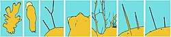
Pseudopods can be classified into several varieties, according to the number of projections (monopodia and polypodia), and according to their appearance:
- Lobopodia (or lobose pseudopods) are bulbous, short, and blunt in form. These finger-like, tubular pseudopodia contain both ectoplasm and endoplasm. They occur in Lobosa and other Amoebozoa, and in some Heterolobosea (Excavata).
- Filopodia (or filose pseudopods) are more slender and filiform with pointed ends, consisting mainly of ectoplasm. These formations are supported by microfilaments. This is observed in some animal cells, in part of Filosa (Rhizaria), in "Testaceafilosia" (an artificial group including Euglyphida), in Vampyrellidae and Pseudosporida (Rhizaria) and in Nucleariida (Opisthokonta).
- Reticulopodia (or reticulose pseudopods),[3] are complex formations where individual pseudopods are blended together and form irregular nets. The primary function of reticulopodia, also known as myxopodia, is the ingestion of food, with locomotion a secondary function. Reticulopods are typical of Foraminifera, Chlorarachnea, Gromia and Filoreta (Rhizaria).
- Axopodia (also known as actinopodia) are thin pseudopods containing complex arrays of microtubules and are enveloped by cytoplasm. Axopodia are mostly responsible for phagocytosis by rapidly retracting in response to physical contacts. This supposedly takes a strain on the helix, for after the sensory action has occurred, it then later on dies. Principally, these pseudopodia are food collecting structures. They are observed in "Radiolaria" (an artificial group inside Rhizaria) and "Heliozoa" (artificial also).
References
- ↑ Schoenwolf, Gary (2009). Larsen's Human Embryology: 4th Edition. Churchill Livingstone Elsevier.
- ↑ David J. Patterson. "Amoebae: Protists Which Move and Feed Using Pseudopodia". Tree of Life Web Project. Retrieved 12 November 2017.
- ↑ "Archived copy". Archived from the original on 2007-07-17. Retrieved 2005-12-30.