Phosphatidylinositol 4,5-bisphosphate
 | |
| Names | |
|---|---|
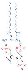
| IUPAC name
1,2-Diacyl-sn-glycero-3-phospho-(1-D-myo-inositol 4,5-bisphosphate) | |
| Identifiers | |
3D model (JSmol) |
|
| ChemSpider | |
PubChem CID |
|
| |
| |
| Properties | |
| C47H80O19P3 | |
| Molar mass | 1042.05 g/mol |
Except where otherwise noted, data are given for materials in their standard state (at 25 °C [77 °F], 100 kPa). | |
| Infobox references | |
Phosphatidylinositol 4,5-bisphosphate or PtdIns(4,5)P2, also known simply as PIP2 or PI(4,5)P2, is a minor phospholipid component of cell membranes. PtdIns(4,5)P2 is enriched at the plasma membrane where it is a substrate for a number of important signaling proteins.[1]
PIP2 is formed primarily by the type I phosphatidylinositol 4-phosphate 5-kinases from PI(4)P. In metazoans, PIP2 can also be formed by type II phosphatidylinositol 5-phosphate 4-kinases from PI(5)P.[2]
The fatty acids of PIP2 are variable in different species and tissues, but the most common fatty acids are stearic in position 1 and arachidonic in 2.[3]
Signaling Pathways
PIP2 is a part of many cellular signaling pathways notably PIP2 cycle, PI3K signalling and PI5P metabolism.[4] Recently it has been found in the nucleus[5] with unknown function.
Functions
Cytoskeleton dynamics near membranes
PIP2 regulates the organization, polymerization, and branching of filamentous actin (F-actin) via direct binding to F-actin regulatory proteins.[6]
Endocytosis and exocytosis
The first evidence that indicated phosphoinositides(PIs) (especially PI(4,5)P2) are important during the exocytosis process was in 1990. Emberhard et al. [7] found that the application of PI-specific phospholipase C into digitonin-permeablized chromaffin cells decreased PI levels, and inhibited calcium-triggered exocytosis. This exocytosis inhibition was preferential for an ATP-dependent stage, indicating PI function was required for secretion. Later studies identified associated proteins necessary during this stage, such as phosphatidylinositol transfer protein ,[8] and phosphoinositol-4-monophosphatase 5 kinase type Iγ (PIPKγ) ,[9] which mediates PI(4,5)P2 restoration in permeable cell incubation in an ATP-dependent way. In these later studies, PI(4,5)P2 specific antibodies strongly inhibited exocytosis, thus providing direct evidence that PI(4,5)P2 plays a pivotal role during the LDCV (Large dense core vesicle) exocytosis process.
Through the use of PI-specific kinase/phosphatase identification and PI antibody/drug/blocker discovery, the role of PI (especially PI(4,5)P2) in secretion regulation was extensively investigated. Studies utilizing PHPLCδ1 domain over-expression (acting as PI(4,5)P2 buffer or blocker) ,[10] PIPKIγ knockout in chromaffin cell [11] and in central nerve system [12] , PIPKIγ knockdown in beta cell lines ,[13] and over-expression of membrane-tethered inositol 5-phosphatase domain of synaptojanin 1 ,[14] all suggested vesicle (synaptic vesicle and LDCV) secretion were severely impaired after PI(4,5)P2 depletion or blockage. Moreover, some studies[14][12][11] showed an impaired/reduced RRP of those vesicles, though the docked vesicle number were not altered[11] after PI(4,5)P2 depletion, indicating a defect at a pre-fusion stage (priming stage). Follow-up studies indicated that PI(4,5)P2 interactions with CAPS,[15] Munc13[16] and synaptotagmin1[17] are likely to play a role in this PI(4,5)P2 dependent priming defect.
IP3/DAG pathway
PIP2 functions as an intermediate in the [IP3/DAG pathway], which is initiated by ligands binding to G protein-coupled receptors activating the Gq alpha subunit. PtdIns(4,5)P2 is a substrate for hydrolysis by phospholipase C (PLC), a membrane-bound enzyme activated through protein receptors such as α1 adrenergic receptors. PIP2 regulates the function of many membrane proteins and ion channels, such as the M-channel. The products of the PLC catalyzation of PIP2 are inositol 1,4,5-trisphosphate (InsP3; IP3) and diacylglycerol (DAG), both of which function as second messengers. In this cascade, DAG remains on the cell membrane and activates the signal cascade by activating protein kinase C (PKC). PKC in turn activates other cytosolic proteins by phosphorylating them. The effect of PKC could be reversed by phosphatases. IP3 enters the cytoplasm and activates IP3 receptors on the smooth endoplasmic reticulum (ER), which opens calcium channels on the smooth ER, allowing mobilization of calcium ions through specific Ca2+ channels into the cytosol. Calcium participates in the cascade by activating other proteins.
Docking phospholipids
Class I PI 3-kinases phosphorylate PtdIns(4,5)P2 forming phosphatidylinositol (3,4,5)-trisphosphate (PtdIns(3,4,5)P3) and PtdIns(4,5)P2 can be converted from PtdIns4P. PtdIns4P, PtdIns(3,4,5)P3 and PtdIns(4,5)P2 not only act as substrates for enzymes but also serve as docking phospholipids that bind specific domains that promote the recruitment of proteins to the plasma membrane and subsequent activation of signaling cascades.[18][19]
- Examples of proteins activated by PtdIns(3,4,5)P3 are AKT, PDPK1, Btk1.
- One mechanism for direct effect of PtdIns(4,5)P2 is opening of Na+ channels as a minor function in growth hormone release by growth hormone-releasing hormone.[20]
Potassium channels
Inwardly rectifying potassium channels have been shown to require docking of PIP2 for channel activity.[21][22]
G protein-coupled receptors
PtdIns(4,5)P2 has been shown to stabilise the active states of Class A G protein-coupled receptors (GPCRs) via direct binding, and enhance their selectivity toward certain G proteins.[23]
Regulation
PIP2 is regulated by many different components. One emerging hypothesis is that PIP2 concentration is maintained locally. Some of the factors involved in PIP2 regulation are:[24]
- Lipid kinases, Lipid Phosphatase
- Lipid Transfer Proteins
- Growth Factors, Small GTPases
- Cell Attachment
- Cell-Cell Interaction
- Change in cell volume
- Cell differentiation state
- Cell stress
References
- ↑ Strachan T, Read AP (1999). Leptospira. In: Human Molecular Genetics (2nd ed.). Wiley-Liss. (via NCBI Bookshelf) ISBN 0-471-33061-2.
- ↑ Rameh, LE; Tolias, K; Duckworth, BC; Cantley, LC (Nov 1997). "A new pathway for synthesis of phosphatydilinositol-4,5-bisphosphate". Nature. 390 (6656): 192–6. doi:10.1038/36621. PMID 9367159.
- ↑ Tanaka T, Iwawaki D, Sakamoto M, Takai Y, Morishige J, Murakami K, Satouchi K (April 2003). "Mechanisms of accumulation of arachidonate in phosphatidylinositol in yellowtail. A comparative study of acylation systems of phospholipids in rat and the fish species Seriola quinqueradiata". Eur J Biochem. 270 (7): 1466–73. doi:10.1046/j.1432-1033.2003.03512.x. PMID 12654002.
- ↑ Bulley SJ, Clarke JH, Droubi A, Giudici ML, Irvine RF. "Exploring phosphatidylinositol 5-phosphate 4-kinase function". Adv Biol Regul. 57: 193–202. doi:10.1016/j.jbior.2014.09.007. PMC 4359101. PMID 25311266.
- ↑ Lewis AE, Sommer L, Arntzen MØ, Strahm Y, Morrice NA, Divecha N, D'Santos CS. "Identification of nuclear phosphatidylinositol 4,5-bisphosphate-interacting proteins by neomycin extraction". Mol Cell Proteomics. 10: M110.003376. doi:10.1074/mcp.M110.003376. PMC 3033679. PMID 21048195.
- ↑ Sun, Hui; Yamamoto, Masaya; Mejillano, Marisan; Yin, Helen (November 19, 1999). "Gelsolin, a Multifunctional Actin Regulatory Protein". The Journal of Biological Chemistry. 274 (47): 33179. doi:10.1074/jbc.274.47.33179. PMID 10559185.
- ↑ Eberhard, David A, et al. (1990). "Evidence that the inositol phospholipids are necessary for exocytosis. Loss of inositol phospholipids and inhibition of secretion in permeabilized cells caused by a bacterial phospholipase C and removal of ATP". Biochemical Journal. 268: 15–25. doi:10.1042/bj2680015. PMC 1131385.
- ↑ Hay, Jesse C, Thomas M (1993). "Phosphatidylinositol transfer protein required for ATP-dependent priming of Ca2+-activated secretion". Nature. 366: 572–575. doi:10.1038/366572a0.
- ↑ Hay, Jesse C, et al. (1995). "ATP-dependent inositide phosphorylation required for Ca2positive-activated secretion". Nature. 374: 173–177. doi:10.1038/374173a0.
- ↑ Holz RW, et al. (2000). "A pleckstrin homology domain specific for phosphatidylinositol 4, 5-bisphosphate (PtdIns-4, 5-P2) and fused to green fluorescent protein identifies plasma membrane PtdIns-4, 5-P2 as being important in exocytosis". J. Biol. Chem. 275: 17878–17885. doi:10.1074/jbc.M000925200.
- 1 2 3 Gong LW, et al. (2005). "Phosphatidylinositol phosphate kinase type Iγ regulates dynamics of large dense-core vesicle fusion". PNAS. 102: 5204–5209. doi:10.1073/pnas.0501412102.
- 1 2 Di Paolo G, et al. (2004). "Impaired PtdIns (4, 5) P2 synthesis in nerve terminals produces defects in synaptic vesicle trafficking". Nature. 431: 415–422. doi:10.1038/nature02896.
- ↑ Waselle L, et al. (2005). "Role of phosphoinositide signaling in the control of insulin exocytosis". Molecular Endocrinology. 19: 3097–3106. doi:10.1210/me.2004-0530.
- 1 2 Milosevic I, et al. (2005). "Plasmalemmal phosphatidylinositol-4, 5-bisphosphate level regulates the releasable vesicle pool size in chromaffin cells". Journal of Neuroscience. 25: 2557–2565. doi:10.1523/JNEUROSCI.3761-04.2005.
- ↑ Grishanin RN, et al. (2004). "CAPS acts at a prefusion step in dense-core vesicle exocytosis as a PIP 2 binding protein". Neuron. 43: 551–562. doi:10.1016/j.neuron.2004.07.028.
- ↑ Kabachinski G, et al. (2014). "CAPS and Munc13 utilize distinct PIP2-linked mechanisms to promote vesicle exocytosis". Molecular Biology of the Cell. 25: 508–521. doi:10.1091/mbc.E12-11-0829. PMC 3923642.
- ↑ Loewen CA, et al. (2006). "C2B polylysine motif of synaptotagmin facilitates a Ca2+-independent stage of synaptic vesicle priming in vivo". Molecular Biology of the Cell. 17: 5211–5226. doi:10.1091/mbc.E06-07-0622.
- ↑ Won DH, et al. (2006). "PI (3, 4, 5) P3 and PI (4, 5) P2 lipids target proteins with polybasic clusters to the plasma membrane". Science. 314 (5804): 1458–1461. doi:10.1126/science.1134389. PMC 3579512.
- ↑ Hammond G, et al. (2012). "PI4P and PI (4, 5) P2 are essential but independent lipid determinants of membrane identity". Science. 337 (6095): 727–730. doi:10.1126/science.1222483. PMC 3646512.
- ↑ GeneGlobe -> GHRH Signaling Retrieved on May 31, 2009
- ↑ Soom, M. "Multiple PtdIns(4,5)P2 binding sites in Kir2.1 inwardly rectifying potassium channels". FEBS Letters. 490 (1–2): 49–53. doi:10.1016/S0014-5793(01)02136-6.
- ↑ Hansen, SB; Tao, X; MacKinnon, R (28 August 2011). "Structural basis of PIP2 activation of the classical inward rectifier K+ channel Kir2.2". Nature. 477 (7365): 495–8. doi:10.1038/nature10370. PMC 3324908. PMID 21874019.
- ↑ Yen, Hsin-Yung; Hoi, Kin Kuan; Liko, Idlir; Hedger, George; Horrell, Michael R.; Song, Wanling; Wu, Di; Heine, Philipp; Warne, Tony (2018-07-11). "PtdIns(4,5)P2 stabilizes active states of GPCRs and enhances selectivity of G-protein coupling". Nature. 559 (7714). doi:10.1038/s41586-018-0325-6. ISSN 0028-0836.
- ↑ Hilgemann, D. W. "The Complex and Intriguing Lives of PIP2 with Ion Channels and Transporters". Science's STKE. 2001 (111): 19re–19. doi:10.1126/stke.2001.111.re19.