Retinoic acid
 | |
 | |
| Names | |
|---|---|
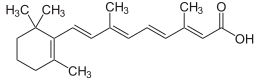
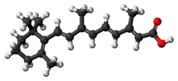
| IUPAC name
(2E,4E,6E,8E)-3,7-dimethyl-9-(2,6,6-trimethylcyclohexen-1-yl)nona-2,4,6,8-tetraenoic acid | |
| Other names
vitamin A acid; RA | |
| Identifiers | |
3D model (JSmol) |
|
| ChEMBL | |
| ChemSpider | |
PubChem CID |
|
| |
| |
| Properties | |
| C20H28O2 | |
| Molar mass | 300.43512 g/mol |
| Appearance | yellow to light orange crystalline powder with characteristic floral odor [1] |
| Melting point | 180 to 182 °C (356 to 360 °F; 453 to 455 K) crystals from ethanol[1] |
| nearly insoluble | |
| Solubility in fat | soluble |
| Related compounds | |
Related compounds |
retinol; retinal; beta-carotene |
Except where otherwise noted, data are given for materials in their standard state (at 25 °C [77 °F], 100 kPa). | |
| Infobox references | |
Retinoic acid is a metabolite of vitamin A (retinol) that mediates the functions of vitamin A required for growth and development. Retinoic acid is required in chordate animals, which includes all higher animals from fish to humans. During early embryonic development, retinoic acid generated in a specific region of the embryo helps determine position along the embryonic anterior/posterior axis by serving as an intercellular signaling molecule that guides development of the posterior portion of the embryo.[2] It acts through Hox genes, which ultimately control anterior/posterior patterning in early developmental stages.[3]
The key role of retinoic acid in embryonic development mediates the high teratogenicity of retinoid pharmaceuticals, such as isotretinoin used for treatment of cancer and acne. Oral megadoses of pre-formed vitamin A (retinyl palmitate), and retinoic acid itself, also have teratogenic potential by this same mechanism.
Mechanism of biological action
Retinoic acid acts by binding to the retinoic acid receptor (RAR), which is bound to DNA as a heterodimer with the retinoid X receptor (RXR) in regions called retinoic acid response elements (RAREs). Binding of the retinoic acid ligand to RAR alters the conformation of the RAR, which affects the binding of other proteins that either induce or repress transcription of a nearby gene (including Hox genes and several other target genes). Retinoic acid receptors mediate transcription of different sets of genes controlling differentiation of a variety of cell types, thus the target genes regulated depend upon the target cells.[4] In some cells, one of the target genes is the gene for the retinoic acid receptor itself (RAR-beta in mammals), which amplifies the response.[5] Control of retinoic acid levels is maintained by a suite of proteins that control synthesis and degradation of retinoic acid.[2][3]
The molecular basis for the interaction between retinoic acid and the Hox genes has been studied by using deletion analysis in transgenic mice carrying constructs of GFP reporter genes. Such studies have identified functional RAREs within flanking sequences of some of the most 3' Hox genes (including Hoxa1, Hoxb1, Hoxb4, Hoxd4), suggesting a direct interaction between the genes and retinoic acid. These types of studies strongly support the normal roles of retinoids in patterning vertebrate embryogenesis through the Hox genes.[6]
Biosynthesis
Retinoic acid can be produced in the body by two sequential oxidation steps that convert retinol to retinaldehyde to retinoic acid, but once produced it cannot be reduced again to retinol. The enzyme that generate retinoic acid for control of gene expression include retinol dehydrogenase (i.e. Rdh10) that metabolize retinol to retinal, and retinal dehydrogenase: RALHD1 (ALDH1A1), RALHD2 (ALDH1A2), and RALHD3 (ALDH1A3)[7] that metabolize retinal to retinoic acid.[2] Enzymes that metabolize excess retinol to prevent toxicity include alcohol dehydrogenase and cytochrome P450(cyp26).
Retinoic acid function in the absence of precursors retinol or retinaldehyde
Retinoic acid is responsible for most of the activity of vitamin A, save visual pigment effects that require retinal (retinaldehyde), and cell metabolism effects that may require retinol itself. Also, some biochemical functions necessary for fertility in vitamin A deficient male and female mammals originally appeared to require retinol for rescue, but this is due to a requirement for local conversion of retinol to retinoic acid, as administered retinoic acid does not reach some critical tissues unless given in high amounts. Thus, if animals are fed only retinoic acid but no vitamin A (retinol or retinal), they suffer none of the growth-stunting or epithelial-damaging effects of lack of vitamin A (including no xerophthalmia—dryness of the cornea). They do suffer retina degeneration and blindness, due to retinal (retinaldehyde) deficiency.
In addition, vitamin A-deprived but retinoic acid-supplemented male rats exhibit hypogonadism and infertility due to lack of local retinoic acid synthesis in the testis; similar treatment of female rats causes infertility due to fetal resorption caused by a lack of local retinoic acid synthesis in the embryo.[8][9] The retinoic acid synthesis in testes is catalyzed primarily by the RALDH2 (ALDH1A2) aldehyde dehydrogenase. Suppressing this enzyme has been proposed as a possible way to make a male contraceptive pill, because retinoic acid is necessary for spermatogenesis in humans, much as in rats.[10]
Retinoic acid function in embryo development
Retinoic acid is a morphogen signaling molecule, which means it is concentration dependent. Other molecules that interact with RA are FGF-8, Cdx and Hox genes, all participating in the development of various structures within the fetus. For example, this molecule plays an important role in hindbrain development. The hindbrain differentiates into the brain stem and serves as a major signaling center during the initial development of the heart.[11] Retinoic acid gradient is vital in somite formation, as well as the formation of the atria in the heart. Malformations can arise when the concentration of RA is in excess or deficient. The hindbrain becomes enlarged when there is excess RA, hindering the growth of other parts of the brain. Other abnormalities that can occur are missing or fused somites and problems with the aorta and large vessels within the heart. With an accumulation of these malformations, an individual can be diagnosed with DiGeorge syndrome.[12] However, since RA partakes in various developmental processes abnormalities are not just limited to sites associated with DiGeorge syndrome. Retinoic acid is essential throughout an individual’s lifetime, but it is critical during pregnancy. Without the proper concentrations of this molecule, severe abnormalities can be present and even fatal to the growing fetus. Knockout experiments have been conducted and results have shown that improper concentrations of RA can lead to abnormal development, most experiments concentrated on hindbrain development.[12]
Related pharmaceuticals
- Tretinoin (Tradename: Retin-A)
- Isotretinoin (Tradename: Accutane(US), Roaccutane)
References
- 1 2 Merck Index, 13th Edition, 8251.
- 1 2 3 Duester, G (September 2008). "Retinoic Acid Synthesis and Signaling during Early Organogenesis". Cell. 134 (6): 921–31. doi:10.1016/j.cell.2008.09.002. PMC 2632951. PMID 18805086.
- 1 2 Holland, Linda Z. (2007). "Developmental biology: A chordate with a difference". Nature. 447 (7141): 153–155. doi:10.1038/447153a. PMID 17495912.
- ↑ Venkatesh K, Srikanth L, Vengamma B, Chandrasekhar C, Sanjeevkumar A, Mouleshwara Prasad BC, Sarma PV. In vitro differentiation of cultured human CD34+ cells into astrocytes. Neurol India 2013;61:383-8
- ↑ Edgar Wingender (1993). "Steroid/Thyroid Hormone Receptors". Gene Regulation in Eukaryotes. New York: VCH. p. 316. ISBN 1-56081-706-2.
- ↑ Marshall, H.; et al. (1996). "Retinoids and Hox genes" (PDF). The FASEB Journal. 10 (9): 969–978. Retrieved 2009-02-19.
- ↑ "ALDH 1 Family". Dr. Vasilis Vasiliou's laboratory at the University of Colorado's Health Sciences Center. Archived from the original on 13 January 2013. Retrieved 22 October 2012.
- ↑ Moore, T.; Holmes, P. D. (1971). "The production of experimental vitamin A deficiency in rats and mice". Lab Anim. 5: 239–250. doi:10.1258/002367771781006492. PMID 5126333.
- ↑ VanPelt, H.M.M.; DeRooij, D.G. (1991). "Retinoic Acid Is Able to Reinitiate Spermatogenesis in Vitamin A-Deficient Rats and High Replicate Doses Support the Full Development of Spermatogenic Cells". Endocrinology. 128 (2): 697–704. doi:10.1210/endo-128-2-697. PMID 1989855.
- ↑ Sam Kean (2012). "Reinventing the Pill: Male Birth Control". Science. 338: 318–320. doi:10.1126/science.338.6105.318. PMID 23087225.
- ↑ Lee, Keun; Skromne, Isaac (2014-11-15). "Retinoic acid regulates size, pattern and alignment of tissues at the head-trunk transition". Development. 141 (22): 4375–4384. doi:10.1242/dev.109603. ISSN 0950-1991. PMID 25371368.
- 1 2 Rhinn, Muriel; Dollé, Pascal (2012-03-01). "Retinoic acid signalling during development". Development. 139 (5): 843–858. doi:10.1242/dev.065938. ISSN 0950-1991. PMID 22318625.