Nasal septum
| Nasal septum | |
|---|---|
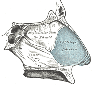 Bones and cartilages of septum of nose. Right side. | |
 MRI image showing nasal septum. | |
| Details | |
| Artery |
Anterior ethmoidal posterior ethmoidal sphenopalatine greater palatine branch of superior labial[1] |
| Nerve | Medial posterosuperior nasal branches of pterygopalatine ganglion |
| Lymph |
Anterior half to submandibular nodes Posterior half to retropharyngeal and deep cervical lymph nodes |
| Identifiers | |
| Latin | Septum nasi |
| MeSH | D009300 |
| TA | A06.1.02.004 |
| FMA | 54375 |
| Anatomical terminology | |
The nasal septum (Latin: septum nasi) separates the left and right airways in the nose, dividing the two nostrils.
It is depressed by the depressor septi nasi muscle.
Structure
The fleshy external end of the nasal septum is sometimes also called the columella nasi. The nasal septum contains bone and hyaline cartilage.[2] It is normally about 2 mm thick.[3]
The nasal septum is composed of five structures:
- perpendicular plate of ethmoid bone
- vomer bone
- septal nasal cartilage
- crest of the maxillary bone
- crest of the palatine bone
Development
At an early period the septum of the nose consists of a plate of cartilage, known as the ethmovomerine cartilage.
The postero-superior part of this cartilage is ossified to form the perpendicular plate of the ethmoid; its antero-inferior portion persists as the septal cartilage, while the vomer is ossified in the membrane covering its postero-inferior part.
Two ossification centers, one on either side of the middle line, appear about the eighth week of fetal life in this part of the membrane, and hence the vomer consists primarily of two lamellae.
About the third month these unite below, and thus a deep groove is formed in which the cartilage is lodged.
As growth proceeds, the union of the lamellae extends upward and forward, and at the same time the intervening plate of cartilage undergoes absorption.
By the onset of puberty the lamellae are almost completely united to form a median plate, but evidence of the bilaminar origin of the bone is seen in the everted alae of its upper border and in the groove on its anterior margin.
Clinical significance
The nasal septum can depart from the centre line of the nose in a condition that is known as a deviated septum caused by trauma. However, it is normal to have a slight deviation to one side. The septum generally stays in the midline until about the age of seven, at which point it will frequently deviate to the right.
A perforated nasal septum can be caused by an ulcer, trauma due to an inserted object, long-term exposure to welding fumes,[4] or cocaine use. There is a procedure that can be of help to those suffering from perforated septum. A silicone button can be inserted in the hole to close the open sore.
An operation to straighten the nasal septum is known as a septoplasty.
The nasal septum can be affected by both benign (fibroma, inflammatory hemangioma of the nasal septum—"bleeding polyp", etc.) and malignant tumors (squamous cell carcinoma, esthesioneuroblastoma, etc.).
Cosmetic procedures
The inferior part of the nasal septum can be pierced, usually through the soft tissue but the piercing can also transverse (partially) the cartilage of the septum.
See also
References
- 1 2 lesson9 at The Anatomy Lesson by Wesley Norman (Georgetown University)
- ↑ Saladin, Kenneth S. (2012). Anatomy and Physiology (6th ed.). New York, NY: McGraw Hill. p. 856. ISBN 9780073378251.
- ↑ Liong, Kyrin; Lee, Shu Jin; Lee, Heow Pueh (2013). "Preliminary Deformational Studies on a Finite Element Model of the Nasal Septum Reveals Key Areas for Septal Realignment and Reconstruction". Journal of Medical Engineering. 2013: 1–8. doi:10.1155/2013/250274. ISSN 2314-5129.
- ↑ Lee, Choong Ryeol; Yoo, Cheol In; Lee, Ji Ho; Kang, Seong Kyu (2002). "Nasal Septum Perforation of Welders". Journal of Industrial Health. 40 (3): 286–289. doi:10.2486/indhealth.40.286.
- This article incorporates text in the public domain from page 993 of the 20th edition of Gray's Anatomy (1918)
External links
| Wikimedia Commons has media related to Nasal septum. |
- Anatomy figure: 33:02-01 at Human Anatomy Online, SUNY Downstate Medical Center - "Diagram of skeleton of medial (septal) nasal wall."
- Diagram at evmsent.org
- lesson9 at The Anatomy Lesson by Wesley Norman (Georgetown University) (nasalseptumbonescarti)