LASIK
| LASIK | |
|---|---|
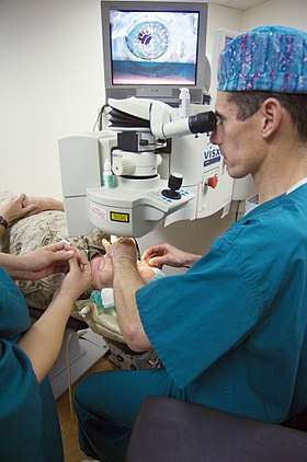 LASIK surgery using an excimer laser at US National Naval Medical Center Bethesda | |
| ICD-9-CM | 11.71 |
| MeSH | D020731 |
| MedlinePlus | 007018 |
LASIK or Lasik (laser-assisted in situ keratomileusis), commonly referred to as laser eye surgery or laser vision correction, is a type of refractive surgery for the correction of myopia, hyperopia, and astigmatism.[1] The LASIK surgery is performed by an ophthalmologist who uses a laser or microkeratome to reshape the eye's cornea in order to improve visual acuity.[2] For most people, LASIK provides a long-lasting alternative to eyeglasses or contact lenses.[3]
LASIK is most similar to another surgical corrective procedure, photorefractive keratectomy (PRK), and LASEK. All represent advances over radial keratotomy in the surgical treatment of refractive errors of vision. For patients with moderate to high myopia or thin corneas which cannot be treated with LASIK and PRK, the phakic intraocular lens is an alternative.[4][5] As of 2018, nearly 10 million LASIK procedures had been performed in the United States[1][6] and, as of 2016, over 40 million have been performed worldwide since 1991.[7][8] However, the procedure seems to be a declining option for many in the United States, dropping more than 50 percent, from about 1.5 million surgeries in 2007 to 604,000 in 2015, according to the eye care data source Market Scope.[9]
Effectiveness
In 2006, the British National Health Service's National Institute for Health and Clinical Excellence (NICE) considered evidence of the effectiveness and the potential risks of the laser surgery stating "current evidence suggests that photorefractive (laser) surgery for the correction of refractive errors is safe and efficacious for use in appropriately selected patients. Clinicians undertaking photorefractive (laser) surgery for the correction of refractive errors should ensure that patients understand the benefits and potential risks of the procedure. Risks include failure to achieve the expected improvement in unaided vision, development of new visual disturbances, corneal infection and flap complications. These risks should be weighed against those of wearing spectacles or contact lenses."[10] The FDA reports "The safety and effectiveness of refractive procedures has not been determined in patients with some diseases."[11]
Satisfaction
Surveys of LASIK surgery find rates of patient satisfaction between 92 and 98 percent.[12][13][14] In March 2008, the American Society of Cataract and Refractive Surgery published a patient satisfaction meta-analysis of over 3,000 peer-reviewed articles from international clinical journals. Data from the prior 10 years revealed a 95.4 percent patient satisfaction rate among LASIK patients.[15]
Dissatisfaction
Some people with poor outcomes from LASIK surgical procedures report a significantly reduced quality of life because of vision problems or physical pain associated with the surgery.[1] A small percentage of patients may need to have another surgery because their condition is over-corrected or under-corrected. Some patients need to wear contact lenses or glasses even after treatment.[16]
The most common reason for dissatisfaction in LASIK patients is chronic severe dry eye. Independent research indicates 95% of patients experience dry eye in the initial post-operative period. This number has been reported to up to 60% after one month. Symptoms begin to improve in the vast majority of patients in the 6 to 12 months following the surgery.[17] However, 30% of post-LASIK referrals to tertiary ophthalmology care centers have been shown to be due to chronic dry eye.[18][19]
Morris Waxler, a former FDA official who was involved in the approval of LASIK, has subsequently criticized its widespread use. In 2010, Waxler made media appearances and claimed that the procedure had a failure rate greater than 50%. The FDA responded that Waxler's information was "filled with false statements, incorrect citations" and "mischaracterization of results".[20]
A 2016 JAMA study indicates that the prevalence of complications from LASIK are higher than indicated, with the study indicating many patients wind up with glare, halos or other visual symptoms.[21]
Presbyopia
A type of LASIK, known as presbyLasik, may be used in presbyopia. Results are, however, more variable and some people have a decrease in visual acuity.[22]
Risks
Higher-order aberrations
Higher-order aberrations are visual problems that require special testing for diagnosis and are not corrected with normal spectacles (eyeglasses). These aberrations include 'starbursts', 'ghosting', 'halos' and others.[23][1] Some patients describe these symptoms post-operatively and associate them with the LASIK technique including the formation of the flap and the tissue ablation.[24]
The advancement of the LASIK technology has reduced the risk of clinically significant visual impairment after surgery. There is a correlation between pupil size and aberrations. This correlation may be the result of irregularity in the corneal tissue between the untouched part of the cornea and the reshaped part. Daytime post-LASIK vision is optimal, since the pupil size is smaller than the LASIK flap. However, at night, the pupil may dilate such that light passes through the edge of the LASIK flap, which gives rise to aberrations. LASIK and PRK may induce spherical aberration if the laser under-corrects as it moves outward from the centre of the treatment zone, especially when major corrections are made.
Others propose that higher-order aberrations are present preoperatively.[25] They can be measured in micrometers (µm) whereas the smallest laser beam size approved by the FDA is about 1000 times larger, at 0.65 mm. In situ keratomileusis effected at a later age increases the incidence of corneal higher-order wavefront aberrations.[26][27] These factors demonstrate the importance of careful patient selection for LASIK treatment.
Dry eyes
95% of patients report dry eye symptoms after LASIK[28][1] Although it is usually temporary, it can develop into chronic and severe dry eye syndrome. Quality of life can be severely affected by dry eye syndrome.[29]
Underlying conditions with dry eye such as Sjögren's syndrome are considered contraindications to Lasik.[30]
Treatments include artificial tears, prescription tears and punctal occlusion. Punctal occlusion is accomplished by placing a collagen or silicone plug in the tear duct, which normally drains fluid from the eye. Some patients complain of ongoing dry eye symptoms despite such treatments and dry eye symptoms may be permanent.[31]
Halos
Some post-LASIK patients see halos and starbursts around bright lights at night.[1] At night, the pupil may dilate to be larger than the flap leading to the edge of the flap or stromal changes causing visual distortion of light that does not occur during the day when the pupil is smaller. The eyes can be examined for large pupils pre-operatively and the risk of this symptom assessed.
Complications due to LASIK have been classified as those that occur due to preoperative, intraoperative, early postoperative, or late postoperative sources:[32] According to the UK National Health Service complications occur in fewer than 5% of cases.[28]
Other complications
- Flap complications – The incidence of flap complications is about 0.244%.[33] Flap complications (such as displaced flaps or folds in the flaps that necessitate repositioning, diffuse lamellar keratitis, and epithelial ingrowth) are common in lamellar corneal surgeries[34] but rarely lead to permanent loss of visual acuity. The incidence of these microkeratome-related complications decreases with increased physician experience.[35]
- Slipped flap – is a corneal flap that detaches from the rest of the cornea. The chances of this are greatest immediately after surgery, so patients typically are advised to go home and sleep to let the flap adhere and heal. Patients are usually given sleep goggles or eye shields to wear for several nights to prevent them from dislodging the flap in their sleep. A short operation time may decrease the chance of this complication, as there is less time for the flap to dry.
- Flap interface particles – are a finding whose clinical significance is undetermined.[36] Particles of various sizes and reflectivity are clinically visible in about 38.7% of eyes examined via slit lamp biomicroscopy and in 100% of eyes examined by confocal microscopy.[36]
- Diffuse lamellar keratitis – an inflammatory process that involves an accumulation of white blood cells at the interface between the LASIK corneal flap and the underlying stroma. It is known colloquially as "sands of Sahara syndrome" because on slit lamp exam, the inflammatory infiltrate appears similar to waves of sand. The USAeyes organisation reports an incidence of 2.3% after LASIK.[37] It is most commonly treated with steroid eye drops. Sometimes it is necessary for the eye surgeon to lift the flap and manually remove the accumulated cells. DLK has not been reported with photorefractive keratectomy due to the absence of flap creation.
- Infection – the incidence of infection responsive to treatment has been estimated at 0.4%.[37]
- Post-LASIK corneal ectasia – a condition where the cornea starts to bulge forwards at a variable time after LASIK, causing irregular astigmatism. the condition is similar to keratoconus.
- Subconjunctival hemorrhage – A report shows the incidence of subconjunctival hemorrhage has been estimated at 10.5%.[37][38]
- Corneal scarring – or permanent problems with cornea's shape making it impossible to wear contact lenses.[16]
- Epithelial ingrowth – estimated at 0.1%.[37]
- Traumatic flap dislocations – Cases of late traumatic flap dislocations have been reported up to seven years after LASIK.[39]
- Retinal detachment: estimated at 0.36 percent.[40]
- Choroidal neovascularization: estimated at 0.33 percent.[40]
- Uveitis: estimated at 0.18 percent.[41]
- For climbers – Although the cornea usually is thinner after LASIK, because of the removal of part of the stroma, refractive surgeons strive to maintain the maximum thickness to avoid structurally weakening the cornea. Decreased atmospheric pressure at higher altitudes has not been demonstrated as extremely dangerous to the eyes of LASIK patients. However, some mountain climbers have experienced a myopic shift at extreme altitudes.[42][43]
- Late postoperative complications – A large body of evidence on the chances of long-term complications is not yet established and may be changing due to advances in operator experience, instruments and techniques.[44][45][46][47]
- Potential best vision loss may occur a year after the surgery regardless of the use of eyewear.[48]
- Eye floaters – ocular mechanical stress created by LASIK have the potential to damage the vitreous, retina, and macula causing floaters as a result.
FDA's position
In October 2009, the FDA, the National Eye Institute (NEI), and the Department of Defense (DoD) launched the LASIK Quality of Life Collaboration Project (LQOLCP) to help better understand the potential risk of severe problems that can result from LASIK[49] in response to widespread reports of problems experienced by patients after LASIK laser eye surgery.[50] This project examined patient-reported outcomes with LASIK (PROWL). The project consisted of three phases: pilot phase, phase I, phase II (PROWL-1) and phase III (PROWL-2).[51] The last two phases were completed in 2014.
The results of the LASIK Quality of Life Study were published in October, 2014.[49]
Based on our initial analyses of our studies:
- Up to 46 percent of participants, who had no visual symptoms before surgery, reported at least one visual symptom at three months after surgery.
- Participants who developed new visual symptoms after surgery, most often developed halos. Up to 40 percent of participants with no halos before LASIK had halos three months following surgery.
- Up to 28 percent of participants with no symptoms of dry eyes before LASIK, reported dry eye symptoms at three months after their surgery.
- Less than 1 percent of study participants experienced "a lot of" difficulty with or inability to do usual activities without corrective lenses because of their visual symptoms (halos, glare, et al.) after LASIK surgery.
- Participants who were not satisfied with the LASIK surgery reported all types of visual symptoms the questionnaire measured (double vision/ghosting, starbursts, glare, and halos).
The FDA's director of the Division of Ophthalmic Devices, said about the LASIK study "Given the large number of patients undergoing LASIK annually, dissatisfaction and disabling symptoms may occur in a significant number of patients".[52] Also in 2014, FDA published an article highlighting the risks and a list of factors and conditions individuals should consider when choosing a doctor for their refractive surgery.[53]
Process
The planning and analysis of corneal reshaping techniques such as LASIK have been standardized by the American National Standards Institute, an approach based on the Alpins method of astigmatism analysis. The FDA website on LASIK states,
- "Before undergoing a refractive procedure, you should carefully weigh the risks and benefits based on your own personal value system, and try to avoid being influenced by friends that have had the procedure or doctors encouraging you to do so."[54]
The procedure involves creating a thin flap on the eye, folding it to enable remodeling of the tissue beneath with a laser and repositioning the flap.
Preoperative procedures
Contact lenses
Patients wearing soft contact lenses are instructed to stop wearing them 5 to 21 days before surgery. One industry body recommends that patients wearing hard contact lenses should stop wearing them for a minimum of six weeks plus another six weeks for every three years the hard contacts have been worn. The cornea is avascular because it must be transparent to function normally. Its cells absorb oxygen from the tear film. Thus, low-oxygen-permeable contact lenses reduce the cornea's oxygen absorption, sometimes resulting in corneal neovascularization—the growth of blood vessels into the cornea. This causes a slight lengthening of inflammation duration and healing time and some pain during surgery, because of greater bleeding. Although some contact lenses (notably modern RGP and soft silicone hydrogel lenses) are made of materials with greater oxygen permeability that help reduce the risk of corneal neovascularization, patients considering LASIK are warned to avoid over-wearing their contact lenses.
Pre-operative examination and education
In the United States, the FDA has approved LASIK for age 18 or 22 and over because the vision has to stabilize. More importantly the patient's eye prescription should be stable for at least one year prior to surgery. The patient may be examined with pupillary dilation and education given prior to the procedure. Before the surgery, the patient's corneas are examined with a pachymeter to determine their thickness, and with a topographer, or corneal topography machine,[2] to measure their surface contour. Using low-power lasers, a topographer creates a topographic map of the cornea. The procedure is contraindicated if the topographer finds difficulties such as keratoconus[2] The preparatory process also detects astigmatism and other irregularities in the shape of the cornea. Using this information, the surgeon calculates the amount and the location of corneal tissue to be removed. The patient is prescribed and self-administers an antibiotic beforehand to minimize the risk of infection after the procedure and is sometimes offered a short acting oral sedative medication as a pre-medication. Prior to the procedure, anaesthetic eye drops are instilled. Factors that may rule out LASIK for some patients include large pupils, thin corneas and extremely dry eyes.[55]
Operative procedure
Flap creation

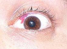
A soft corneal suction ring is applied to the eye, holding the eye in place. This step in the procedure can sometimes cause small blood vessels to burst, resulting in bleeding or subconjunctival hemorrhage into the white (sclera) of the eye, a harmless side effect that resolves within several weeks. Increased suction causes a transient dimming of vision in the treated eye. Once the eye is immobilized, a flap is created by cutting through the corneal epithelium and Bowman's layer. This process is achieved with a mechanical microkeratome using a metal blade, or a femtosecond laser that creates a series of tiny closely arranged bubbles within the cornea. A hinge is left at one end of this flap. The flap is folded back, revealing the stroma, the middle section of the cornea. The process of lifting and folding back the flap can sometimes be uncomfortable.
Laser remodeling
The second step of the procedure uses an excimer laser (193 nm) to remodel the corneal stroma. The laser vaporizes the tissue in a finely controlled manner without damaging the adjacent stroma. No burning with heat or actual cutting is required to ablate the tissue. The layers of tissue removed are tens of micrometers thick.
Performing the laser ablation in the deeper corneal stroma provides for more rapid visual recovery and less pain than the earlier technique, photorefractive keratectomy (PRK).[56]
During the second step, the patient's vision becomes blurry, once the flap is lifted. They will be able to see only white light surrounding the orange light of the laser, which can lead to mild disorientation. The excimer laser uses an eye tracking system that follows the patient's eye position up to 4,000 times per second, redirecting laser pulses for precise placement within the treatment zone. Typical pulses are around 1 millijoule (mJ) of pulse energy in 10 to 20 nanoseconds.[57]
Repositioning of the flap
After the laser has reshaped the stromal layer, the LASIK flap is carefully repositioned over the treatment area by the surgeon and checked for the presence of air bubbles, debris, and proper fit on the eye. The flap remains in position by natural adhesion until healing is completed.
Postoperative care
Patients are usually given a course of antibiotic and anti-inflammatory eye drops. These are continued in the weeks following surgery. Patients are told to rest and are given dark eyeglasses to protect their eyes from bright lights and occasionally protective goggles to prevent rubbing of the eyes when asleep and to reduce dry eyes. They also are required to moisturize the eyes with preservative-free tears and follow directions for prescription drops. Occasionally after the procedure a bandage contact lens is placed to aid the healing, and typically removed after 3–4 days. Patients should be adequately informed by their surgeons of the importance of proper post-operative care to minimize the risk of complications.[58]
Wavefront-guided
Wavefront-guided LASIK is a variation of LASIK surgery in which, rather than applying a simple correction of only long/short-sightedness and astigmatism (only lower order aberrations as in traditional LASIK), an ophthalmologist applies a spatially varying correction, guiding the computer-controlled excimer laser with measurements from a wavefront sensor. The goal is to achieve a more optically perfect eye, though the final result still depends on the physician's success at predicting changes that occur during healing and other factors that may have to do with the regularity/irregularity of the cornea and the axis of any residual astigmatism. Another important factor is whether the excimer laser can correctly register eye position in 3 dimensions, and to track the eye in all the possible directions of eye movement. If a wavefront guided treatment is performed with less than perfect registration and tracking, pre-existing aberrations can be worsened. In older patients, scattering from microscopic particles (cataract or incipient cataract) may play a role that outweighs any benefit from wavefront correction. Therefore, patients expecting so-called "super vision" from such procedures may be disappointed.[59][60][61][62]
When treating a patient with preexisting astigmatism, most wavefront-guided LASIK lasers are designed to treat regular astigmatism as determined externally by corneal topography. In patients who have an element of internally induced astigmatism, therefore, the wavefront-guided astigmatism correction may leave regular astigmatism behind (a cross-cylinder effect). If the patient has preexisting irregular astigmatism, wavefront-guided approaches may leave both regular and irregular astigmatism behind. This can result in less-than-optimal visual acuity compared with a wavefront-guided approach combined with vector planning, as shown in a 2008 study.[63] Thus, vector planning offers a better alignment between corneal astigmatism and laser treatment, and leaves less regular astigmatism behind on the cornea, which is advantageous whether irregular astigmatism coexists or not.
The "leftover" astigmatism after a purely surface-guided laser correction can be calculated beforehand, and is called ocular residual astigmatism (ORA). ORA is a calculation of astigmatism due to the noncorneal surface (internal) optics. The purely refraction-based approach represented by wavefront analysis actually conflicts with corneal surgical experience developed over many years.[62]
The pathway to "super vision" thus may require a more customized approach to corneal astigmatism than is usually attempted, and any remaining astigmatism ought to be regular (as opposed to irregular), which are both fundamental principles of vector planning overlooked by a purely wavefront-guided treatment plan.[62] This was confirmed by the 2008 study mentioned above, which found a greater reduction in corneal astigmatism and better visual outcomes under mesopic conditions using wavefront technology combined with vector analysis than using wavefront technology alone, and also found equivalent higher-order aberrations (see below).[63] Vector planning also proved advantageous in patients with keratoconus.[64]
No good data can be found that compare the percentage of LASIK procedures that employ wavefront guidance versus the percentage that do not, nor the percentage of refractive surgeons who have a preference one way or the other. Wavefront technology continues to be positioned as an "advance" in LASIK with putative advantages;[65][66][67][68] however, it is clear that not all LASIK procedures are performed with wavefront guidance.[69]
Still, surgeons claim patients are generally more satisfied with this technique than with previous methods, particularly regarding lowered incidence of "halos," the visual artifact caused by spherical aberration induced in the eye by earlier methods. A meta-analysis of eight trials showed a lower incidence of these higher order aberrations in patients who had wavefront-guided LASIK compared to non-wavefront-guided LASIK.[70] Based on their experience, the United States Air Force has described WFG-Lasik as giving "superior vision results".[71]
Topography-assisted
Topography-assisted LASIK is intended to be an advancement in precision and reduce night vision side effects. The first topography-assisted device received FDA approval September 13, 2013.[72][73]
History
Barraquer's early work
In the 1950s, the microkeratome and keratomileusis technique were developed in Bogotá, Colombia, by the Spanish ophthalmologist Jose Barraquer. In his clinic, he would cut thin (one hundredth of a mm thick) flaps in the cornea to alter its shape. Barraquer also investigated how much of the cornea had to be left unaltered in order to provide stable long-term results.[74] This work was followed by that of the Russian scientist, Svyatoslav Fyodorov, who developed radial keratotomy (RK) in the 1970s and designed the first posterior chamber implantable contact lenses (phakic intraocular lens) in the 1980s.
Laser refractive surgery
In 1980, Rangaswamy Srinivasan, at the IBM Research laboratory, discovered that an ultraviolet excimer laser could etch living tissue, with precision and with no thermal damage to the surrounding area. He named the phenomenon "ablative photo-decomposition" (APD).[75] Five years later, in 1985, Steven Trokel at the Edward S. Harkness Eye Institute, Columbia University in New York City, published his work using the excimer laser in radial keratotomy. He wrote,
- "The central corneal flattening obtained by radial diamond knife incisions has been duplicated by radial laser incisions in 18 enucleated human eyes. The incisions, made by 193 nm far-ultraviolet light radiation emitted by the excimer laser, produced corneal flattening ranging from 0.12 to 5.35 diopters. Both the depth of the corneal incisions and the degree of central corneal flattening correlated with the laser energy applied. Histopathology revealed the remarkably smooth edges of the laser incisions."[76]
Together with his colleagues, Charles Munnerlyn and Terry Clapham, Trokel founded VISX USA inc.[77] Marguerite B. MacDonald MD performed the first human VISX refractive laser eye surgery in 1989.[78]
Patent
A number of patents have been issued for several techniques related to LASIK. Stuart I. Brown and Josef F. Bille filed a patent on surgical lasers in 1988.[79] Samuel E. Blum, Rangaswamy Srinivasan and James Wynne filed a patent application on the ultraviolet excimer laser, in 1982, issued in 1988.[80] In 1989, Gholam A. Peyman was granted a US patent for using an excimer laser to modify corneal curvature.[81] It was,
- "A method and apparatus for modifying the curvature of a live cornea via use of an excimer laser. The live cornea has a thin layer removed therefrom, leaving an exposed internal surface thereon. Then, either the surface or thin layer is exposed to the laser beam along a predetermined pattern to ablate desired portions. The thin layer is then replaced onto the surface. Ablating a central area of the surface or thin layer makes the cornea less curved, while ablating an annular area spaced from the center of the surface or layer makes the cornea more curved. The desired predetermined pattern is formed by use of a variable diaphragm, a rotating orifice of variable size, a movable mirror or a movable fiber optic cable through which the laser beam is directed towards the exposed internal surface or removed thin layer."[80]
The patents related to so-called broad-beam LASIK and PRK technologies were granted to US companies including Visx and Summit during 1990-1995 based on the fundamental US patent issued to IBM (1983) which claimed the use of UV laser for the ablation of organic tissues.[80]
Implementation in the U.S.
The LASIK technique was implemented in the U.S. after its successful application elsewhere. The Food and Drug Administration (FDA) commenced a trial of the excimer laser in 1989. The first enterprise to receive FDA approval to use an excimer laser for photo-refractive keratectomy was Summit Technology (founder and CEO, Dr. David Muller).[82] In 1992, under the direction of the FDA, Greek ophthalmologist Ioannis Pallikaris introduced LASIK to ten VISX centres. In 1998, the "Kremer Excimer Laser", serial number KEA 940202, received FDA approval for its singular use for performing LASIK.[83] Subsequently, Summit Technology was the first company to receive FDA approval to mass manufacture and distribute excimer lasers. VISX and other companies followed.[83]
Pallikaris suggested a flap of cornea could be raised by microkeratome prior to the performing of PRK with the excimer laser. The addition of a flap to PRK became known as LASIK.
Further research
Since 1991, there have been further developments such as faster lasers; larger spot areas; bladeless flap incisions; intraoperative corneal pachymetry; and "wavefront-optimized" and "wavefront-guided" techniques. The goal of refractive surgery is to avoid permanently weakening the cornea with incisions and to deliver less energy to the surrounding tissues.
Experimental techniques
- "plain" LASIK: LASEK, Epi-LASIK,
- Sub-Bowman’s keratomileusis (thin flap LASIK),
- Wavefront-guided PRK,
- advanced intraocular lenses.
- Femtosecond laser intrastromal vision correction: using all-femtosecond correction, for example, Femtosecond Lenticule EXtraction, FLIVC, or IntraCOR),
- Keraflex: a thermobiochemical solution which has received the CE Mark for refractive correction.[84] and is in European clinical trials for the correction of myopia and keratoconus.[85]
- Technolas FEMTEC laser: for incisionless IntraCOR ablation for presbyopia,[86] with trials ongoing for myopia and other conditions.[87]
- LASIK with the IntraLase femtosecond laser: early trials comparing to the «LASIK with microkeratomes for the correction of myopia suggest no significant differences in safety or efficacy. However, the femtosecond laser has a potential advantage in predictability, although this finding was not significant».[88]
Comparison to photorefractive keratectomy
A systematic review that compared PRK and LASIK concluded that LASIK has shorter recovery time and less pain.[89] The two techniques after a period of one year have similar results.[89]
A 2017 systematic review found uncertainty in visual acuity, but found that in one study, those receiving PRK were less likely to achieve a refractive error, and were less likely to have an over-correction than compared to LASIK.[90]
References
- 1 2 3 4 5 6 Rabin, Roni Caryn (June 11, 2018). "Lasik's Risks Are Coming Into Sharper Focus - Some patients who undergo the eye surgery report a variety of side effects. They may persist for years, studies show". The New York Times. Retrieved June 11, 2018.
- 1 2 3 Finn, Peter (20 December 2012). "Medical Mystery: Preparation for surgery revealed cause of deteriorating eyesight". The Washington Post.
- ↑ Maguire, Stephen. "Laser Eye Surgery". The Irish Times.
- ↑ Lovisolo CF, Reinstein DZ; Reinstein (Nov–Dec 2005). "Phakic intraocular lenses". Survey of ophthalmology. 50 (6): 549–87. doi:10.1016/j.survophthal.2005.08.011. PMID 16263370.
- ↑ Sanders DR, Vukich JA; Vukich (May 2003). "Comparison of Implantable Contact Lens and Laser Assisted In Situ Keratomileusis for Moderate to High Myopia". Cornea. 22 (4): 324–331. doi:10.1097/00003226-200305000-00009. PMID 12792475.
- ↑ Lindfield, Dan; Poole, Tom. "Nd:YAG Treatment of Epithelial Ingrowth". Cataract and Refractive Surgery Today. Retrieved 12 September 2013.
- ↑ Stodola, Ellen (April 1, 2016). "LASIK worldwide". EyeWorld.org. Retrieved June 12, 2018.
- ↑ "A Look at LASIK Past, Present and Future". EyeNet Magazine. Archived from the original on 31 July 2013. Retrieved 12 September 2013.
- ↑ Schoenberg, Nara (May 23, 2016). "Lasik surgery falling out of favor with patients". Chicago Tribune.
- ↑ "Photorefractive (laser) surgery for the correction of refractive errors" (pdf). National Health Service. March 2006.
- ↑ http://www.fda.gov/medicaldevices/productsandmedicalprocedures/surgeryandlifesupport/lasik/ucm061366.htm
- ↑ Saragoussi D, Saragoussi JJ; Saragoussi (September 2004). "["Lasik, PRK and quality of vision: a study of prognostic factors and a satisfaction survey"]". J Fr Ophtalmol (in French). 27 (7): 755–64. doi:10.1016/S0181-5512(04)96210-9. PMID 15499272.
- ↑ Bailey MD, Mitchell GL, Dhaliwal DK, Boxer Wachler BS, Zadnik K; Mitchell; Dhaliwal; Boxer Wachler; Zadnik (July 2003). "Patient satisfaction and visual symptoms after laser in situ keratomileusis". Ophthalmology. 110 (7): 1371–8. doi:10.1016/S0161-6420(03)00455-X. PMID 12867394.
- ↑ McGhee CN, Craig JP, Sachdev N, Weed KH, Brown AD; Craig; Sachdev; Weed; Brown (April 2000). "Functional, psychological, and satisfaction outcomes of laser in situ keratomileusis for high myopia". J Cataract Refract Surg. 26 (4): 497–509. doi:10.1016/S0886-3350(00)00312-6. PMID 10771222.
- ↑ "Study On Post-Lasik Quality Of Life" http://www.medicalnewstoday.com/articles/103194.php
- 1 2 "LASIK Eye Surgery". The New York Times - Health Guide. Retrieved 10 September 2013.
- ↑ Shtein, Roni M (1 October 2011). "Post-LASIK dry eye". Expert review of ophthalmology. 6 (5): 575–582. doi:10.1586/eop.11.56. PMC 3235707. PMID 22174730.
- ↑ "Referrals to the Wills Eye Institute Cornea Service after laser in situ keratomileusis: reasons for patient dissatisfaction". PMID 18165078.
- ↑ "Survey of complications and recommendations for management in dissatisfied patients seeking a consultation after refractive surgery". PMID 15342048.
- ↑ Rodemich, Karen (2010). "Former FDA official warns of LASIK risks: the man who OK'd LASIK now warns of an "epidemic" of eye problems". Review of Optometry. 147 (10): 4.
- ↑ Cha, Ariana Eunjung (2016-11-23). "Many LASIK patients may wind up with glare, halos or other visual symptoms, study suggests". Washington Post. ISSN 0190-8286. Retrieved 2018-04-04.
- ↑ Pallikaris, IG; Panagopoulou, SI (July 2015). "PresbyLASIK approach for the correction of presbyopia". Current Opinion in Ophthalmology. 26 (4): 265–72. doi:10.1097/icu.0000000000000162. PMID 26058023.
- ↑ Pop M, Payette Y; Payette (January 2004). "Risk factors for night vision complaints after LASIK for myopia". Ophthalmology. 111 (1): 3–10. doi:10.1016/j.ophtha.2003.09.022. PMID 14711706.
- ↑ Padmanabhan P, Basuthkar SS, Joseph R; Basuthkar, Subams; Joseph, Roy (Jul–Aug 2010). "Ocular aberrations after wavefront optimized LASIK for myopia". Indian Journal of Ophthalmology. 58 (4): 307–312. doi:10.4103/0301-4738.64139. PMC 2907032. PMID 20534921.
- ↑ "Individual Risk Factors of Halos, Loss of Contrast Sensitivity, Glare and Starbursts after LASIK." operationauge.com
- ↑ Yamane N, Miyata K, Samejima T, Hiraoka T, Kiuchi T, Okamoto F, Hirohara Y, Mihashi T, Oshika T (November 2004). "Ocular higher-order aberrations and contrast sensitivity after conventional laser in situ keratomileusis". Invest. Ophthalmol. Vis. Sci. 45 (11): 3986–90. doi:10.1167/iovs.04-0629. PMID 15505046.
- ↑ Oshika T, Miyata K, Tokunaga T, Samejima T, Amano S, Tanaka S, Hirohara Y, Mihashi T, Maeda N, Fujikado T; Miyata; Tokunaga; Samejima; Amano; Tanaka; Hirohara; Mihashi; Maeda; Fujikado (June 2002). "Higher order wavefront aberrations of cornea and magnitude of refractive correction in laser in situ keratomileusis". Ophthalmology. 109 (6): 1154–8. doi:10.1016/S0161-6420(02)01028-X. PMID 12045059.
- 1 2 "Laser eye surgery". NHS Choices. 5 March 2012. Retrieved 26 October 2013.
- ↑ "LASIK – What are the risks and how can I find the right doctor for me?". Food and Drug Administration. 12 September 2011. Retrieved 26 October 2013.
- ↑ Simpson RG, Moshirfar M, Edmonds JN, Christiansen SM, Behunin N; Moshirfar; Edmonds; Christiansen; Behunin (2012). "Laser in situ keratomileusis in patients with collagen vascular disease: A review of the literature". Clinical ophthalmology (Auckland, N.Z.). 6: 1827–37. doi:10.2147/OPTH.S36690. PMC 3497460. PMID 23152662.
- ↑ "LASIK". Fda.gov. 2008-11-11. Retrieved 2011-12-10.
- ↑ Majmudar, PA. "LASIK Complications." Focal Points: Clinical Modules for Ophthalmologists. American Academy of Ophthalmology. September, 2004. Archived March 11, 2006, at the Wayback Machine.
- ↑ Carrillo C, Chayet AS, Dougherty PJ, Montes M, Magallanes R, Najman J, Fleitman J, Morales A; Chayet; Dougherty; Montes; Magallanes; Najman; Fleitman; Morales (2005). "Incidence of complications during flap creation in LASIK using the NIDEK MK-2000 microkeratome in 26,600 cases". J Refract Surg. 21 (5 Suppl): S655–7. PMID 16212299.
- ↑ "Eye Surgery Education Council". Lasikinstitute.org. Retrieved 2011-12-10.
- ↑ Tham VM, Maloney RK; Maloney (May 2000). "Microkeratome complications of laser in situ keratomileusis". Ophthalmology. 107 (5): 920–4. doi:10.1016/S0161-6420(00)00004-X. PMID 10811084.
- 1 2 Vesaluoma M, Pérez-Santonja J, Petroll WM, Linna T, Alió J, Tervo T; Pérez-Santonja; Petroll; Linna; Alió; Tervo (1 February 2000). "Corneal stromal changes induced by myopic LASIK". Invest. Ophthalmol. Vis. Sci. 41 (2): 369–76. PMID 10670464.
- 1 2 3 4 Sun L, Liu G, Ren Y, Li J, Hao J, Liu X, Zhang Y; Liu; Ren; Li; Hao; Liu; Zhang (2005). "Efficacy and safety of LASIK in 10,052 eyes of 5081 myopic Chinese patients". J Refract Surg. 21 (5 Suppl): S633–5. PMID 16212294.
- ↑ "Ectasia After LASIK". American Academy of Ophthalmology.
- ↑ Cheng AC, Rao SK, Leung GY, Young AL, Lam DS; Rao; Leung; Young; Lam (May 2006). "Late traumatic flap dislocations after LASIK". J Refract Surg. 22 (5): 500–4. PMID 16722490.
- 1 2 Ruiz-Moreno JM, Alió JL; Alió (2003). "Incidence of retinal disease following refractive surgery in 9,239 eyes". J Refract Surg. 19 (5): 534–47. PMID 14518742.
- ↑ Suarez E, Torres F, Vieira JC, Ramirez E, Arevalo JF; Torres; Vieira; Ramirez; Arevalo (October 2002). "Anterior uveitis after laser in situ keratomileusis". J Cataract Refract Surg. 28 (10): 1793–8. doi:10.1016/S0886-3350(02)01364-0. PMID 12388030.
- ↑ Boes DA, Omura AK, Hennessy MJ; Omura; Hennessy (December 2001). "Effect of high-altitude exposure on myopic laser in situ keratomileusis". J Cataract Refract Surg. 27 (12): 1937–41. doi:10.1016/S0886-3350(01)01074-4. PMID 11738908.
- ↑ Dimmig JW, Tabin G; Tabin (2003). "The ascent of Mount Everest following laser in situ keratomileusis". J Refract Surg. 19 (1): 48–51. PMID 12553606.
- ↑ Hammer T, Heynemann M, Naumann I, Duncker GI; Heynemann; Naumann; Duncker (March 2006). "Correction and induction of high-order aberrations after standard and wavefront-guided LASIK and their influence on the postoperative contrast sensitivity". Klin Monatsbl Augenheilkd (in German). 223 (3): 217–24. doi:10.1055/s-2005-858864. PMID 16552654.
- ↑ Alió JL, Montés-Mico R; Montés-Mico (February 2006). "Wavefront-guided versus standard LASIK enhancement for residual refractive errors". Ophthalmology. 113 (2): 191–7. doi:10.1016/j.ophtha.2005.10.004. PMID 16378639.
- ↑ Caster AI, Hoff JL, Ruiz R; Hoff; Ruiz (2005). "Conventional vs wavefront-guided LASIK using the LADARVision4000 excimer laser". J Refract Surg. 21 (6): S786–91. PMID 16329381.
- ↑ http://www.fda.gov/MedicalDevices/ProductsandMedicalProcedures/SurgeryandLifeSupport/LASIK/ucm061354.htm
- ↑ "LASIK laser eye surgery". WebMD Boots. Retrieved 2016-05-06.
- 1 2 "LASIK Quality of Life Collaboration Project". U.S Food and Drug Administration. Retrieved 28 November 2014.
- ↑ "Latest on FDA's LASIK Program". U.S Food and Drug Administration.
- ↑ Malvina B. Eydelman, LASIK Quality of Life Collaboration Project (LQOLCP) (pdf), U.S. Food and Drug Administration
- ↑ LASIK Quality of Life Collaboration Project: Study Results Presented at the Refractive Surgery Subspecialty Day of the American Academy of Ophthalmology (AAO) on October 17, 2014 (PDF - 1.8MB)
- ↑ "What are the risks and how can I find the right doctor for me?". U.S. Food and Drug Administration. Retrieved 2015-12-03.
- ↑ "US FDA/CDRH: LASIK - What are the risks and how can I find the right doctor for me?". Fda.gov. June 9, 2014. Retrieved December 23, 2016.
- ↑ http://www.tlcvision.com/learn-about-laser-eye-surgery/am-i-a-lasik-surgery-candidate/
- ↑ Shortt, Alex J.; Allan, Bruce D. S.; Evans, Jennifer R. (1 January 2013). "Laser-assisted in-situ keratomileusis (LASIK) versus photorefractive keratectomy (PRK) for myopia". The Cochrane Database of Systematic Reviews. 1 (1): CD005135. doi:10.1002/14651858.CD005135.pub3. ISSN 1469-493X. PMID 23440799.
There was evidence that LASIK gives a faster visual recovery than PRK and is a less painful technique. Results at one year after surgery were comparable: most analyses favoured LASIK but they were not statistically significant.
- ↑ "Patent: ultraviolet solid state laser". Freepatentsonline.com. Retrieved 2011-12-10.
- ↑ Dimitri T. Azar; Damien Gatinel (2007). Refractive surgery (2nd ed.). Philadelphia: Mosby Elsevier. ISBN 9780323035996.
- ↑ Walsh MJ. Is the future of refractive surgery based on corneal topography or wavefront? "Ocular Surgery News". August 1, 2000, page 26.
- ↑ Walsh MJ. Wavefront is showing signs of success, but can it do it alone? Ocular Surgery News. September 1, 2000, page 41.
- ↑ EW Dialogue: the future of wavefront refraction as a diagnostic tool. "EyeWorld". May 2000, pages 64 and 65.
- 1 2 3 Alpins NA (2002). "Wavefront technology: A new advance that fails to answer old questions on corneal vs. Refractive astigmatism correction". Journal of refractive surgery. 18 (6): 737–9. PMID 12458868.
- 1 2 Alpins N, Stamatelatos G; Stamatelatos (2008). "Clinical outcomes of laser in situ keratomileusis using combined topography and refractive wavefront treatments for myopic astigmatism". Journal of cataract and refractive surgery. 34 (8): 1250–9. doi:10.1016/j.jcrs.2008.03.028. PMID 18655973.
- ↑ Alpins N, Stamatelatos G; Stamatelatos (2007). "Customized photoastigmatic refractive keratectomy using combined topographic and refractive data for myopia and astigmatism in eyes with forme fruste and mild keratoconus". Journal of cataract and refractive surgery. 33 (4): 591–602. doi:10.1016/j.jcrs.2006.12.014. PMID 17397730.
- ↑ American Academy of Ophthalmology. "Refractive Laser Surgery: An In-Depth Look at LASIK and Brief Overview of PRK, Epi-LASIK, and LASEK: A Science Writer’s Guide". Accessed January 29, 2012.
- ↑ Abbott Medical Optics website. "WaveScan WaveFront System". Accessed August 15, 2012.
- ↑ Emory Healthcare website. "Wavefront technology". Accessed August 15, 2012.
- ↑ Croes K. AllAboutVision website. "Custom LASIK or wavefront LASIK: individualized vision correction". Accessed August 15, 2012.
- ↑ Liz Segre. "Cost of LASIK eye surgery and other corrective procedures". allaboutvision.com. Retrieved 2012-08-15.
- ↑ Fares U, Suleman H, Al-Aqaba MA, Otri AM, Said DG, Dua HS; Suleman; Al-Aqaba; Otri; Said; Dua (2011). "Efficacy, predictability, and safety of wavefront-guided refractive laser treatment: Metaanalysis". Journal of Cataract & Refractive Surgery. 37 (8): 1465–1475. doi:10.1016/j.jcrs.2011.02.029. PMID 21782089.
- ↑ Sue Campbell. "Air Force aims for 'weapons-grade' vision". Af.mil. Archived from the original on 2012-07-28. Retrieved 2011-12-10.
- ↑ "Nidek EC-5000 Excimer Laser System - P970053/S011". Food and Drug Administration. 2013-10-13. Retrieved 2016-05-01.
- ↑ Doyle Stulting, MD (2014-04-28). "Topography-guided LASIK: A paradigm shift in refractive laser treatment" (PDF). EyeWorld Daily News. Retrieved 2016-05-01.
- ↑ Troutman RC, Swinger C; Swinger (1978). "Refractive keratoplasty: keratophakia and keratomileusis". Trans Am Ophthalmol Soc. 76: 329–39. PMC 1311630. PMID 382579.
- ↑ "Prize for the Industrial Application of Physics Winner - American Institute of Physics". Aip.org. Archived from the original on 2011-09-28. Retrieved 2011-12-10.
- ↑ Cotliar AM, Schubert HD, Mandel ER, Trokel SL; Schubert; Mandel; Trokel (Feb 1985). "Excimer laser radial keratotomy". Ophthalmology. 92 (2): 206–8. doi:10.1016/s0161-6420(85)34052-6. PMID 3982798.
- ↑ VISX USA Technology (Last accessed 4th June, 2012)
- ↑ McDonald M.B., Kaufman H.E., Frantz J.M., Shofner S, Salmeron B, Klyce S.D.; Kaufman; Frantz; Shofner; Salmeron; Klyce (1989). "Excimer laser ablation - human eye". Arch Ophthalmol. 107 (5): 641–642. doi:10.1001/archopht.1989.01070010659013. PMID 2719572.
- ↑ US4881808, Josef F. Bille, Stuart I. Brown, "Imaging system for surgical lasers", issued 1989-11-21
- 1 2 3 US4784135, Samuel E. Blum, Rangaswamy Srinivasan, James J. Wynne, "Far ultraviolet surgical and dental procedures", issued 1988-11-15
- ↑ US4840175, Gholam A. Peyman, "Method for modifying corneal curvature", issued 1988-6-20
- ↑ "FDA-Approved Lasers for PRK and Other Refractive Surgeries". Fda.gov. Retrieved 2011-12-10.
- 1 2 "List of FDA-Approved Lasers for LASIK". Fda.gov. Retrieved 2011-12-10.
- ↑
- ↑ "CRSTodayEurope.com > May 2010 > Industry interview: Aiming to change the face of refractive surgery—again". Bmctoday.net. 2010-04-16. Retrieved 2011-12-10.
- ↑ IntraCOR for presbyopia.
- ↑ IntraCOR for myopia
- ↑ Chen, Shihao; Feng, Yifan; Stojanovic, Aleksandar; Jankov, Mirko R.; Wang, Qinmei (2012). "IntraLase Femtosecond Laser vs Mechanical Microkeratomes in LASIK for Myopia: A Systematic Review and Meta-analysis" (PDF). Journal of Refractive Surgery. 28 (1): 15–24. doi:10.3928/1081597x-20111228-02. PMID 22233436. Retrieved 1 November 2015.
- 1 2 Shortt, AJ; Allan, BD; Evans, JR (31 January 2013). "Laser-assisted in-situ keratomileusis (LASIK) versus photorefractive keratectomy (PRK) for myopia". The Cochrane Database of Systematic Reviews. 1 (1): CD005135. doi:10.1002/14651858.CD005135.pub3. PMID 23440799.
- ↑ Kuryan J, Cheema A, Chuck RS (2017). "Laser-assisted subepithelial keratectomy (LASEK) versus laser-assisted in-situ keratomileusis (LASIK) for correcting myopia". Cochrane Database Syst Rev. 2 (2): CD011080. doi:10.1002/14651858.CD011080.pub2. PMC 5408355. PMID 28197998.
External links
- What is LASIK? — Food and Drug Administration
- Laser Eye Surgery — United States National Library of Medicine
- Ectasia After LASIK on EyeWiki