Cuneate fasciculus
| Cuneate fasciculus | |
|---|---|
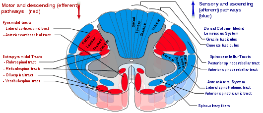 Cuneate fasciculus is labeled in blue at upper right. | |
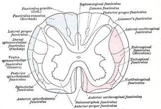 Diagram of the principal fasciculi of the spinal cord. | |
| Details | |
| Identifiers | |
| Latin | fasciculus cuneatus medullae spinalis |
| TA |
A14.1.02.244 A14.1.04.016 A14.1.04.108 |
| FMA | 73941 |
| Anatomical terminology | |
The cuneate fasciculus, fasciculus cuneatus, cuneate tract, (tract of Burdach, named for Karl Friedrich Burdach) is a tract of nerves in the dorsal column of the spinal cord that primarily transmits information from the upper part of the body (the neck, trunk, and arms). It is part of the dorsal column-medial lemniscus pathway.
Structure
The fasciculus cuneatus is triangular on transverse section, and lies between the fasciculus gracilis and the posterior column, its base corresponding with the surface of the medulla spinalis.
Its fibers, larger than those of the fasciculus gracilis, are mostly derived from the same source, viz., the posterior nerve roots.
Some ascend for only a short distance in the tract, and, entering the gray matter, come into close relationship with the cells of the dorsal nucleus, while others can be traced as far as the medulla oblongata, where they end in the gracile nucleus and cuneate nucleus.
Function
The fasciculus cuneatus transmits fine touch, fine pressure, vibration, and proprioception information from spinal nerves located in dermatomes C1 through T6.
Neurons
The fasciculus cuneatus tract is composed of first-order neurons that synapse onto second-order neurons in the brain stem.
The second-order neurons decussate in the brainstem and continue on to the thalamus where the second-order neurons synapse onto third-order neurons.
The third-order neurons carry the received signals to the somatosensory cortex, where the signals, in the form of action potentials, are interpreted.
Additional images
 Decussation of pyramids.
 Superficial dissection of brain-stem. Lateral view.
 The sensory tract.
 Superior terminations of the posterior fasciculi of the medulla spinalis.
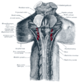 Upper part of medulla spinalis and hind- and mid-brains; posterior aspect, exposed in situ.
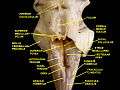 Fourth ventricle. Posterior view. Deep dissection.
See also
External links
- Illustration and text: sc97/text/p1/Pathway.htm at the University of Wisconsin-Madison Medical school