Corpora arenacea
| Brain sand | |
|---|---|
| Details | |
| Identifiers | |
| Latin | Corpora arenacea |
| TH | H3.08.02.3.00007 |
| Anatomical terminology | |
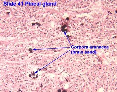
Corpora arenacea (or brain sand or acervuli[1][2] or corpus arenaceum[3]) are calcified structures in the pineal gland and other areas of the brain such as the choroid plexus. Older organisms have numerous corpora arenacea, whose function, if any, is unknown. Concentrations of "brain sand" increase with age, so the pineal gland becomes increasingly visible on X-rays over time, usually by the third or fourth decade. They are sometimes used as anatomical landmarks in radiological examinations.[4]
Chemical analysis shows that they are composed of calcium phosphate (later characterized as hydroxyapatite[5]), calcium carbonate, magnesium phosphate, and ammonium phosphate.[6] Recently, calcite deposits have been described as well.[7]
- ↑ Vígh, B; Szél, A; Debreceni, K; Fejér, Z; Manzano e Silva, MJ; Vígh-Teichmann, I (1998). "Comparative histology of pineal calcification". Histology and Histopathology. 13 (3): 851–70. PMID 9690142.
- ↑ Kim, Jinkyung; Kim, Hyun-Wook; Chang, Soeun; Kim, Jee Woong; Je, Jung Ho; Rhyu, Im Joo (2012). "Growth patterns for acervuli in human pineal gland". Scientific Reports. 2: 984. Bibcode:2012NatSR...2E.984K. doi:10.1038/srep00984. PMC 3523289. PMID 23248747.
- ↑ Tomonari, Yuki; Sato, Junko; Wako, Yumi; Tsuchitani, Minoru (2012). "Age-related Histological Findings in the Pineal Gland of Crl:CD(SD) Rats". Journal of Toxicologic Pathology. 25 (4): 287–91. doi:10.1293/tox.25.287. PMC 3517926. PMID 23345933.
- ↑ "thesis:The Effect of Fluoride on the Physiology of the Pineal Gland" (PDF). Retrieved 2016-08-04.
The calcified pineal is used as a landmark in skull X- rays because of its radio-opacity
- ↑ Angervall, Lennart; Berger, Sven; Röckert, Hans (2009). "A Microradiographic and X-Ray Crystallographic Study of Calcium in the Pineal Body and in Intracranial Tumours". Acta Pathologica Microbiologica Scandinavica. 44 (2): 113–119. doi:10.1111/j.1699-0463.1958.tb01060.x.
- ↑ Bocchi, Giancarlo; Valdre, Giovanni; Valdre, Giovanni (1993). "Physical, chemical, and mineralogical characterization of carbonate-hydroxyapatite concretions of the human pineal gland". Journal of Inorganic Biochemistry. 49 (3): 209–20. doi:10.1016/0162-0134(93)80006-U. PMID 8381851.
- ↑ Baconnier, Simon; Lang, Sidney B.; Polomska, Maria; Hilczer, Bozena; Berkovic, Garry; Meshulam, Guilia (2002). "Calcite microcrystals in the pineal gland of the human brain: First physical and chemical studies". Bioelectromagnetics. 23 (7): 488–95. doi:10.1002/bem.10053. PMID 12224052.
Further reading
- Garma-Aviña, A. (2000). "Excretory Plugs from the Choroid Plexus in the Cerebrospinal Fluid of Dogs with Neurological Disease: Possible Role in the Formation of Corpora Arenacea". Journal of Comparative Pathology. 123 (2–3): 146–51. doi:10.1053/jcpa.2000.0405. PMID 11032668.
- Reiter, Russel J.; Welsh, Marcia G.; Vaughan, Mary K. (1976). "Age-related changes in the intact and sympathetically denervated gerbil pineal gland". American Journal of Anatomy. 146 (4): 427–31. doi:10.1002/aja.1001460405. PMID 941859.
- Klein, Pavel; Rubinstein, Lucien J (1989). "Benign symptomatic glial cysts of the pineal gland: a report of seven cases and review of the literature". Journal of Neurology, Neurosurgery & Psychiatry. 52 (8): 991–5. doi:10.1136/jnnp.52.8.991. PMC 1031840. PMID 2677249.
- Vigh, B; Vigh-Teichmann, I; Aros, B (1989). "Pineal corpora arenacea produced by arachnoid cells in the bat Myotis blythi oxygnathus". Zeitschrift für Mikroskopisch-anatomische Forschung. 103 (1): 36–45. PMID 2756745.
External links
- Histology image: 14401loa – Histology Learning System at Boston University
- Histology image: 41_03 at the University of Oklahoma Health Sciences Center - "Pineal gland"
This article is issued from
Wikipedia.
The text is licensed under Creative Commons - Attribution - Sharealike.
Additional terms may apply for the media files.