Blood–air barrier
| Blood–air barrier | |
|---|---|
 Gaseous exchange in the lung | |
| Details | |
| System | Respiratory system |
| Location | Lungs |
| Identifiers | |
| MeSH | D015824 |
| TH | H3.05.02.0.00040 |
| Anatomical terminology | |
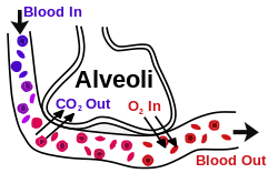
The blood–air barrier (alveolar–capillary barrier or membrane) exists in the gas exchanging region of the lungs. It exists to prevent air bubbles from forming in the blood, and from blood entering the alveoli. It is formed by the type 1 pneumocytes of the alveolar wall, the endothelial cells of the capillaries and the basement membrane between the two cells. The barrier is permeable to molecular oxygen, carbon dioxide, carbon monoxide and many other gases.[1]
Structure
This blood gas barrier is extremely thin (approximately 600nm-2μm; in some places merely 200 nm) to allow sufficient oxygen diffusion, yet it is extremely strong. This strength comes from the type IV collagen in between the endothelial and epithelial cells. Damage can occur to this barrier at a pressure difference of around 40 millimetres of mercury (0.053 bar).
Clinical significance
Failure of the barrier may occur in a pulmonary barotrauma. This can be a result of several possible causes, including blast injury, swimming-induced pulmonary edema, and breathing gas entrapment or retention in the lung during depressurization, which can occur during ascent from underwater diving or loss of pressure from a pressurized vehicle, habitat or pressure suit.
Possible consequences of rupture of the blood–air barrier include arterial gas embolism and hemoptysis.
Other animals
Failure of the barrier is often seen in racehorses and other domesticated horses due to exercise induced blood pressure rising above normal.
See also
References
- ↑ Sheenan Kindlen (2003). Physiology for Health Care and Nursing. Elsevier Health Sciences. p. 130. ISBN 0-443-07116-0.
External links
- Anatomy photo: Respiratory/lung/vasc4/vasc - Comparative Organology at University of California, Davis – "Mammal, lung vasculature (EM, High)"
- Swiss embryology (from UL, UB, and UF) rrespiratory/phasen05