Silver stain
Silver staining is the use of silver to selectively alter the appearance of a target in microscopy of histological sections; in temperature gradient gel electrophoresis; and in polyacrylamide gels.
History
Camillo Golgi perfected silver staining for the study of the nervous system. Although the exact chemical mechanism by which this occurs is unknown,[1] Golgi's method stains a limited number of cells at random in their entirety.[2]
Silver staining was introduced by Kerenyi and Gallyas as a sensitive procedure to detect trace amounts of proteins in gels.[3] The technique has been extended to the study of other biological macromolecules that have been separated in a variety of supports.[4]
Classical Coomassie Brilliant Blue staining can usually detect a 50 ng protein band; silver staining increases the sensitivity typically 50 times.
Many variables can influence the colour intensity and every protein has its own staining characteristics; clean glassware, pure reagents, and water of highest purity are the key points to successful staining.[5]
Chemistry
Some cells are argentaffin. These reduce silver solution to metallic silver after formalin fixation. Other cells are argyrophilic. These reduce silver solution to metallic silver after being exposed to the stain that contains a reductant, for example hydroquinone or formalin.
Silver nitrate forms insoluble silver phosphate with phosphate ions; this method is known as the Von Kossa Stain. When subjected to a reducing agent, usually hydroquinone, it forms black elementary silver. This is used for study of formation of calcium phosphate particles during bone growth.
Applications
Histological characterisation
Silver staining aids the visualization of targets of interest, namely intracellular and extracellular cellular components such as DNA and proteins, such as type III collagen and reticulin fibres by the deposition of metallic silver particles on the targets of interest.[6]
Diagnostic microbiology
Pseudomonas,[7] Legionella, Leptospira, H. pylori and Treponema, and fungi such as Pneumocystis, Cryptococcus, and Candida are organisms that are stained with silver.
Karyotype analysis
Silver staining is used in karyotyping. Silver nitrate stains the nucleolar organization region (NOR)-associated protein, producing a dark region wherein the silver is deposited and denoting the activity of rRNA genes within the NOR. Human chromosomes 13, 14, 15, 21, and 22 have NORs, which increase the silver stain activity by at least 50 times.
Genomic and proteomic analysis
Silver staining is used to stain gels. The silver stain of proteins in Agarose gels was developed in 1973 by Kerenyi and Gallyas.[8] Later it was adapted to polyacrylamide gels used in SDS-PAGE,[9][10][11][12][13] and also for staining DNA or RNA.[14] The glycosylations of glycoproteins and polysaccharides can be oxidised by a 1 hour pre-treatment with 0.1 % periodic acid at 4 °C, which improves the binding of silver ions and the staining result.[15]
First, the proteins are denatured in the gel by a fixative solution of 10 % acetic acid and 30% ethanol and precipitated, at the same time the detergent (mostly SDS) is extracted. The diffusion of the proteins is thus significantly reduced. After repeated washing with water, the gel is incubated in a silver nitrate solution. Silver ions bind to negatively charged side chains of the proteins. Excess silver ions are then washed off with water. In the final development step, the silver ions are reduced to elemental silver by addition of alkaline formaldehyde]. This stains the sites where proteins are present, brown to black.
The intensity of the staining depends on the primary structure of the protein. Furthermore, the cleanliness of the vessels used and the purity of the reagents influence the silver stain.[16] Common artifacts in silver stained gels are bands of keratin in the ranges of 54-57 kDa and 65-68 kDa[17] as a contamination of the sample prior to the electrophoresis.
In art
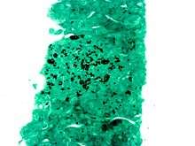
Silver staining is also a technique in traditional stained glass to produce the yellow, brown, or amber shading when painting on glass. It is a technique that is often used for realistic hair colors. It was discovered in the 14th Century but was not originally used very frequently.
Variations
Methenamine silver stains
There are several silver stains incorporating methenamine, including:
- Grocott's methenamine silver stain, used widely as a screen for fungal organisms.
- Jones' stain, a methenamine silver-Periodic acid-Schiff that stains for basement membrane, availing to view the "spiked" GBM associated with membranous glomerulonephritis.
Gallery
 A silver stain (GMS) demonstrating the fungus Histoplasma (black round balls) in a liver biopsy.
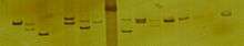 A number of DNA samples from specimens of Littorina plena amplified using PCR with primers targeting a variable simple sequence repeat (SSR, a.k.a. microsatellite) locus. Samples have been run on a 5% polyacrylamide gel and visualized using silver staining.
References
- ↑ Golgi C (1873). "Sulla struttura della sostanza grigia del cervello". Gazzetta Medica Italiana (Lombardia). 33: 244–246.
- ↑ Grant G (Oct 2007). "How the 1906 Nobel Prize in Physiology or Medicine was shared between Golgi and Cajal". Brain Res Rev. 55 (2): 490–498. doi:10.1016/j.brainresrev.2006.11.004. PMID 17306375.
- ↑ Kerenyi L, Gallyas F (1973). "Über Probleme der quantitiven Auswertung der mit physikalischer Entwicklung versilberten Agarelektrophoretogramme". Clin. Chim. Acta. 47 (3): 425–436. doi:10.1016/0009-8981(73)90276-3. PMID 4744834.
- ↑ Switzer RC 3rd, Merril CR, Shifrin S (Sep 1979). "A highly sensitive silver stain for detecting proteins and peptides in polyacrylamide gels". Anal. Biochem. 98 (1): 231–237. doi:10.1016/0003-2697(79)90732-2. PMID 94518.
- ↑ Hempelmann E, Schulze M, Götze O (1984). "Free SH-groups are important for the polychromatic staining of proteins with silver nitrate". Neuhof V (ed)Electrophoresis '84, Verlag Chemie Weinheim 1984: 328–330.
- ↑ Schwint OA, Labraga M, Cervino CO, Haffar M, Sequeiros PH, Marcos HJ (2004). "A modification of the staining technique of reticular fibres for image analysis of the cardiac collagen network". Cardiovasc. Pathol. 13 (4): 213–20. doi:10.1016/S1054-8807(03)00153-4. PMID 15210137.
- ↑ Barnini S, Dodi C, Campa M (2004). "Enhanced resolution of random amplified polymorphic DNA genotyping of Pseudomonas aeruginosa". Lett. Appl. Microbiol. 39 (3): 274–7. doi:10.1111/j.1472-765X.2004.01576.x. PMID 15287874.
- ↑ L. Kerényi, F. Gallyas: [Errors in quantitative estimations on agar electrophoresis using silver stain]. In: Clinica chimica acta; international journal of clinical chemistry. Band 47, Nummer 3, September 1973, S. 425–436, PMID 4744834.
- ↑ C. R. Merril, R. C. Switzer, M. L. Van Keuren: Trace polypeptides in cellular extracts and human body fluids detected by two-dimensional electrophoresis and a highly sensitive silver stain. In: Proc Natl Acad Sci U S A. 76(9), 1979, S. 4335–4339. PMID 92027.
- ↑ R. C. Switzer, C. R. Merril, S. Shifrin (1979-09), "A highly sensitive silver stain for detecting proteins and peptides in polyacrylamide gels" (in German), Anal Biochem 98 (1): pp. 231–237, doi:10.1016/0003-2697(79)90732-2, PMID 94518
- ↑ T. Rabilloud et. al.: Improvement and simplification of low-background silver staining of proteins by using sodium dithionite. In: Electrophoresis. 9(6), 1998, S. 288–291. PMID 2466660.
- ↑ T. Rabilloud: A comparison between low background silver diammine and silver nitrate protein stains. In: Electrophoresis. 13, 1992, S. 429–439. PMID 1425556.
- ↑ C. Lelong, M. Chevallet, S. Luche, T. Rabilloud: Silver staining of proteins in 2DE gels. In: Methods Mol Biol. 519, 2009, S. 339–350. PMID 19381593.
- ↑ H. Blum, H. Beier, H. J. Gross: Improved silver staining of plant protein, RNA & DNA in PAA gels. In: Electrophoresis. 8, 1987, S. 93–99.
- ↑ G. Dubray, G. Bezard: A highly sensitive periodic acid-silver stain for 1,2-diol groups of glycoproteins and polysaccharides in polyacrylamide gels. In: Anal. Biochem.. 119(2), 1982, S. 325–329. PMID 6176144.
- ↑ E. Hempelmann, M. Schulze, O. Götze: Free SH-groups are important for the polychromatic staining of proteins with silver nitrate. In: V. Neuhof (Editor): Electrophoresis. Verlag Chemie, Weinheim, 1984, pp. 328–330.
- ↑ D. Ochs: Protein contaminants of sodium dodecyl sulfate polyacrylamide gels. In: Analytical biochemistry. Vol. 135, Number 2, December 1983, pp. 470-474, PMID 6197906.
- ↑ Hempelmann E, Götze O (1984). "Characterization of membrane proteins by polychromatic silver staining". Hoppe-Seyler's Z Physiol Chem. 365: 241–242.
External links
- MedEd at Loyola Histo/practical/stains/hp2-55.html
- Hempelmann E. SDS-Protein PAGE and protein detection by silverstaining and immunoblotting of Plasmodium falciparum proteins. in: Moll K, Ljungström J, Perlmann H, Scherf A, Wahlgren M (eds) Methods in Malaria Research, 5th edition, 2008, 263-266