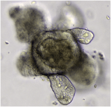
Organoid
An organoid is a miniaturized and simplified version of an organ produced in vitro in three dimensions that shows realistic micro-anatomy. They are derived from one or a few cells from a tissue, embryonic stem cells or induced pluripotent stem cells, which can self-organize in three-dimensional culture owing to their self-renewal and differentiation capacities. The technique for growing organoids has rapidly improved since the early 2010s, and it was named by The Scientist as one of the biggest scientific advancements of 2013.[1] Organoids are used by scientists to study disease and treatments in a laboratory.
History
Attempts to create organs in vitro started with one of the first dissociation-reaggregation experiment[2] where Henry Van Peters Wilson demonstrated that mechanically dissociated sponge cells can reaggregate and self-organize to generate a whole organism.[3] In the subsequent decades, multiple labs were able to generate different types of organs[2] in vitro through the dissociation and reaggregation of organ tissues obtained from amphibians[4] and embryonic chicks.[5] The phenomena of mechanically dissociated cells aggregating and reorganizing to reform the tissue they were obtained from subsequently led to the development of the differential adhesion hypothesis by Malcolm Steinberg.[2] With the advent of the field of stem cell biology, the potential of stem cells to form organs in vitro was realized early on with the observation that when stem cells form teratomas or embryoid body, the differentiated cells can organize into different structures resembling those found in multiple tissue types.[2] The advent of the field of organoids, started with a shift from culturing and differentiating stem cells in 2D media, to 3D media to allow for the development of the complex 3-dimensional structures of organs.[2] Since 1987, researchers have devised different methods for 3-D culturing, and were able to utilize different types of stem cells to generate organoids resembling a multitude of organs.[2] In 2008, Yoshiki Sasai and his team at RIKEN institute demonstrated that stem cells can be coaxed into balls of neural cells that self-organize into distinctive layers.[6] In 2009 the Laboratory of Hans Clevers at Hubrecht Institute and University Medical Center Utrecht, The Netherlands showed that single LGR5 stem cells build crypt-villus structures in vitro without a mesenchymal niche.[7] In 2010, Mathieu Unbekandt & Jamie A. Davies demonstrated the production of renal organoids from murine fetus-derived renogenic stem cells:[8] subsequent reports showed significant physiological function of these organoids in vitro[9] and in vivo.[10]
In 2013, Madeline Lancaster at the Austrian Academy of Sciences established a protocol for culturing cerebral organoids derived from stem cells that mimic the developing human brain's cellular organization.[11] In 2014, Artem Shkumatov et al. at the University of Illinois at Urbana-Champaign demonstrated that cardiovascular organoids can be formed from ES cells through modulation of the substrate stiffness, to which they adhere. Physiological stiffness promoted three-dimensionality of EBs and cardiomyogenic differentiation.[12]
Takebe et al. demonstrate a generalized method for organ bud formation from diverse tissues by combining pluripotent stem cell-derived tissue-specific progenitors or relevant tissue samples with endothelial cells and mesenchymal stem cells. They suggested that the less mature tissues, or organ buds, generated through the self-organized condensation principle might be the most efficient approach toward the reconstitution of mature organ functions after transplantation, rather than condensates generated from cells of a more advanced stage.[13]
Properties
Lancaster and Knoblich[2] define an organoid as a collection of organ-specific cell types that develops from stem cells or organ progenitors, self-organizes through cell sorting and spatially restricted lineage commitment in a manner similar to in vivo, and exhibits the following properties:
- it has multiple organ-specific cell types;
- it is capable of recapitulating some specific function of the organ (e.g. contraction, neural activity, endocrine secretion, filtration, excretion);
- its cells are grouped together and spatially organized, similar to an organ.
Process
Organoid formation generally requires culturing the stem cells or progenitor cells in a 3D medium.[2] The 3D medium can be made using an extracellular matrix hydrogel Matrigel, which is a laminin-rich extracellular matrix that is secreted by the Engelbreth-Holm-Swarm tumor line.[14] Organoid bodies can then be made through embedding stem cells in the 3D medium.[2] When pluripotent stem cells are used for the creation of the organoid, the cells are usually, but not all the time, allowed to form embryoid bodies.[2] Those embryoid bodies are then pharmacologically treated with patterning factors to drive the formation of the desired organoid identity.[2] Organoids have also been created using adult stem cells extracted from the target organ, and cultured in 3D media.[15]
Types
A multitude of organ structures have been recapitulated using organoids.[2] This section aims to outline the state of the field as of now through providing an abridged list of the organoids that have been successfully created, along with a brief outline based on the most recent literature for each organoid, and examples of how it has been utilized in research.
Cerebral organoid
A Cerebral organoid describes artificially grown, in vitro, miniature organs resembling the brain. Cerebral organoids are created by culturing human pluripotent stem cells in a three-dimensional rotational bioreactor and develop over a course of months.[16]
Gut organoid
Gut organoids refer to organoids that recapitulate structures of the gastrointestinal tract. The gastrointestinal tract arises from the endoderm, which during development forms a tube that can be divided in three distinct regions, which give rise to, along with other organs, the following sections of the gastrointestinal tract:[2]
- The Foregut gives rise to the oral cavity and the stomach
- The Midgut gives rise to the small intestines and the ascending colon
- The Hindgut gives rise to the rectum and the rest of the colon
Organoids have been created for the following structures of the gastrointestinal tract:
Intestinal organoid
Intestinal organoids[7] have thus far been among the gut organoids to be generated directly from pluripotent stem cells.[2] One way human pluripotent stem cells can be driven to form intestinal organoids is through first the application of activin A to drive the cells into a mesoendodermal identity, followed by the pharmacological upregulation of Wnt3a and Fgf4 signaling pathways as they have been demonstrated to promote posterior gut fate.[2] Intestinal organoids have also been generated from intestinal stem cells, extracted from adult tissue and cultured in 3D media.[15] Intestinal organoids recapitulate the crypt-Villus structure of the intestine, by recapitulating its function, physiology and organization, and maintaining all the cell types found normally in the structure including intestinal stem cells.[2] Intestinal organoids recapitulate the crypt-Villus structure to high degree of fidelity that they have been successfully transplanted to mouse intestines, and are hence highly regarded as a valuable model for research.[2] One of the fields of research that intestinal organoids have been utilized is that of stem cell niche. Intestinal organoids were used to study the nature of the intestinal stem cell niche, and research done with them demonstrated the positive role IL-22 has in maintaining in intestinal stem cells,[17] along with demonstrating the roles of other cell types like neurons and fibroblasts in maintenance of intestinal tem cells.[15] Intestinal organoids have also demonstrated therapeutic potential. A 2014 study published in Nature,[18] for example, reported that FOXO1 inhibition in intestinal organoids resulted in the formation of glucose responsive insulin producing cells. The data from this study thus acts as a proof of concept of the use of intestinal organoids as a therapeutic approach for diabetes.
Stomach or gastric organoid
Gastric organoids recapitulate at least partly the physiology of the stomach. Gastric organoids have been generated directly from pluripotent stem cells through the temporal manipulation of the FGF, WNT, BMP, retinoic acid and EGF signalling pathways in three-dimensional culture conditions.[19] Gastric organoids have also been generated using LGR5 expressing stomach adult stem cells.[20] Gastric organoids have been used as model for the study of cancer[21][22] along with human disease[19] and development.[19] For example one study[22] investigated the underlying genetic alterations behind a patient's metastatic tumor population, and identified that unlike the patient's primary tumor, the metastasis had both alleles of the TGFBR2 gene mutated. To further assess the role of TGFBR2 in the metastasis, the investigators created organoids where TGFBR2 expression is knocked down, through which they were able to demonstrate that reduced TGFBR2 activity leads to invasion and metastasis of cancerous tumors both in vitro and in vivo.
Lingual organoid
Lingual organoids are organoids that recapitulate, at least partly, aspects of the tongue physiology. Epithelial lingual organoids have been generated using BMI1 expressing epithelial stem cells in three-dimensional culture conditions through the manipulation of EGF, WNT, and TGF-β.[23] This organoid culture, however, lacks taste receptors, as these cells do not arise from Bmi1 expressing epithelial stem cells.[23] Lingual taste bud organoids containing taste cells, however, have been created using the LGR5+ or CD44+ stem/progenitor cells of circumvallate (CV) papilla tissue.[24] These taste bud organoids have been successfully created both directly from isolated Lgr5- or LGR6-expressing taste stem/progenitor cells.[25] and indirectly, through the isolation, digestion, and subsequent culturing of CV tissue containing Lgr5+ or CD44+ stem/progenitor cells.[24]
Other types of organoids
- Thymic organoids recapitulate at least partly the architecture and stem-cell niche functionality of the thymus,[28] which is a lymphoid organ where T cells mature. Thymic organoids have been generated through the seeding of thymic stromal cells in 3-dimensional culture.[28] Thymic organoids seem to successfully recapitulate the thymus’s function, as co-culturing human hematopoietic or bone marrow stem cells with mouse thymic organoids resulted in the production of T-cells.[28]
- Testicular organoid
- Hepatic organoid[29]
- Pancreatic organoid[30]
- Epithelial organoid[7][31]
- Lung organoid[32]
- Kidney organoid[33][34][35][36]
- Gastruloid (embryonic organoid)[37][38][39][40] - Generates all embryonic axes and fully implements the collinear Hox gene expression patterns along the anteroposterior axis[40].
- Cardiac organoid[41] - In 2018 hollow cardiac organoids were made to beat, and to respond to stimuli to beat faster or slower.[42]
Basic research
Organoids are an excellent tool to study basic biological processes. Organoids enable to study how cells interact together in an organ, their interaction with their environment, how diseases affect them and the effect of drugs. In vitro culture makes this system easy to manipulate and facilitates their monitoring. While organs are difficult to culture because their size limits the penetration of nutrients, the small size of organoids limits this problem. On the other hand, they don't exhibit all organ features and interactions with other organs are not recapitulated in vitro. While research on stem cells and regulation of stemness was the first field of application of intestinal organoids,[7] they are now also used to study e.g. uptake of nutrients, drug transport and secretion of incretin hormones.[43] This is of great relevance in the context of malabsorption diseases as well as metabolic diseases such as obesity, insulin resistance, and diabetes.
Models of disease
Organoids provide an opportunity to create cellular models of human disease, which can be studied in the laboratory to better understand the causes of disease and identify possible treatments. In one example, the genome editing system called CRISPR was applied to human pluripotent stem cells to introduce targeted mutations in genes relevant to two different kidney diseases, polycystic kidney disease and focal segmental glomerulosclerosis.[35] These CRISPR-modified pluripotent stem cells were subsequently grown into human kidney organoids, which exhibited disease-specific phenotypes. Kidney organoids from stem cells with polycystic kidney disease mutations formed large, translucent cyst structures from kidney tubules. When cultured in the absence of adherent cues (in suspension), these cysts reached sizes of 1 cm in diameter over several months.[44] Kidney organoids with mutations in a gene linked to focal segmental glomerulosclerosis developed junctional defects between podocytes, the filtering cells affected in that disease.[45] Importantly, these disease phenotypes were absent in control organoids of identical genetic background, but lacking the CRISPR mutations.[35][44][45] Comparison of these organoid phenotypes to diseased tissues from mice and humans suggested similarities to defects in early development.[44][45] These experiments demonstrate how organoids can be utilized to create complex models of human disease in the laboratory, which recapitulate tissue-level phenotypes in a petri dish.
Personalised medicine
Intestinal organoids grown from rectal biopsies using culture protocols established by the Clevers group have been used to model cystic fibrosis,[46] and led to the first application of organoids for personalised treatment.[47] Cystic fibrosis is an inherited disease that is caused by gene mutations of the cystic fibrosis transmembrane conductance regulator gene that encodes an epithelial ion channel necessary for healthy epithelial surface fluids. Studies by the laboratory of Jeffrey Beekman (Wilhelmina Children's Hospital, University Medical Center Utrecht, The Netherlands) described in 2013 that stimulation of colorectal organoids with cAMP-raising agonists such as forskolin or cholera toxin induced rapid swelling of organoids in a fully CFTR dependent manner.[46] Whereas organoids from non-cystic fibrosis subjects swell in response to forskolin as a consequence of fluid transport into the organoids' lumens, this is severely reduced or absent in organoids derived from people with cystic fibrosis. Swelling could be restored by therapeutics that repair the CFTR protein (CFTR modulators), indicating that individual responses to CFTR modulating therapy could be quantitated in a preclinical laboratory setting. Schwank et al. also demonstrated that the intestinal cystic fibrosis organoid phenotype could be repaired by CRISPR-Cas9 gene editing in 2013.[48]
Follow-up studies by Dekkers et al. in 2016 revealed that quantitative differences in forskolin-induced swelling between intestinal organoids derived from people with cystic fibrosis associate with known diagnostic and prognostic markers such as CFTR gene mutations or in vivo biomarkers of CFTR function.[47] In addition, the authors demonstrated that CFTR modulator responses in intestinal organoids with specific CFTR mutations correlated with published clinical trial data of these treatments. This led to preclinical studies where organoids from patients with extremely rare CFTR mutations for who no treatment was registered were found to respond strongly to a clinically available CFTR modulator. The suggested clinical benefit of treatment for these subjects based on the preclinical organoid test was subsequently confirmed upon clinical introduction of treatment by members of the clinical CF center under supervision of Kors van der Ent (Department of Paediatric Pulmonology, Wilhelmina Children's Hospital, University Medical Center Utrecht, The Netherlands). These studies show for the first time that organoids can be used for the individual tailoring of therapy or personalised medicine.
As a model for developmental biology
Organoids offer researchers an exceptional model to study developmental biology.[49] Since the identification of pluripotent stem cells, there have been great advancements in directing pluripotent stem cells fate in vitro using 2D cultures.[49] These advancements in PSC fate direction, coupled with the advancements in 3D culturing techniques allowed for the creation of organoids that recapitulate the properties of various specific subregions of a multitude of organs.[49] The use of these organoids has thus greatly contributed to expanding our understanding of the processes of organogenesis, and the field of developmental biology.[49] In central nervous system development, for example, organoids have contributed to our understanding of the physical forces that underlie retinal cup formation,[49][50][51] the species-specific dimensionality of the eye,[49][50][51] and the factors underlying the control of progenitor cells in the cortex.[49][52][53]
References
- ↑ Grens, Kerry (December 24, 2013). "2013's Big Advances in Science". The Scientist. Retrieved 26 December 2013.
- 1 2 3 4 5 6 7 8 9 10 11 12 13 14 15 16 17 Lancaster, M. A.; Knoblich, J. A. (2014). "Organogenesis in a dish: Modeling development and disease using organoid technologies". Science. 345 (6194): 1247125. doi:10.1126/science.1247125. PMID 25035496.
- ↑ Wilson, H. V. (1907). "A new method by which sponges may be artificially reared". Science. 25 (649): 912–915. Bibcode:1907Sci....25..912W. doi:10.1126/science.25.649.912. PMID 17842577.
- ↑ J. Holtfreter, Experimental studies on the development of the pronephros. Rev. Can. Biol. 3, 220–250 (1944).
- ↑ Weiss, P.; Taylor, A. C. (1960). "Reconstitution of complete organs from single-cell suspensions of chick embryos in advanced stages of differentiation". Proc. Natl. Acad. Sci. U.S.A. 46 (9): 1177–1185. Bibcode:1960PNAS...46.1177W. doi:10.1073/pnas.46.9.1177. PMC 223021. PMID 16590731.
- ↑ Yong, Ed (August 28, 2013). "Lab-Grown Model Brains". The Scientist. Retrieved 26 December 2013.
- 1 2 3 4 Sato, Toshiro; Vries, Robert G.; Snippert, Hugo J.; Van De Wetering, Marc; Barker, Nick; Stange, Daniel E.; Van Es, Johan H.; Abo, Arie; Kujala, Pekka; Peters, Peter J.; Clevers, Hans (2009). "Single Lgr5 stem cells build cryptvillus structures in vitro without a mesenchymal niche". Nature. 459 (7244): 262–5. Bibcode:2009Natur.459..262S. doi:10.1038/nature07935. PMID 19329995.
- ↑ Unbekandt, M.; Davies, J.A. (2010). "Dissociation of embryonic kidneys followed by reaggregation allows the formation of renal tissues". Kidney International. 77 (5): 407–416. doi:10.1038/ki.2009.482. PMID 20016472.
- ↑ Lawrence, M.L.; Chang, C.H.; Davies, J.A. (2015). "Transport of organic anions and cations in murine embryonic kidney development and in serially-reaggregated engineered kidneys". Scientific Reports. 5: 9092. Bibcode:2015NatSR...5E9092L. doi:10.1038/srep09092. PMC 4357899. PMID 25766625.
- ↑ Xinaris, C.; Benedetti, V.; Rizzo, P.; Abbate, M.; Corna, D.; Azzolini, N.; Conti, S.; Unbekandt, M.; Davies, J.A.; Morigi, M.; Begnini, A.; Remuzzi, G. (2012). "In vivo maturation of functional renal organoids formed from embryonic cell suspensions". J. Am. Soc. Nephrol. 23 (11): 1857–1868. doi:10.1681/ASN.2012050505. PMC 3482737. PMID 23085631.
- ↑ Chambers, Stuart M.; Tchieu, Jason; Studer, Lorenz (October 2013). "Build-a-Brain". Cell Stem Cell. 13 (4): 377–8. doi:10.1016/j.stem.2013.09.010. PMID 24094317.
- ↑ Shkumatov, A; Baek, K; Kong, H (2014). "Matrix Rigidity-Modulated Cardiovascular Organoid Formation from Embryoid Bodies". PLoS ONE. 9 (4): e94764. Bibcode:2014PLoSO...994764S. doi:10.1371/journal.pone.0094764. PMC 3986240. PMID 24732893.
- ↑ Takebe, T.; Enomura, M.; Yoshizawa, E.; Kimura, M.; Koike, H.; Ueno, Y.; Taniguchi, H. (2015). "Vascularized and Complex Organ Buds from Diverse Tissues via Mesenchymal Cell-Driven Condensation". Cell Stem Cell. 16 (5): 556–565. doi:10.1016/j.stem.2015.03.004. PMID 25891906.
- ↑ M. L. Li et al., Influence of a reconstituted basement membrane and its components on casein gene expression and secretion in mouse mammary epithelial cells" Proc. Natl. Acad. Sci. U.S.A 1987; 84, 136–140 doi: 10.1073/ pnas.84.1.136; PMID 3467345
- 1 2 3 Pastuła, A., Middelhoff, M., Brandtner, A., Tobiasch, M., Höhl, B., Nuber, A. H., . . . Quante, M. (2016). Three-Dimensional Gastrointestinal Organoid Culture in Combination with Nerves or Fibroblasts: A Method to Characterize the Gastrointestinal Stem Cell Niche. Stem Cells International, 2016, 1-16. doi:10.1155/2016/3710836
- ↑ Lancaster, M. A.; Renner, M.; Martin, C. A.; Wenzel, D.; Bicknell, L. S.; Hurles, M. E.; Homfray, T.; Penninger, J. M.; Jackson, A. P.; Knoblich, J. A. (2013). "Cerebral organoids model human brain development and microcephaly". Nature. 501 (7467): 373–379. doi:10.1038/nature12517. PMC 3817409. PMID 23995685.
- ↑ Lindemans, C.; Mertelsmann, A.; Dudakov, J. A.; Velardi, E.; Hua, G.; O'connor, M.; Hanash, A. M. (2014). "IL-22 Administration Protects Intestinal Stem Cells from Gvhd". Biology of Blood and Marrow Transplantation. 20 (2): S53–S54. doi:10.1016/j.bbmt.2013.12.056.
- ↑ Bouchi, R., Foo, K. S., Hua, H., Tsuchiya, K., Ohmura, Y., Sandoval, P. R., . . . Accili, D. (2014). FOXO1 inhibition yields functional insulin-producing cells in human gut organoid cultures. Nature Communications, 5. doi:10.1038/ncomms5242
- 1 2 3 Mccracken, K. W.; Catá, E. M.; Crawford, C. M.; Sinagoga, K. L.; Schumacher, M.; Rockich, B. E.; Wells, J. M. (2014). "Modelling human development and disease in pluripotent stem-cell-derived gastric organoids". Nature. 516 (7531): 400–404. Bibcode:2014Natur.516..400M. doi:10.1038/nature13863. PMC 4270898. PMID 25363776.
- ↑ Barker, N.; Huch, M.; Kujala, P.; Wetering, M. V.; Snippert, H. J.; Es, J. H.; Clevers, H. (2010). "Lgr5 ve Stem Cells Drive Self-Renewal in the Stomach and Build Long-Lived Gastric Units In Vitro". Cell Stem Cell. 6 (1): 25–36. doi:10.1016/j.stem.2009.11.013. PMID 20085740.
- ↑ Li, X.; Nadauld, L.; Ootani, A.; Corney, D. C.; Pai, R. K.; Gevaert, O.; Kuo, C. J. (2014). "Oncogenic transformation of diverse gastrointestinal tissues in primary organoid culture". Nature Medicine. 20 (7): 769–777. doi:10.1038/nm.3585. PMC 4087144. PMID 24859528.
- 1 2 Nadauld, L. D.; Garcia, S.; Natsoulis, G.; Bell, J. M.; Miotke, L.; Hopmans, E. S.; Ji, H. P. (2014). "Metastatic tumor evolution and organoid modeling implicate TGFBR2as a cancer driver in diffuse gastric cancer". Genome Biology. 15 (8): 8. doi:10.1186/s13059-014-0428-9. PMC 4145231. PMID 25315765.
- 1 2 Hisha, H., Tanaka, T., Kanno, S., Tokuyama, Y., Komai, Y., Ohe, S., . . . Ueno, H. (2013). Establishment of a Novel Lingual Organoid Culture System: Generation of Organoids Having Mature Keratinized Epithelium from Adult Epithelial Stem Cells. Scientific Reports, 3. doi:10.1038/srep03224
- 1 2 Aihara, E.; Mahe, M. M.; Schumacher, M. A.; Matthis, A. L.; Feng, R.; Ren, W.; Montrose, M. H. (2015). "Characterization of stem/progenitor cell cycle using murine circumvallate papilla taste bud organoid". Scientific Reports. 5: 17185. Bibcode:2015NatSR...517185A. doi:10.1038/srep17185. PMC 4665766. PMID 26597788.
- ↑ Ren, W.; Lewandowski, B. C.; Watson, J.; Aihara, E.; Iwatsuki, K.; Bachmanov, A. A.; Jiang, P. (2014). "Single Lgr5- or Lgr6-expressing taste stem/progenitor cells generate taste bud cells ex vivo". Proceedings of the National Academy of Sciences. 111 (46): 16401–16406. Bibcode:2014PNAS..11116401R. doi:10.1073/pnas.1409064111. PMC 4246268. PMID 25368147.
- ↑ Martin, Andreas; Barbesino, Giuseppe; Davies, Terry F. (1999). "T-Cell Receptors and Autoimmune Thyroid Disease—Signposts for T-Cell-Antigen Driven Diseases". International Reviews of Immunology. 18 (1–2): 111–40. doi:10.3109/08830189909043021. PMID 10614741.
- ↑ Bredenkamp, Nicholas; Ulyanchenko, Svetlana; O’Neill, Kathy Emma; Manley, Nancy Ruth; Vaidya, Harsh Jayesh; Blackburn, Catherine Clare (2014). "An organized and functional thymus generated from FOXN1-reprogrammed fibroblasts". Nature Cell Biology. 16 (9): 902–908. doi:10.1038/ncb3023. PMC 4153409. PMID 25150981.
- 1 2 3 Vianello, F., & Poznansky, M. C. (n.d.). Generation of a Tissue-Engineered Thymic Organoid. Immunological Tolerance, 163-170. doi:10.1385/1-59745-395-1:163
- ↑ Huch, M; Gehart, H; Van Boxtel, R; Hamer, K; Blokzijl, F; Verstegen, M. M.; Ellis, E; Van Wenum, M; Fuchs, S. A.; De Ligt, J; Van De Wetering, M; Sasaki, N; Boers, S. J.; Kemperman, H; De Jonge, J; Ijzermans, J. N.; Nieuwenhuis, E. E.; Hoekstra, R; Strom, S; Vries, R. R.; Van Der Laan, L. J.; Cuppen, E; Clevers, H (2015). "Long-Term Culture of Genome-Stable Bipotent Stem Cells from Adult Human Liver". Cell. 160 (1–2): 299–312. doi:10.1016/j.cell.2014.11.050. PMC 4313365. PMID 25533785.
- ↑ Huch, M; Bonfanti, P; Boj, S. F.; Sato, T; Loomans, C. J.; Van De Wetering, M; Sojoodi, M; Li, V. S.; Schuijers, J; Gracanin, A; Ringnalda, F; Begthel, H; Hamer, K; Mulder, J; Van Es, J. H.; De Koning, E; Vries, R. G.; Heimberg, H; Clevers, H (2013). "Unlimited in vitro expansion of adult bi-potent pancreas progenitors through the Lgr5/R-spondin axis". The EMBO Journal. 32 (20): 2708–2721. doi:10.1038/emboj.2013.204. PMC 3801438. PMID 24045232.
- ↑ Barker, Nick; Van Es, Johan H.; Kuipers, Jeroen; Kujala, Pekka; Van Den Born, Maaike; Cozijnsen, Miranda; Haegebarth, Andrea; Korving, Jeroen; Begthel, Harry; Peters, Peter J.; Clevers, Hans (2007). "Identification of stem cells in small intestine and colon by marker gene Lgr5". Nature. 449 (7165): 1003–7. Bibcode:2007Natur.449.1003B. doi:10.1038/nature06196. PMID 17934449.
- ↑ Lee, Joo-Hyeon; Bhang, Dong Ha; Beede, Alexander; Huang, Tian Lian; Stripp, Barry R.; Bloch, Kenneth D.; Wagers, Amy J.; Tseng, Yu-Hua; Ryeom, Sandra (2014). "Lung Stem Cell Differentiation in Mice Directed by Endothelial Cells via a BMP4-NFATc1-Thrombospondin-1 Axis". Cell. 156 (3): 440–455. doi:10.1016/j.cell.2013.12.039. ISSN 0092-8674. PMC 3951122. PMID 24485453.
- ↑ Unbekandt, M.; Davies, J.A. (2010). "Dissociation of embryonic kidneys followed by reaggregation allows the formation of renal tissues". Kidney International. 77 (5): 407–416. doi:10.1038/ki.2009.482. PMID 20016472.
- ↑ Takasato, Minoru; Er, Pei X.; Chiu, Han S.; Maier, Barbara; Baillie, Gregory J.; Ferguson, Charles; Parton, Robert G.; Wolvetang, Ernst J.; Roost, Matthias S. (2015). "Kidney organoids from human iPS cells contain multiple lineages and model human nephrogenesis". Nature. 526 (7574): 564–568. Bibcode:2015Natur.526..564T. doi:10.1038/nature15695. PMID 26444236.
- 1 2 3 Freedman, BS; Brooks, CR; Lam, AQ; Fu, H; Morizane, R; Agrawal, V; Saad, AF; Li, MK; Hughes, MR; Werff, RV; Peters, DT; Lu, J; Baccei, A; Siedlecki, AM; Valerius, MT; Musunuru, K; McNagny, KM; Steinman, TI; Zhou, J; Lerou, PH; Bonventre, JV (23 October 2015). "Modelling kidney disease with CRISPR-mutant kidney organoids derived from human pluripotent epiblast spheroids". Nature Communications. 6: 8715. Bibcode:2015NatCo...6E8715F. doi:10.1038/ncomms9715. PMC 4620584. PMID 26493500.
- ↑ Morizane, Ryuji; Lam, Albert; Freedman, Benjamin; Kishi, Seiji; Valerius, Todd; Bonventre, Joseph (2015). "Nephron organoids derived from human pluripotent stem cells model kidney development and injury". Nature Biotechnology. 33 (11): 1193–1200. doi:10.1038/nbt.3392. PMC 4747858. PMID 26458176.
- ↑ van den Brink, Susanne C.; Baillie-Johnson, Peter; Balayo, Tina; Hadjantonakis, Anna-Katerina; Nowotschin, Sonja; Turner, David A.; Martinez Arias, Alfonso (2014-11-01). "Symmetry breaking, germ layer specification and axial organisation in aggregates of mouse embryonic stem cells". Development. 141 (22): 4231–4242. doi:10.1242/dev.113001. ISSN 1477-9129. PMC 4302915. PMID 25371360.
- ↑ Turner, David A.; Baillie-Johnson, Peter; Martinez Arias, Alfonso (2016-02-01). "Organoids and the genetically encoded self-assembly of embryonic stem cells". BioEssays: News and Reviews in Molecular, Cellular and Developmental Biology. 38 (2): 181–191. doi:10.1002/bies.201500111. ISSN 1521-1878. PMC 4737349. PMID 26666846.
- ↑ Turner, David A.; Girgin, Mehmet; Alonso-Crisostomo, Luz; Trivedi, Vikas; Baillie-Johnson, Peter; Glodowski, Cherise R.; Hayward, Penelope C.; Collignon, Jérôme; Gustavsen, Carsten (2017-11-01). "Anteroposterior polarity and elongation in the absence of extra-embryonic tissues and of spatially localised signalling in gastruloids: mammalian embryonic organoids". Development. 144 (21): 3894–3906. doi:10.1242/dev.150391. ISSN 0950-1991. PMC 5702072. PMID 28951435.
- 1 2 Beccari, Leonardo; Moris, Naomi; Girgin, Mehmet; Turner, David A.; Baillie-Johnson, Peter; Cossy, Anne-Catherine; Lutolf, Matthias P.; Duboule, Denis; Arias, Alfonso Martinez (2018-10). "Multi-axial self-organization properties of mouse embryonic stem cells into gastruloids". Nature. 562 (7726): 272–276. doi:10.1038/s41586-018-0578-0. ISSN 0028-0836. Check date values in:
|date=(help) - ↑ Lee, Eun Jung; Kim, Do Eun; Azeloglu, Evren U.; Costa, Kevin D. (February 2008). "Engineered cardiac organoid chambers: toward a functional biological model ventricle". Tissue Engineering. Part A. 14 (2): 215–225. doi:10.1089/tea.2007.0351. ISSN 1937-3341. PMID 18333774.
- ↑ MOLTENI, MEGAN (2018-06-27). "These Beating Mini-Hearts Could Save Big Bucks—And Maybe Lives". WIRED. Retrieved 2018-06-30.
- ↑ Zietek, Tamara; Rath, Eva; Haller, Dirk; Daniel, Hannelore (2015). "Intestinal organoids for assessing nutrient transport, sensing and incretin secretion". Scientific Reports. 5: 16831. Bibcode:2015NatSR...516831Z. doi:10.1038/srep16831. PMC 4652176. PMID 26582215.
- 1 2 3 Cruz, NM; Song, X; Czerniecki, SM; Gulieva, RE; Churchill, AJ; Kim, YK; Winston, K; Tran, LM; Diaz, MA; Fu, H; Finn, LS; Pei, Y; Himmelfarb, J; Freedman, BS (2017). "Organoid cystogenesis reveals a critical role of microenvironment in human polycystic kidney disease". Nat Mater. 16 (11): 1112–1119. Bibcode:2017NatMa..16.1112C. doi:10.1038/nmat4994. PMC 5936694. PMID 28967916.
- 1 2 3 Kim, YK; Refaeli, I; Brooks, CR; Jing, P; Gulieva, RE; Hughes, MR; Cruz, NM; Liu, Y; Churchill, AJ; Wang, Y; Fu, H; Pippin, JW; Lin, LY; Shankland, SJ; Vogl, AW; McNagny, KM; Freedman, BS (2017). "Gene-Edited Human Kidney Organoids Reveal Mechanisms of Disease in Podocyte Development". Stem Cells. 35 (12): 2366–2378. doi:10.1002/stem.2707. PMC 5742857. PMID 28905451.
- 1 2 Dekkers, JF; Wiegerinck, CL; de Jonge, HR; Bronsveld, I; Janssens, HM; de Winter-de Groot, KM; Brandsma, AM; de Jong, NW; Bijvelds, MJ; Scholte, BJ; Nieuwenhuis, EE; van den Brink, S; Clevers, H; van der Ent, CK; Middendorp, S; Beekman, JM (20 December 2012). "A functional CFTR assay using primary cystic fibrosis intestinal organoids". Nature Medicine. 19 (7): 939–945. doi:10.1038/nm.3201. PMID 23727931.
- 1 2 Dekkers, JF; Berkers, G; Kruisselbrink, E; Vonk, A; de Jonge, HR; Janssens, HM; Bronsveld, I; van de Graaf, EA; Nieuwenhuis, EES; Houwen, RH; Vleggaar, FP; Escher, JC; deRijke, YB; Majoor, CJ; Heijerman, HG; de Winter-de Groot, KM; Clevers, H; van der Ent, CK; Beekman, JM (22 June 2016). "Characterizing responses to CFTR-modulating drugs using rectal organoids derived from subjects with cystic fibrosis". Science Translational Medicine. 8 (344): 344ra84. doi:10.1126/scitranslmed.aad8278. PMID 27334259.
- ↑ Schwank, G; Koo, BK; Dekkers, JF; Heo, I; Demircan, T; Sasaki, N; Boymans, S; Cuppen, E; van der Ent, CK; Nieuwenhuis, EE; Beekman, JM; Clevers, H (5 December 2013). "Functional repair of CFTR by CRISPR/Cas9 in intestinal stem cell organoids of cystic fibrosis patients". Cell Stem Cell. 13 (6): 635–638. doi:10.1016/j.stem.2013.11.002. PMID 24315439.
- 1 2 3 4 5 6 7 Ader, M.; Tanaka, E. M. (2014). "Modeling human development in 3D culture". Current Opinion in Cell Biology. 31: 23–28. doi:10.1016/j.ceb.2014.06.013. PMID 25033469.
- 1 2 T. Nakano, S. Ando, N. Takata, M. Kawada, K. Muguruma, K. Sekiguchi, K. Saito, S. Yonemura, M. Eiraku, Y. Sasai Self-formation of optic cups and storable stratified neural retina from human ESCs Cell Stem Cell, 10 (2012), pp. 771–785
- 1 2 M. Eiraku, N. Takata, H. Ishibashi, M. Kawada, E. Sakakura, S. Okuda, K. Sekiguchi, T. Adachi, Y. Sasai Self-organizing optic-cup morphogenesis in three-dimensional culture Nature, 472 (2011), pp. 51–56
- ↑ M. Eiraku, K. Watanabe, M. Matsuo-Takasaki, M. Kawada, S. Yonemura, M. Matsumura, T. Wataya, A. Nishiyama, K. Muguruma, Y. Sasai Self-organized formation of polarized cortical tissues from ESCs and its active manipulation by extrinsic signals Cell Stem Cell, 3 (2008), pp. 519–532
- ↑ J. Mariani, M.V. Simonini, D. Palejev, L. Tomasini, G. Coppola, A.M. Szekely, T.L. Horvath, F.M. Vaccarino Modeling human cortical development in vitro using induced pluripotent stem cells Proc Natl Acad Sci U S A, 109 (2012), pp. 12770–12775
Further reading
- Willyard, Cassandra (2015). "The boom in mini stomachs, brains, breasts, kidneys and more". Nature. 523 (7562): 520–522. Bibcode:2015Natur.523..520W. doi:10.1038/523520a. PMID 26223610.
- Kelly Rae Chi (2015). Orchestrating Organoids. A guide to crafting tissues in a dish that reprise in vivo organs. The Scientist.
- Takebe, T.; Enomura, M.; Yoshizawa, E.; Kimura, M.; Koike, H.; Ueno, Y.; Taniguchi, H. (2015). "Vascularized and Complex Organ Buds from Diverse Tissues via Mesenchymal Cell-Driven Condensation". Cell Stem Cell. 16 (5): 556–565. doi:10.1016/j.stem.2015.03.004. PMID 25891906.
- Turner, David A.; Baillie-Johnson, Peter; Martinez Arias, Alfonso (February 2016). "Organoids and the genetically encoded self-assembly of embryonic stem cells". BioEssays. 38 (2): 181–91. doi:10.1002/bies.201500111. PMC 4737349. PMID 26666846.