Kidney tumour
| Kidney tumour | |
|---|---|
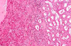 | |
| Micrograph of a renal oncocytoma, a type of benign kidney tumour. H&E stain. | |
| Classification and external resources |
Kidney tumours (or kidney tumors), also known as renal tumours, are tumours, or growths, on or in the kidney. These growths can be benign or malignant (kidney cancer).
Presentation
Kidney tumours may be discovered on medical imaging incidentally (i.e. an incidentaloma), or may be present in patients as an abdominal mass or kidney cyst, hematuria, abdominal pain, or manifest first in a paraneoplastic syndrome that seems unrelated to the kidney.[1]
Workup
A CT scan is the first choice modality for workup of solid masses in the kidneys. Nevertheless, hemorrhagic cysts can resemble renal cell carcinomas on CT, but they are easily distinguished with Doppler ultrasonography (Doppler US). In renal cell carcinomas, Doppler US often shows vessels with high velocities caused by neovascularization and arteriovenous shunting. Some renal cell carcinomas are hypovascular and not distinguishable with Doppler US. Therefore, renal tumors without a Doppler signal, which are not obvious simple cysts on US and CT, should be further investigated with contrast-enhanced ultrasound, as this is more sensitive than both Doppler US and CT for the detection of hypovascular tumors.[2]
Renal ultrasonography
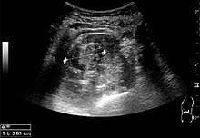

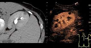
On renal ultrasonography, a solid renal mass appears in the US exam with internal echoes, without the well-defined, smooth walls seen in cysts, often with Doppler signal, and is frequently malignant or has a high malignant potential. The most common malignant renal parenchymal tumor is renal cell carcinoma (RCC), which accounts for 86% of the malignancies in the kidney. RCCs are typically isoechoic and peripherally located in the parenchyma, but can be both hypo- and hyper-echoic and are found centrally in medulla or sinus. The lesions can be multifocal and have cystic elements due to necrosis, calcifications and be multifocal (Figure 8 and Figure 9). RCC is associated with von Hippel–Lindau disease, and with tuberous sclerosis, and US has been recommended as a tool for assessment and follow-up of renal masses in these patients.[2]
Classification
Diagnoses
There are many forms of kidney tumours:
Malignant (cancerous)
- The most frequent, malignant, primary kidney cancer is renal cell carcinoma (RCC) - which has several subtypes:
- Clear cell RCC.
- Papillary RCC
- Chromophobe RCC
- Collecting duct RCC
- Mesoblastic nephroma, a congenital tumor of the kidney's mesenchyme (i.e. connective tissue cells) detected prenatally or, more typically, during the first <4 years of life.[3]
- Metastatic tumour, e.g. ovarian carcinoma.
Benign
Surgical complexity
The RENAL Nephrometry Scoring System is used to measure the complexity of kidney tumors for surgical excision, and is estimated by CT scan as follows:[4]
| Score | |||
|---|---|---|---|
| Component | 1 point | 2 points | 3 points |
| R (radius, maximal diameter) | ≤ 4 cm | > 4 and < 7 cm | ≥ 7 cm |
| E (exophytic/endophytic) | ≥ 50% exophytic | < 50% exophytic | Completely endophytic |
| N (nearness to collecting system/renal sinus) | ≥ 7 mm | > 4 and < 7 mm | ≤ 4 mm |
| A (anterior/posterior locator) | A descriptor of “a,” “p,” or “x” is assigned for tumour location. | ||
| L (location relative to polar lines) | Entirely below lower polar or above upper polar line | Mass crosses polar line | 50% of the mass is across polar line, or mass is entirely between polar lines, or mass crosses axial midline |
A higher score indicates a higher difficulty in removing the tumor surgically, potentially making nephrectomy necessary.
References
- ↑ Gill IS, Aron M, Gervais DA, Jewett MA (February 2010). "Small renal mass". N. Engl. J. Med. 362 (7): 624–34. doi:10.1056/NEJMcp0910041. PMID 20164486.
- 1 2 3 4 Content initially copied from: Hansen, Kristoffer; Nielsen, Michael; Ewertsen, Caroline (2015). "Ultrasonography of the Kidney: A Pictorial Review". Diagnostics. 6 (1): 2. doi:10.3390/diagnostics6010002. ISSN 2075-4418. (CC-BY 4.0)
- ↑ Gooskens SL, Houwing ME, Vujanic GM, Dome JS, Diertens T, Coulomb-l'Herminé A, Godzinski J, Pritchard-Jones K, Graf N, van den Heuvel-Eibrink MM (2017). "Congenital mesoblastic nephroma 50 years after its recognition: A narrative review". Pediatric Blood & Cancer. 64 (7). doi:10.1002/pbc.26437. PMID 28124468.
- ↑ Parsons, Rosaleen B.; Canter, Daniel; Kutikov, Alexander; Uzzo, Robert G. (2012). "RENAL Nephrometry Scoring System: The Radiologist's Perspective". American Journal of Roentgenology. 199 (3): W355–W359. doi:10.2214/AJR.11.8355. ISSN 0361-803X.