KIAA1551
| KIAA1551 | |||||||||||||||||||||||||
|---|---|---|---|---|---|---|---|---|---|---|---|---|---|---|---|---|---|---|---|---|---|---|---|---|---|
| Identifiers | |||||||||||||||||||||||||
| Aliases | KIAA1551, C12orf35, UTA2-1, GET | ||||||||||||||||||||||||
| External IDs | MGI: 1914496 HomoloGene: 19251 GeneCards: KIAA1551 | ||||||||||||||||||||||||
| |||||||||||||||||||||||||
| |||||||||||||||||||||||||
| Orthologs | |||||||||||||||||||||||||
| Species | Human | Mouse | |||||||||||||||||||||||
| Entrez | |||||||||||||||||||||||||
| Ensembl | |||||||||||||||||||||||||
| UniProt | |||||||||||||||||||||||||
| RefSeq (mRNA) | |||||||||||||||||||||||||
| RefSeq (protein) | |||||||||||||||||||||||||
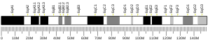
| Location (UCSC) | Chr 12: 31.96 – 31.99 Mb | Chr 6: 149.31 – 149.34 Mb | |||||||||||||||||||||||
| PubMed search | [3] | [4] | |||||||||||||||||||||||
| Wikidata | |||||||||||||||||||||||||
| |||||||||||||||||||||||||


KIAA1551 in Homo sapiens is a novel protein with a function not yet well understood in the scientific community. It is encoded by the KIAA1551 gene. KIAA1551 is broadly expressed in the lymph nodes, ovaries, appendix and spleen.[5] KIAA1551 shows characteristics of being a minor histocompatibility antigen, as well as tumor suppressor capabilities.[6][7] The high epression in the lymph nodes and spleen indicate function in the immune system.
Gene
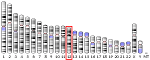
KIAA1551 is a protein coding gene found on Chromosome 12 and maps to 12p11.21.[8] Alternative names for this gene include Gonad Expressed Transcript (GET), UTA2-1 and C12orf35.[5] KIAA1551 has 7 exons, 3 of which occur before the start codon.[5]
Expression
Normal Tissue Expression
A study of normal human tissue expression profiling shows that KIAA1551 is highly expressed in the thymus, spleen, bone marrow and liver.[9] This is interesting as it relates to common organs associated with the Immune system.
Gene tissue expression patterns found through the National Center for Biotechnology Information UniGene EST Profile showed that there was also high expression of KIAA1551 in the lymph nodes, uterus, mouth, thyroid, larynx and blood.[10]
Expression by Health State
An evaluation of KIAA1551 expression in health states was performed using NCBI Unigene’s EST Profile.[11] Although KIAA1551 is highly expressed in uterine tumors, it is also highly expressed in the uterus, suggesting that it is unlikely the gene is associated closely with uterine cancer. However, KIAA1551 may be related to adrenal tumors, as there was lower expression of this gene within normal kidney tissue.
Transcript
Predicted Transcription Factor Binding Sites
Transcription factor binding sites within the promoter of KIAA1551 included mainly transcription factors that were associated with bone marrow cells, antibody- producing cells, and blood cells.[12] This supports the association of KIAA1551 with the functioning immune system.
Protein
KIAA1551 is 1747 amino acids in length and has one domain of unknown function, DUF4617.[13] The Molecular Weight of KIAA1551 is 194.9 kdal.[14] The basal isoelectric point is 8.95.[15] A localization prediction suggests that KIAA1551 is likely a nuclear protein.[15]
Protein Structure
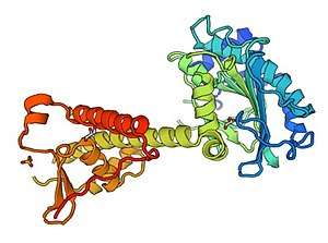
The secondary structure of KIAA1551 consists of mainly random coil structures (approximately 59.2%), few alpha helices (24% of residues) and fewer extended strands (15.8% of residues).[16]
A predicted 3-D structure was created using Swiss model work space, shown above.[17]
Protein Interactions
KIAA1551 interacts with NANOG, MDM2, EXOC1 and CALML3. These interactions further suggest KIAA1551 is a nuclear protein, and that it may be associated with tumor-suppressor proteins and immune system proteins.[18]
EXOC1 was involved in a schizophrenia study, relating a schizophrenia risk gene (DISC1) to a network of protein-protein interactions.[19] This study used a two-hybrid assay as evidence to the protein interaction between KIAA1551 and EXOC1. EXOC1 functions as a response to microbial infections, which reduces viral RNA synthesis and protein translation.[20]
NANOG was predicted to interact with KIAA1551 based on an affinity capture-MS, which linked NANOG to proteins involved with the cell cycle. This study used affinity purification combined with high accuracy mass spectrometry to find specific protein interactions.[21] NANOG was also found to be an essential transcription factor in embryonic stem cells, specifically involved in gene expression to affect cell fate.[22]
MDM2 is a gene that interacts with others to affect the cell cycle and apoptosis, and is located in tissues common to KIAA1551, such as the uterus and lymph node.[23] MDM2 was found to interact with KIAA1551 through the use of a phage display library. This interaction further suggests that KIAA1551 is a nuclear protein, as MDM2 and its splice variants contain nuclear localization signals for nucleoplasmic distribution.[24]
CALML3 was found to interact with KIAA1551 based on affinity capture-MS assay, similar to how NANOG was found to interact with KIAA1551.[25] A study on CALML3 expression in epidermal development showed that CALML3 was useful marker for development, and loss of CALML3 expression correlated with malignant phenotypes.[26]
Evolutionary Relationships
Orthologs
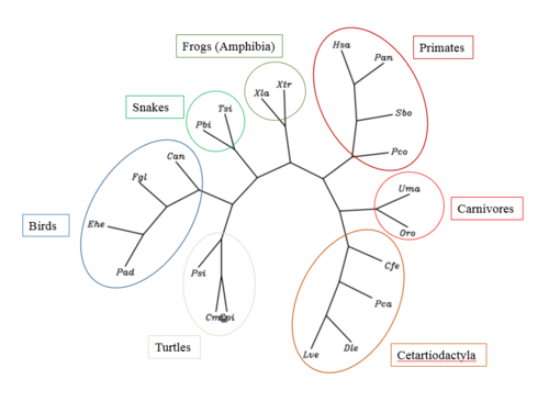
The closest orthologs to KIAA1551 are primates, however, conserved sequences can be found in whales, bears, snakes, birds, turtles, and frogs. Orthologs of KIAA1551 diverged as long ago as 353 million years ago (Xenopus laevis), while the closest evolutionary ortholog is Papio anubis, which diverged approximately 28.1 million years ago.
| Scientific Name | Name | Accession | Sequence Similarity % | Date of Divergence (MYA) |
|---|---|---|---|---|
| Papio anubis | Olive Baboon | XP_003906231.2 | 28.1 | 28.1 |
| Propithecus coquereli | Crowned Sifaka | XP_012496506 | 81 | 73 |
| Physeter Catadon | Sperm whale | XP_007116796.1 | 74 | 94 |
| Delphinapterus leucas | Beluga whale | XP_022433618.1 | 74 | 94 |
| Calypte anna | Anna's Hummingbird | XP_008493940 | 55 | 312 |
| Chrysemys picta belli | Western Painted Turtle | XP_008175485.1 | 43 | 312 |
| Python bivittatus | Burmese Python | XP_007443900.1 | 43 | 312 |
| Xenopus laevis | African Clawed Frog | XP_018107375 | 43 | 353 |
Phylogenetic Tree
An unrooted phylogenetic tree of KIAA1551 was created of 20 orthologs and the human KIAA1551 gene.
Molecular Phylogeny
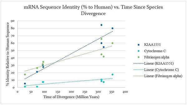
A graph shown below of the molecular evolution of KIAA1551 shows that it evolved relatively quickly compared to both cytochrome C, a slowly evolving protein, and fibrinogen alpha, which evolved more quickly than cytochrome C. The comparison shows that KIAA1551 is fairly quickly diverging, which suggests that it could be a gene that changes quickly in response to its environment, such as the introduction of a pathogen.
References
- 1 2 3 GRCh38: Ensembl release 89: ENSG00000174718 - Ensembl, May 2017
- 1 2 3 GRCm38: Ensembl release 89: ENSMUSG00000032712 - Ensembl, May 2017
- ↑ "Human PubMed Reference:".
- ↑ "Mouse PubMed Reference:".
- 1 2 3 "uncharacterized protein KIAA1551 [Homo sapiens] - Protein - NCBI". www.ncbi.nlm.nih.gov. Retrieved 2018-02-04.
- ↑ Roopenian, D., Choi, E. Y. and Brown, A. (2002), The immunogenomics of minor histocompatibility antigens. Immunological Reviews, 190: 86–94. doi:10.1034/j.1600-065X.2002.19007.x
- ↑ Oostvogels, R et al. (2013). Towards effective and safe immunotherapy after allogeneic stem cell transplantation: identification of hematopoietic-specific minor histocompatibility antigen UTA2-1. Leukemia. 27, 642-649.
- ↑ Gene Cards: KIAA1551 gene. http://www.genecards.org/cgi-bin/carddisp.pl?gene=KIAA1551
- ↑ Itai Yanai, Hila Benjamin, Michael Shmoish, Vered Chalifa-Caspi, Maxim Shklar, Ron Ophir, Arren Bar-Even, Shirley Horn-Saban, Marilyn Safran, Eytan Domany, Doron Lancet, Orit Shmueli (2005). Genome-wide midrange transcription profiles reveal expression level relationships in human tissue specification, Bioinformatics, Volume 21, Issue 5: Pages 650–659.
- ↑ "Home - UniGene - NCBI". www.ncbi.nlm.nih.gov. Retrieved 2018-04-23.
- ↑ "Home - UniGene - NCBI". www.ncbi.nlm.nih.gov. Retrieved 2018-05-06.
- ↑ Genomatix: ElDorado. http://www.genomatix.de/cgi-bin/eldorado/eldorado.pl?s=7f922effeb0f738f6055dd84d1d5ea02 Accessed 4/18/18.
- ↑ NCBI (National Center for Biotechnology Information) Protein entry on KIAA1551. [https://www.ncbi.nlm.nih.gov/protein/NP_060639.3] ]
- ↑ EMBL-EBI. "SAPS < Sequence Statistics < EMBL-EBI". www.ebi.ac.uk. Retrieved 2018-04-23.
- 1 2 "Welcome to psort.org!!". psort.org. Retrieved 2018-04-23.
- ↑ NPS@: Network Protein Sequence Analysis TIBS 2000 March Vol. 25, No 3 [291]:147-150 Combet C., Blanchet C., Geourjon C. and Deléage G.
- ↑ Biasini, M., Bienert, S., Waterhouse, A., Arnold, K., Studer, G., Schmidt, T., Kiefer, F., Cassarino, T.G., Bertoni, M., Bordoli, L., Schwede, T. SWISS-MODEL: modelling protein tertiary and quaternary structure using evolutionary information. Nucleic Acids Res. 42, W252-W258 (2014).
- ↑ KIAA1551. BioGrid 3.4 https://thebiogrid.org/120881/summary/homo-sapiens/exoc1.html?sort=evidence Accessed 4/19/18
- ↑ Camargo, L. M., V. Collura, J.-C. Rain, K. Mizuguchi, H. Hermjakob, S. Kerrien, T. P. Bonnert, P. J. Whiting, and N. J. Brandon. (2007)“Disrupted in schizophrenia 1 interactome: evidence for the close connectivity of risk genes and a potential synaptic basis for schizophrenia.” Molecular Psychiatry 12, no. 1: 74–86.
- ↑ Gene Cards. EXOC1. http://www.genecards.org/cgi-bin/carddisp.pl?gene=EXOC1 Accessed 5/5/18
- ↑ Oliviero G, Munawar N, Watson A, et al. The variant Polycomb Repressor Complex 1 component PCGF1 interacts with a pluripotency sub-network that includes DPPA4, a regulator of embryogenesis. Scientific Reports. 2015;5:18388.
- ↑ Blinka, S., & Rao, S. (2017). Nanog expression in embryonic stem cells – an ideal model system to dissect enhancer function. BioEssays : News and Reviews in Molecular, Cellular and Developmental Biology, 39(12), 10.1002/bies.201700086.
- ↑ Guo, Z., Wang, X., Li, H., & Gao, Y. (2013). Screening E3 substrates using a live phage display library. PLoS ONE, 8(10), e76622.
- ↑ Schuster, Katja, Liying Fan, and Linda C. Harris. “MDM2 splice variants predominantly localize to the nucleoplasm mediated by a COOH-terminal nuclear localization signal.” Molecular Cancer Research : MCR 5, no. 4 (April 2007): 403–12. https://doi.org/10.1158/1541-7786.MCR-06-0146.
- ↑ Huttlin EL, Ting L, Bruckner RJ, et al. The BioPlex Network: A systematic exploration of the human interactome. Cell. 2015;162(2):425-440. doi:10.1016/j.cell.2015.06.043.
- ↑ Bennett, R. D., Pittelkow, M. R., & Strehler, E. E. (2013). Immunolocalization of the tumor-sensitive calmodulin-like protein CALML3 in normal human skin and hyperproliferative skin disorders. PLoS ONE, 8(4), e62347.